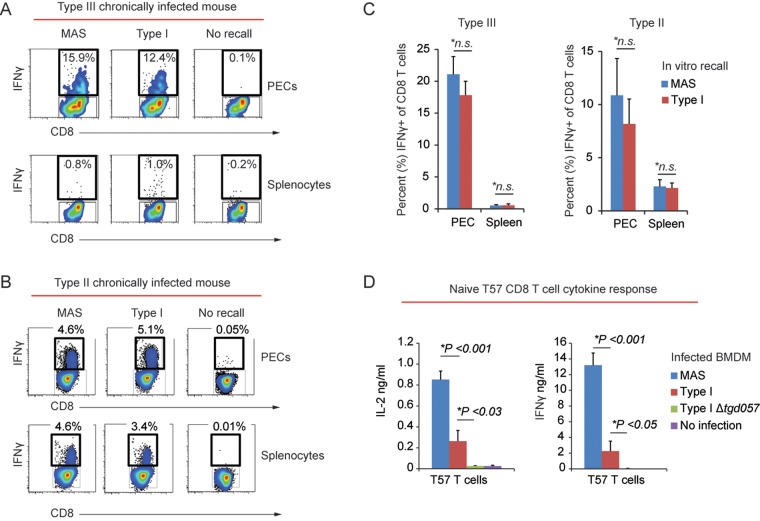
FIG 2 .
The CD8 T cell IFN-γ response to the atypical strain MAS is unimpaired. (A) PECs and splenocytes were obtained from an individual C57BL/6 mouse chronically infected with the type III (CEP) strain and were then infected in vitro (i.e., “recall” infections) with the indicated parasite strain (MOI, 0.2). Sixteen hours later, CD8 T cells were analyzed for intracellular IFN-γ by FACS analysis. The percentage of CD8+ (CD3+ CD19−) T cells that were positive for IFN-γ within the indicated gate following recall infection is shown. (B) Results of an experiment similar to that of panel A, except recall infections were performed using PECs and splenocytes from a type II (Pru) chronically infected mouse. (C) Cumulative data showing the average frequency (+ SEM) of IFN-γ+ CD8+ (CD3+ CD19−) T cells from PECs (3 experiments; n = 7 mice) and spleens (1 experiment; n = 3 mice) from type III (CEP) chronically infected C57BL/6 mice and PECs (2 experiments; n = 5 mice) and spleens (2 experiments; n = 6 mice) from type II (Pru) chronically infected C57BL/6 mice. Average frequencies obtained from recall infections with the type I RH strain and MAS strain are plotted. Significant differences in the average frequencies between RH and MAS recall infections were calculated with Student’s t test; *n.s., not significant (P > 0.05). (D) Naive TGD05796–103-specific T57 transnuclear CD8 T cells (77) were cocultured with BMDMs infected with the indicated parasite strain (MOI, 0.2) in triplicate. The average amounts (+ standard deviation) of IFN-γ and IL-2 detected in the supernatants via an ELISA 48 h later are plotted. *, P < 0.05 (Student’s t test). Results are representative of 5 independent experiments.

