Abstract
Background:
Cancer is one of the major fatal human diseases. Natural products have been used in the treatment of cancer for long time. Bee products including honey and propolis have been introduced for malignancy treatment in recent decades. In this study cytotoxicity of bee products and their effects on the expression of proapoptotic genes have been investigated.
Materials and Methods:
Cytotoxic effects of Astragalus honey, ethanol extract of propolis and a sugar solution (as control) against HepG2, 5637 and L929 cell lines have been evaluated by the MTT assay. Total RNAs of treated cells were isolated and p53 and Bcl-2 gene expression were evaluated, using real-time PCR.
Results:
Propolis IC50 values were 58, 30 and 15 μg/ml against L929, HepG2 and 5637, respectively. These values for honey were 3.1%, 2.4% and 1.9%, respectively. Propolis extract has increased the expression of the Bcl-2 gene in all cell lines whereas the honey decreased that significantly (P < 0.05). Also, we found that honey and propolis decreased p53 gene expression in HepG2 and 5637 significantly but not in L929 cells. The sugar solution increased the expression of p53 in two cancer cell lines but no significant changes were observed in the expression of this gene in L929 as normal mouse cell.
Conclusion:
By downregulation of Bcl-2 expression it could be concluded that the cytotoxicity of honey was more than two fold against tested cancer cells compared with the sugar solution. No significant changes were observed in the expression of p53 in honey-treated cells. Propolis had no significant effect on Bcl-2 and p53 gene expressions (P > 0.05).
Keywords: Apoptosis, honey, MTT assay, propolis, real time PCR
INTRODUCTION
Cancer is one of the major fatal diseases to human. Chemotherapy is the most widely used approaches to cancer treatment. Long-term use of chemotherapy can lead to drug resistance via several different mechanisms, such as gene mutation, DNA methylation and histone modification. These resistance mechanisms have been reported to play important roles in the resistance of patients with cancer to antineoplastic agents including 5-fluorouracil, taxol, doxorubicin, cisplatin, camptothecin and etc.[1,2] Due to this resistance and also severe side effects of synthetic chemotherapeutic agents, it is important to find new natural anticancer agents in order to circumvent or moderate these adverse effects.
Although more than 1400 years ago, Allah in the holly Qur’an said, honey is a “healing for mankind,”[3] one thousand years later in Europe, honey and propolis were accepted as an official drug due to its antibacterial effects.[4] Honey is reported to contain about 181 substances[5] and is considered as part of traditional medicine. Caffeic acid, caffeic acid phenethyl ester and flavonoid glycons are between its most biologically active compounds, providing to have an inhibitory effect on tumor cell proliferation by different mechanisms including protein tyrosine kinase, cyclooxygenase and ornithine decarboxylase pathways.[6] The therapeutic potential of honey is gradually growing and scientific evidences for the effectiveness of honey in several experimental and clinical conditions are beginning to emerge. One of the intrinsic features of honey is its antibiotic properties, where it can be kept for long periods of time without becoming deteriorated. In addition, it has high osmotic pressure which increases the resistance to spoilage by microorganisms.[7] Honey also possessed moderate antitumor and antimetastatic effects in some different strains of rat and mouse tumors.[8] Bee honey has introduced as an inhibitor of the growth of T24, RT4, 253J and MBT-2 bladder cancer cell lines in vitro. It was also effective when administered orally in the MBT-2 bladder cancer implantation models in vivo.[9] Furthermore, honey potentiated the antitumor effects of chemotherapeutics drugs such as 5-FU and cyclophosphamide.[10]
Propolis is a sticky resin and varies in color, including brown, green and red amongst others; based upon the plant exudates that the bees have selectively collected from flower buds, leaf buds and tree barks. These plant resins are mixed with waxes and other bee excretions, including enzymes,[11] to form the final propolis product. Propolis has been extensively used in the traditional medicine for its antimicrobial activities.[12] The chemical composition of each type of propolis and its associated bioactivities mainly depend on the macro- and micro-geographical regions, due to the differences in the plant resin compositions or available plant species,[13,14] and on the bee species, due to the different preference for food and resin plants and for aging distances between bee species.[15] Since honey from different sources and plant species showed different activities e.g. active manuka honey exhibited good antimicrobial effects, in this study the cytotoxicity and cytotoxic mechanisms of Astragalus honey (endemic to central part of Iran in Isfahan province), propolis and a modified sugar solution were evaluated against human bladder cancer cell (5637), hepatic cancer cell (HepG2) and normal mice connective tissue cell (L929). This research was done because honey's composition and effects are different according to its source. On the other hand, to the best of our knowledge the effect of Astragalus honey has not been evaluated against cancer cell lines yet.
MATERIALS AND METHODS
Sample collection
Honey of Astragalus spp. and propolis was collected from Fereydoonshahr, Isfahan province, Iran and was kept in the dark at 4°C until used.
Preparing of ethanolic extract of propolis
Propolis extraction was prepared as reported by Najafi et al.,[16] with slight modifications. Briefly, propolis (10 g) was cut into small pieces and then extracted by 95% (v/v) ethanol (100 ml) at 15°C by shaking at 100 rpm for 24 hours. The suspension was then centrifuged to remove the residual propolis solids. The supernatant was harvested and kept while the pellet was re-extracted and then clarified as above. Two extracts were mixed and evaporated at 60°C. The obtained residue (crude ethanolic extract) was stored in a dark bottle before use.
Sample preparation
Honey was diluted in complete media to give concentrations of 6.25, 5, 4, 3, 2, 1.5, and 0.8% w/v. 50 mg of the ethanolic extract of propolis was dissolved in dimetylsulfoxide (DMSO) (1ml) and diluted with media so that the final concentrations of propolis were 125,100, 60, 50, 40, 30, 20, 15, 12.5, 10, 6.25 μg/ml (DMSO concentration in each well was kept less than 0.5%). The sugar solution was prepared by dissolving a mixture of sugars including glucose, fructose, maltose and sucrose (31%, 39%, 8% and 3%, respectively) in H2O.[17] Prepared syrup was very similar to the honey base and was used as a control. Above-mentioned dilutions were prepared by adding complete media (Roswell park Memorial Institute; RPMI 1640, Invitrogen) and sterilized. Dilute solutions were sterilized by filtration (0.22 μm) and viscose solutions under UV radiation for 20 minutes.
Cell culture
Human hepatic carcinoma cell line (HepG2), human bladder carcinoma (5637) and mice fibroblast normal cell line (L929) were purchased from Pasteur Institute of Iran. All cell lines were cultured in RPMI 1640 (Invitrogen) medium containing fetal bovine serum (FBS; 10% (v/v); Gibco) and 1% antibiotic (penicillin 100 μ/ml and streptomycin 100 μg/ml; PAA, Austria). Cells were incubated at 37°C in a humidified air atmosphere containing 5% CO2.
Cell viability
The trypan blue exclusion assay was used to measure viable cells. Briefly, 200 μl of cell suspension (5 × 10⁴ cell/ml) were mixed with 200 μl of trypan blue solution (Merck, 0.4% w/v) then viable and non-viable cells were counted using an inverted microscope (Ceti; Belgium) and a hemocytometer. Cell suspensions containing more than 85% viable cells were used in this experiment.
Cytotoxicity assay
180 μl of each cell suspension (5 × 10⁴ cells/ml) in complete media were transferred into each well of a U-bottom 96 well microplate (Orange; Belgium) and then incubated at 37°C for 24 hours. Old media were replaced with fresh one containing 20 μl of different concentrations of propolis extract, honey or sugar solution (4 wells for each concentration). 20 μl of doxorubicin solution (EBEWE Pharma, Austria) was added to the wells which assumed as positive control. The plate was incubated for 48 h at the same conditions and cell survival was determined by the MTT (3-(4,5-dimethylthiazol-2-yl)-2,5-diphenyltetrazolium bromide)assay as mentioned before.[18] Percentage of cell survival was calculated using following equation:
% cell survival = [(mean absorbance of treated cells - mean absorbance of blank)/(mean absorbance of control - mean absorbance of blank)] × 100
To determine IC50, concentration–response curves were generated relative to negative control. IC50 values were calculated from the linear portion of the obtained curves.
Total RNA isolation and RT-PCR
Total cellular RNAs were isolated from 106 cells of each cell line using RNase plus mini kit (QIAGEN) according to the manufacturer's protocol. First-strand cDNA was made from total RNA using QuantiTech reverse transcription kit (QIAGEN) with oligo (dT) 15 primers (QIAGEN). Expression of p53, Bcl-2 and glyceraldehyde -3-phosphate dehyrogenase (GAPDH) gene were examined in three cell lines. The primers’ sequences of the genes are as follows:
Bcl-2 forward primer: 5’-ATCAGGAAGGCTAGAGTT-3’ Bcl-2 reverse primer: 5’-TCGGTCTCCTAAAAGCAGGC-3’
GAPDH forward primer: 5’-GAAGGTGAAGGTCGG AGTCA-3’
GAPDH reverse primer: 5’-GAAGATGGTGATGGG ATTTC-3’
p53 forward primer: 5’-TAAGAGTTCCTGCAT GGGCGGC-3’.
p53 reverse primer: 5’-AGGACAGGCACAAACA CGCACC-3’
Real-time PCR
Real-time PCR was performed using the ABI step one system (Applied Biosystem, USA). The SYBR Green method (ABI, U.K) was used for measuring the expression of Bcl-2 and p53 genes. The GAPDH gene was considered as endogenous control. Real-time PCR was performed with the following cycling profile: Initial denaturation at 94°C for 10 min; followed by 50 cycles of 15 sec denaturation at 94°C, annealing at 60°C for 15 sec and extension at 72°C for 20 seconds. The melt curve cycle was: Denaturation step at 94°C for 15 sec, 72°C for 2 min and 94°C for 15 seconds. Analysis of the expression levels of the above-mentioned genes was done using the comparative (ΔΔCT) method.
RESULTS
Antiproliferation studies
Propolis extracts
Propolis was obtained as a sticky brownish to dark brown resin with a distinctive smell. Its cytotoxic effects were evaluated against tested cell lines, using the MTT assay. IC50 values were 58, 30 and 15 μg/ml against L929, HepG2 and 5637, respectively [Figure 1].
Figure 1.
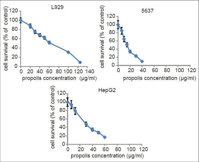
Cytotoxic effects of different concentrations of propolis ethanolic extract on L929, 5637 and HepG2 cell lines. Cell viability was assessed using the MTT assay. Data are presented as mean ± SD of cell survival compared with negative control (cell survival assumed 100%). Significant differences (P < 0.05) were seen in all concentrations ≥ 6 μg/ml (as shown in graphs).
Astragalus honey and sugar solutions
Cytotoxic effects of various concentrations of Astragalus honey were evaluated against tested cell lines after an incubation of 48 hours. IC50s were measured in concentrations containing 3.1%, 2.4%, and 1.9% w/v of honey for L929, 5637 and HepG2, respectively. A sugar solution at different concentrations has also been tested against cells, using the same conditions. Dose–response curves showed that sugars have higher IC50s compared to honey and propolis [Figure 2]. Data showed that bee products exhibited their cytotoxic effects against tested cell lines in a concentration-dependent manner.
Figure 2.
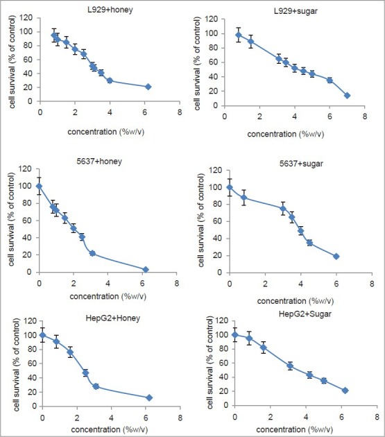
Cytotoxic effects of different concentrations of honey and sugar solution on L929, 5637 and HepG2 cell lines. Cell viability was assessed using the MTT assay. Data are presented as mean ± SD of cell survival compared with negative control (cell survival assumed 100%). In L929 and 5637 treated with honey, significant differences (P < 0.05) were observed for concentrations of ≤ 0.8 % w/v (as seen in graphs). Significant differences were seen when HepG2 exposed to honey (P < 0.05), whereas no significance was observed in the presence of sugar solutions (P ≥ 0.05)
Gene expression profiles
Bcl-2 mRNA expression
HepG2 cells were treated with honey, propolis and sugar and the expression of the Bcl-2 gene were measured. Results showed a significant decrease in Bcl-2 mRNA expression [Figure 3]. In the case of 5637cells, honey and sugar decreased the expression of the Bcl-2 gene significantly while propolis increased Bcl-2 gene expression compared to the untreated cells [Figure 4]. Interestingly when L929 as a normal mouse cell line treated with honey and sugar solution, Bcl-2 gene expression remained unchanged and again propolis-treated cells showed more than seven-fold increase in Bcl-2 gene expression [Figure 5].
Figure 3.
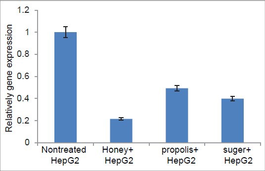
HepG2 Cell exposed to honey, sugar and propolis (see the material and methods section for details)
Figure 4.
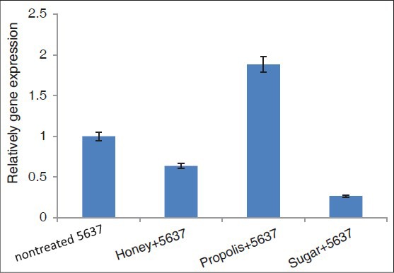
5637 cells treated with honey, propolis and sugar (For detail see material and methods section)
Figure 5.
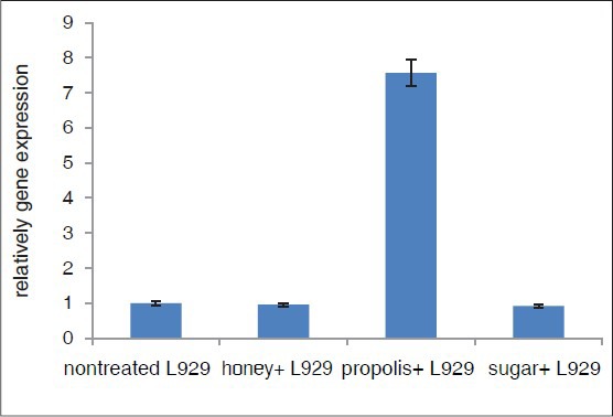
L929 cells treated with honey, propolis and sugar (For detail see material and methods section)
p53 mRNA expression
Expressions of the p53 gene in tested cell lines were measured after exposure to honey, propolis and sugar. Results showed that honey and propolis similarly decreased the expression of the p53 gene in HepG2 cells while sugar increased that more than 2.5-fold compared to untreated cells [Figure 6]. p53 gene expressions in L929 cells were decreased to half by honey and no changes were seen when cells treated with propolis and sugar solution [Figure 7]. 5637 cells showed a different pattern so that honey and propolis decreased the expression of the p53 gene by 80% and 40%, respectively, while the sugar solution increased that by two-fold [Figure 8].
Figure 6.
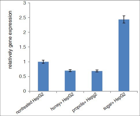
HepG2 cells treated with honey, propolis and sugar solution (For detail see material and methods section)
Figure 7.
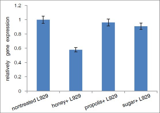
L929 cells treated with honey, propolis and sugar solution (For detail see material and methods section)
Figure 8.
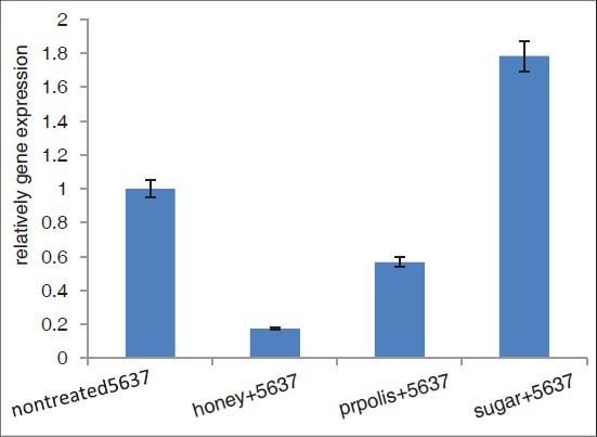
5637cells treated with honey, sugar solutions and propolis (For detail see material and methods section)
DISCUSSION
Honey as a traditional medicine and dietary natural product has recently become the focus of attention in the treatment of certain diseases as well as promoting overall health and well-being. There is strong evidence supporting the positive role of natural food and food product on the induction of apoptosis in different tumor cells.[19] In this regard, we investigated the antiproliferative and apoptotic activities (in gene expression level) of Astragalus honey and propolis on human 5637, HepG2 and mice L929 cell lines and compared with a sugar solution. We found that honey was about two-fold more cytotoxic toward cancer cells than L929 as a normal cell line. The IC50 for honey was 3.12, 2.4 and 1.9% against L929, HepG2 and 5637, respectively. These effects were dose dependent. In the oral health setting, honey has been found to be effective for the treatment of radiation-induced oral mucositis[20] and is also found to be anticarcinogenic,[21] antiproliferative and induces apoptosis in prostate cancer cells.[19] This is consistent with results of the present study. The honey tested in this study has been originally produced in Iran, Isfahan province, and sold as “natural honey of Astragalus” in the local markets. According to our best knowledge, Astragalus honey and its propolis have not been reported to test against cancer cells. While most of the previous studies on honey have focused on its anti-microbial and wound-healing properties, only few studies have investigated the anticarcinogenic properties of honey. Gribel and Pashinskii reported that honey revealed moderate antitumor and pronounced antimetastatic effects. Their results also showed that the antitumor activity of 5-fluorouracil and cyclophosphamide has been increased in combination with honey.[8] Honey was inhibited the growth of bladder cancer cell lines in vitro, and bladder cancer implanted mice models in vivo.[22] The present study showed that unfractionated Astragalus honey and propolis had antiproliferative effects against HepG2, 5637 cancer cells and normal L929 cell line in a time- and concentration-dependent manner. Antiproliferative activity of honey may relate to its low pH (3.2-4.6). It was suggested that the polyphenols found in honey, including Caffeic acid, and its phenyl esters, present in natural honey at the levels of 20-25%, to be promising pharmacological agents in the treatment of cancer by reviewing their antiproliferative and molecular mechanisms.[23] These compounds are thought to exhibit a broad spectrum of activity including tumor inhibition[6] by downregulation of many cellular enzymatic pathway including protein tyrosine kinas cycloxygenase and ornitine decarboxylase pathways.[24] Jungle honey obtained from the tropical forest of Nigeria showed chemotactic activity for neutrophils, which were found to possess potent antitumor activity.[25] Moreover, the expression of various proapoptotic and antiapoptotic proteins was found to be altered during apoptosis.[26] Unfractionated honey was induced cell-growth arrest, resulting in cell cycle blockage at the sub-G1 phase.[27]
Bcl-2 was first identified as an oncogene translocated from chromosome 18 to the heavy chain immunoglobin locus of chromosome 14 in B-cell follicular lymphoma.[28] Overexpression of Bcl-2 inhibits apoptosis induced by a variety of circumstances including growth factor such as tumor necrosis factor Alfa (TNF α) receptor ligation, or by conflicting sub-cellular signaling events.[29] Our study showed that expression of Bcl-2 was decreased in HepG2 and 5637 cancer cell but not in L929 normal cells [Figures 3–5]. This phenomenon may be responsible for the increasing cytotoxicity of cancer cells probably via apoptosis. Recent studies show that Bcl-2 is over expressed in many types of tumors, especially in relapsed or chemo resistant malignancies[30,31] therefore it was an important target for a selective novel cancer therapy.[32] In support of these data, several researchers have shown that honey components can induce apoptosis through a large number of mechanisms, including Bcl-2 regulation.[33]
The p53 (phosphoprotein 53-kDa) is a tumor-suppressor gene located at the short arm of the chromosome 17 and act as a transcription factor. Its important role in tumor development has been attested by numerous reports stating that p53 is the most commonly altered gene in human cancer. More than 50% of human cancers have a defect in the p53 gene.[34] Our data demonstrated that although Bcl-2 expression was decreased significantly in cells treated with honey but the expression of the p53 gene was not upregulated.
It has been already reported that cell cytotoxicity observed by treatment of cells with propolis was not related to gene expression.[35] In this regard we found that gene expression level was not changed significantly in cancer and normal cells while exposed to propolis extract. It seems that some other mechanisms are involved.
CONCLUSION
By downregulation of Bcl-2 expression it could be concluded that the cytotoxicity of honey was more than two-fold against tested cancer cells when compared with a sugar solution. No significant changes were observed in the expression of p53 in honey-treated cells. Propolis had no significant effect on Bcl-2 and p53 gene expressions (P > 0.05).
ACKNOWLEDGEMENTS
The authors would like to thank Mrs. Moazzen for her help and technical support.
Footnotes
Source of Support: Nil
Conflict of Interest: None declared.
REFERENCES
- 1.Ma J, Dong C, Ji C. MicroRNA and drug resistance. Cancer Gene Ther. 2010;17:523–31. doi: 10.1038/cgt.2010.18. [DOI] [PubMed] [Google Scholar]
- 2.Sarkar FH, Banerjee S, Li Y. Pancreatic cancer: Pathogenesis, prevention and treatment. Toxicol Appl Pharmacol. 2007;224:326–36. doi: 10.1016/j.taap.2006.11.007. [DOI] [PMC free article] [PubMed] [Google Scholar]
- 3.The holy Quran. Chapter 16. Surat-al-Nahl, ayat 69 [Google Scholar]
- 4.Castaldo S, Capasso F. Propolis, an old remedy used in modern medicine. Fitoterapia. 2002;73(Suppl 1):S1–6. doi: 10.1016/s0367-326x(02)00185-5. [DOI] [PubMed] [Google Scholar]
- 5.White JW, Crane E. London: Heinemann Publisher; 1979. Honey: A Comprehensive Survey; pp. 157–207. [Google Scholar]
- 6.Rao CV, Desai D, Simi B, Kulkarni N, Amin S, Reddy BS. Inhibitory effect of caffeic acid esters on azoxymethane-induced biochemical changes and aberrant crypt foci formation in rat colon. Cancer Res. 1993;53:4182–8. [PubMed] [Google Scholar]
- 7.Kamran Azim M, Ahmed Mesaik M, Simjee S, Auranzeb M, Nazimuddi, Arshad Saeed S. Medicinal properties of honey from northern Pakistan. Malays J Med Sci. 2007;14:110. [Google Scholar]
- 8.Gribel NV, Pashiniski VG. Antitumor properties of honey. Vopr Onkol. 1990;36:704–9. [PubMed] [Google Scholar]
- 9.Swellam T, Miyanaga N, Onozawa M, Hattori K, Kawai K, Shimazui T, et al. Antineoplastic activity of honey in an experimental bladder cancer implantation model: In vivo and in vitro studies. Int J Urol. 2003;10:213–9. doi: 10.1046/j.0919-8172.2003.00602.x. [DOI] [PubMed] [Google Scholar]
- 10.Wattenberg LW. Chemoprevention of cancer by naturally occurring and synthetic compounds. In: Wattenberg M, Lipkin CW, Boone CW, Kelloff GJ, editors. Cancer Chemoprevention. Boca Raton, Florida: CRC Press; 1992. pp. 19–39. [Google Scholar]
- 11.Bankova VS, De Castro SL, Marcucci MC. Propolis: Recent advances in chemistry and plant origin. Apidologie. 2000;31:3–15. [Google Scholar]
- 12.Banskota AH, Tezuka Y, Kadota S. Recent progress in pharmacological research of propolis. Phytother Res. 2001;15:561–71. doi: 10.1002/ptr.1029. [DOI] [PubMed] [Google Scholar]
- 13.Kucuk M, Kolayli S, Karaoglu S, Ulusoy E, Baltaci C, Candan F. Biological activities and chemical composition of three honeys of different types from Anatolia. Food Chem. 2007;100:526–34. [Google Scholar]
- 14.Silici S, Kutluca S. Chemical composition, antibacterial activity of propolis collected by three different races of honeybees in the same region. J Ethnopharmacol. 2005;99:69–73. doi: 10.1016/j.jep.2005.01.046. [DOI] [PubMed] [Google Scholar]
- 15.Pratsinis H, Kletsas D, Melliou E, Chinou I. Antiproliferative activity of Greek propolis. J Med Food. 2010;13:286–90. doi: 10.1089/jmf.2009.0071. [DOI] [PubMed] [Google Scholar]
- 16.Najafi MF, Vahedy F, Seyyedin M, Jomehzadeh HR, Bozary K. Effect of the water extracts of propolis on stimulation and inhibition of different cells. Cytotechnology. 2007;54:49–56. doi: 10.1007/s10616-007-9067-2. [DOI] [PMC free article] [PubMed] [Google Scholar]
- 17.Bera A, Almeida M, Sabato SF. Effect of gamma radiation on honey quality control. Radiat Phys Chem. 2009;78:583–4. [Google Scholar]
- 18.Sadeghi-aliabadi H, Aliasgharluo M, Mirian M, Ghanadian M. In vitro cytotoxic evaluation of some synthesized COX-2 inhibitor derivatives against a panel of human cancer cell lines. Res Pharm Sci. 2013;8:299–304. [PMC free article] [PubMed] [Google Scholar]
- 19.Samarghandian S, Afshari JT, Davoodi S. Chrysin reduces proliferation and induces apoptosis in human prostate cancer cell line PC3. Clinics (Sao Paulo) 2011;66:1073–9. doi: 10.1590/S1807-59322011000600026. [DOI] [PMC free article] [PubMed] [Google Scholar]
- 20.Biswal BM, Zakaria A, Ahmad NM. Topical application of honey in the management of radiation mucositis. a preliminary study. Support Care Cancer. 2003;11:242–8. doi: 10.1007/s00520-003-0443-y. [DOI] [PubMed] [Google Scholar]
- 21.Sela M, Maroz D, Gedalia I. Streptococcus mutans in saliva of normal subjects and neck and head irradiated cancer subjects after consumption of honey. J Oral Rehabil. 2000;27:269–70. doi: 10.1046/j.1365-2842.2000.00504.x. [DOI] [PubMed] [Google Scholar]
- 22.Swellam T, Miyanaga N, Onozawa M, Hattori K, Kawai K, Shimazui T, et al. Antineoplastic activity of honey in an experimental bladder cancer implantation model. In vivo and in vitro studies. Int J Urol. 2003;10:213–9. doi: 10.1046/j.0919-8172.2003.00602.x. [DOI] [PubMed] [Google Scholar]
- 23.Jaganathan SK, Mandal M. Antiproliferative effects of honey and of its polyphenols. A review. J Biomed Biotechnol 2009. 2009 doi: 10.1155/2009/830616. 830616. [DOI] [PMC free article] [PubMed] [Google Scholar]
- 24.Rao CV, Desai D, Simi B, Kulkarni N, Amin S, Reddy BS. Inhibitory effect of caffeic acid esters on azoxymethane-induced biochemical changes and aberrant crypt foci formation in rat colon. Cancer Res. 1993;53:4182–8. [PubMed] [Google Scholar]
- 25.Fukuda M, Kobayashi K, Hirono Y, Miyagawa M, Ishida T, Ejiogu EC, et al. Jungle Honey Enhances Immune Function and Antitumor Activity. Evid Based Complement Alternat Med 2011. 2011 doi: 10.1093/ecam/nen086. 908743. [DOI] [PMC free article] [PubMed] [Google Scholar]
- 26.Ghashm AA, Othman NH, Khattak MN, Ismail NM, Saini R. Antiproliferative effect of Tualang honey on oral squamous cell carcinoma and osteosarcoma cell lines. BMC Complement Altern Med. 2010;10:49. doi: 10.1186/1472-6882-10-49. [DOI] [PMC free article] [PubMed] [Google Scholar]
- 27.Jaganathan SK, Mandal M. Involvement of non-protein thiols, mitochondrial dysfunction, reactive oxygen species and p53 in honey induced apoptosis. Invest New Drugs. 2010;28:624–33. doi: 10.1007/s10637-009-9302-0. [DOI] [PubMed] [Google Scholar]
- 28.Tsujimoto Y, Croce CM. Analysis of the structure, transcripts, and protein products of Bcl-2, the gene involved in human follicular lymphoma. Proc Natl Acad Sci U S A. 1986;83:5214–8. doi: 10.1073/pnas.83.14.5214. [DOI] [PMC free article] [PubMed] [Google Scholar]
- 29.Yang E, Korsmeyer SJ. Molecular thanatopsis: A discourse on the Bcl-2 family and cell death. Blood. 1996;88:386–401. [PubMed] [Google Scholar]
- 30.Reed JC, Miyashita T, Krajewski S, Takayama S, Aime-Sempe C, Kitada S, et al. Bcl-2 family proteins and the regulation of programmed cell death in leukemia and lymphoma. Cancer Treat Res. 1996;84:31–72. doi: 10.1007/978-1-4613-1261-1_3. [DOI] [PubMed] [Google Scholar]
- 31.Borner MM, Brousset P, Pfanner-Meyer B, Bacchi M, Vonlanthen S, Hotz MA, et al. Expression of apoptosis regulatory proteins of the Bcl-2 family and p53 in primary resected non-small-cell lung cancer. Br J Cancer. 1999;79:952–8. doi: 10.1038/sj.bjc.6690152. [DOI] [PMC free article] [PubMed] [Google Scholar]
- 32.An J, Chen Y, Huang Z. Critical upstream signals of cytochrome C release induced by a novel Bcl-2 inhibitor. J Biol Chem. 2004;279:19133–40. doi: 10.1074/jbc.M400295200. [DOI] [PubMed] [Google Scholar]
- 33.Lin JG, Chen GW, Li TM, Chouh ST, Tan TW, Chung JG. Aloe-emodin induces apoptosis in T24 human bladder cancer cells through the p53 dependent apoptotic pathway. J Urol. 2006;175:343–7. doi: 10.1016/S0022-5347(05)00005-4. [DOI] [PubMed] [Google Scholar]
- 34.Bai L, Zhu WG. p53: Structure, Function and therapeutic applications. J Cancer Mol. 2006;2:141–53. [Google Scholar]
- 35.Supawadee U, Preecha Ph, Songchan P, Chanpen CH. In vitro antiproliferative activity of partially purified Trigona laeviceps propolis from Thailand on human cancer cell lines. BMC Complement Altern Med. 2011;11:37. doi: 10.1186/1472-6882-11-37. [DOI] [PMC free article] [PubMed] [Google Scholar]


