Abstract
Context:
Glandular odontogenic cyst (GOC) is a rare cyst occurring in the middle-age people with mandibular anterior as the common site of occurrence.
Case Report:
We report a case of massive GOC in a 65-year-old female with an emphasis on its clinical course, histological features, and treatment modalities.
Conclusion:
The aggressiveness and recurrences of GOC warrants clinicians for the careful examination, treatment, and long-term follow-up.
Keywords: Aggressive, Anterior, Glandular, Mandibular, Odontogenic
Introduction
Glandular odontogenic cyst (GOC) is an uncommon developmental cyst.[1] Padayachee and Van Wykin 1987,[2] reported the first two cases of this cyst as a “sialo-odontogenic cyst”. Later Gardner et al.,[3] in 1988 established this cyst as a distinct entity under the term “glandular odontogenic cyst”. The GOC is included in the World Health Organization (WHO) histology typing of odontogenic tumors under the terms “glandular odontogenic cyst or sialo-odontogenic cyst”. The term “polymorphous odontogenic cyst” was introduced by High et al., in 1996 because of its varied histological appearances.[4]
GOCs are rare, with just over 111 cases reported in the English language literature.[5] Between 1977 and 1995, Magnusson et al., analyzed 5,800 biopsies of jaw cysts and observed that only seven cases fulfilled the criteria for GOC, which comprised 0.012% of the total.[6,7] Later, Shen et al., (2006) found a similar frequency of 12 cases in a series of 7,023 jaw cysts (0.17%) over a 37-year period and Jones et al., (2006) listed 11 cases (0.2%) in their sample of 7,121 odontogenic cysts over a 30-year period.[8]
GOC predominantly occurs in the anterior mandible of middle-aged patients with slight male predilection. It can be asymptomatic or may cause pain, slow-growing swelling leading to tooth displacement.[9,10] Radiographic appearance shows a well-defined unilocular or multilocular radiolucency, often with scalloped margins.[11] The microscopic features of GOC, particularly the morphology of the epithelium, strongly suggest an origin from the remains of dental laminawhich showspapillary projections and focal thickenings (plaques) within it along withmucous cells, interepithelial gland-like structures, and lack of inflammation in the connective tissue.[2,3,11] Treatment of GOC is still controversial and varies from curettage, enucleation to wide surgical resection; but due to the tendency to recur after curettage or enucleation, most of the authors prefer marginal resection.[12]
In the present study, a case of GOC is reported, emphasizing the origin, epidemiology, clinical, radiographic, and histopathological aspects along with a note on its treatment, potential for recurrence, and the importance of long-term follow-up.
Case Presentation
A 65-year-old female reported to the Department of Oral and Maxillofacial Surgery with the chief complaint of pain due to decayed tooth in right lower back region since 2 months. The intraoral examination revealed partial edentulous lower jaw with 33, 34, 35, 36, 37, 43, 44, and 45 teeth present. Careful examination revealed swelling in the anterior mandible, which was not obvious for the patient to take treatment. The swelling was present from past 9 years and was asymptomatic with slow growth. There was no associated history of trauma. The anterior teeth 31, 32, 41, and 42 were extracted 4 years back by the dental surgeon due to loosening [Figure 1a and b]. Patient was not advised for the intraoral radiographs before extraction. Patient was wearing removable partial denture in the lower anterior mandible, which impinged the underlying alveolar mucosa and appeared erythematous.
Figure 1.
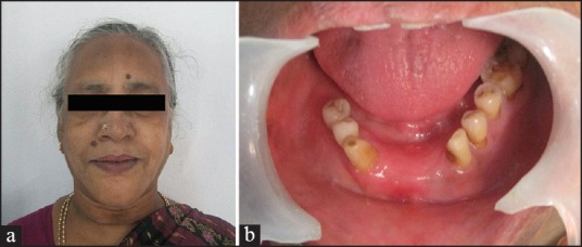
(a) Frontal profile of the patient. (b) Intraoral photograph
The medical history of the patient revealed her to be hypertensive and diabetic from past 2 years and is under medication for the same. The swelling appeared hard without any change in the color of overlying mucosa except in denture region. The extraoral examination showed mild swelling of the lower anterior mandible. The submandibular glands were palpable, nontender but hard, and appeared fixed to the underlying structures.
Introral periapical radiograph and panoramic radiographs were advised, which revealed well-defined lytic multilocular radiolucency involving the whole of the anterior mandible with its extension in the anteroposterior direction involving right and left body of the mandible [Figures 2 and 3]. Based on the clinical and radiographic finding, a differential diagnosis of odontogenic keratocyst and ameloblastoma were made. Fine-needle aspiration cytology (FNAC) was advised, which revealed only blood cells. Incisional biopsy was carried out, which on gross examination appeared as blood clots; but on histopathology, it showed ciliated cells with intraepithelial microcyst formation and mucous cells with periodic acid-Schiff (PAS) positive material. The connective tissue was filled with blood cells and few inflammatory cells [Figure 4]. The diagnosis of GOC was made.
Figure 2.
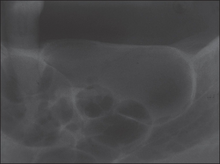
Intraoral periapical radiograph showing honey-combed appearance
Figure 3.
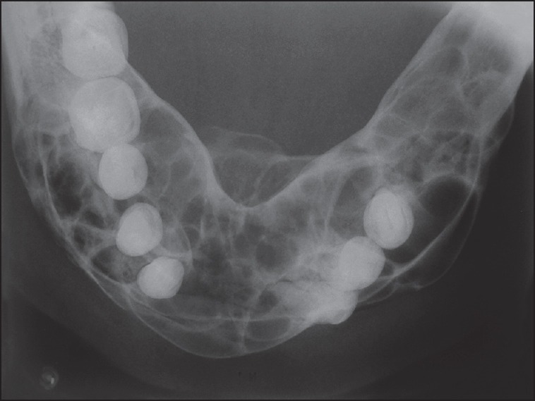
Occlusal radiograph showing multilocular lesion involving the body of the mandible
Figure 4.
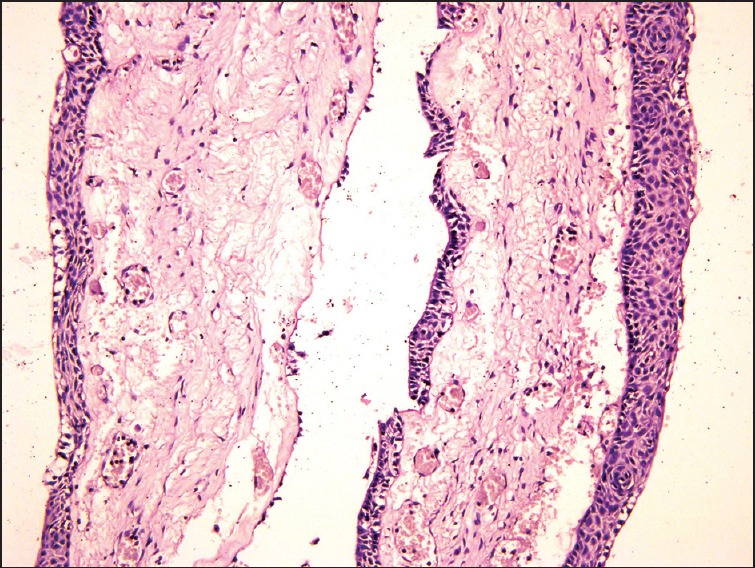
Hematoxylin and eosin (H and E) stained section showing ciliated cells with intraepithelial microcyst and mucous cells
The patient was explained about the lesion, the surgical treatment plan, and informed consent was taken. Segmental resection of the mandible was advised, which was carried out under general anesthesia. A bilateral extended submandibular incision was made and the mandible was exposed [Figure 5]. The osteotomy cuts were performed from right antegonial to left antegonial region including the surgical margin of 1cm. The submandibular lymph nodes were also removed. The angle of the mandible was spared along with the ramus andcondyle. Reconstruction plate was adapted along with the curvature of mandible across the midline and fixed to the remaining mandible [Figure 6]. Closure of the surgical site was done in layers. Postoperative healing was good with no complications except for the paresthesia. The patient is kept under long-term follow-up and no recurrence was observed in 2 years of duration.
Figure 5.
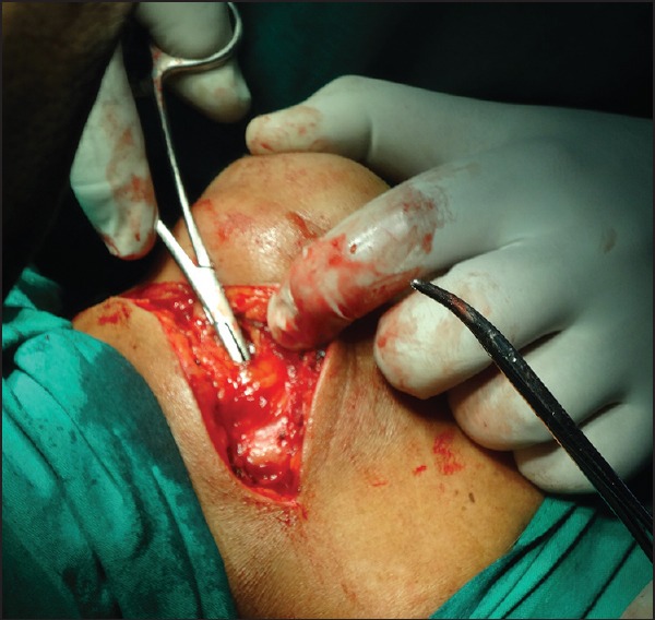
Submandibular incision
Figure 6.
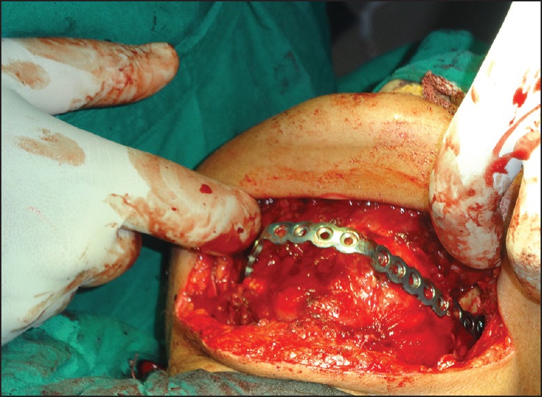
Reconstruction plate
The excisional biopsy of the mandible was sent for the confirmatory histopathological diagnosis and margin clearance. The final diagnosis was in correlation with the incisional diagnosis and the margins were also free of the lesion.
Discussion
GOC is a very rare pathology and has so far been documented just over 111 cases. The clinical aspects of the present case are consistent with those reported for GOC in the literature.[5,6] There is a slight predilection for men and the condition occurs mostly in middle-aged patients, but the present case reported is in female patient of elderly age. The anterior mandible is the most common site of occurrence; however in the current case report, the lesion extended to the right and left molars. The pain reported by the patient was probably due to the inflammation because of impingement and pressure by the removable partial denture in anterior region.
GOC does not display specific or pathognomonic radiographical features. It may present as a multilocular or unilocular radiolucency with well-defined borders. The recognition of this cyst on the base of clinical and radiological examination is practically impossible. Only the histopathological examination allow for certain diagnosis of the cyst.[5] GOC is an intraosseous developmental cyst which may occur in the apical region of the tooth and may show interdental extension. On the basis of clinical and radiographic examination, the diagnosis of the dentigerous cyst, odontogenic keratocyst cyst, radicular cyst, myxoma, ameloblastoma, central giant cell lesion, fibrous dysplasia, and central mucoepidermoid carcinoma (MEC) can be made.
The aspirated fluid in the present case showed compatibility with the peripheral blood, perhaps due to inflammation. Literature suggests the low viscous, colorless fluid content may indicate clinically GOC.[6] The characteristic feature of GOC histopathologically strongly suggests its origin from the dental lamina. The study reported by Koppang and Matthews and a case report by Ide et al., suggested the odontogenic nature of GOC.[13,14]
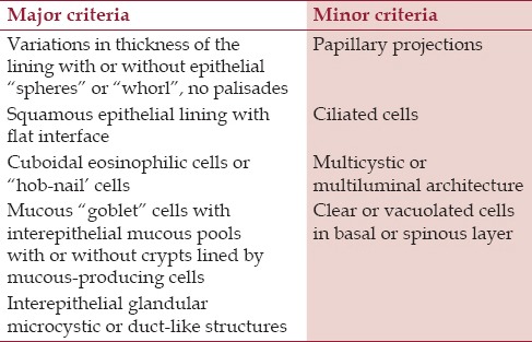
The microscopic features of the GOC shares its similarity with the lateral periodontal cyst (LPC), botroid odontogenic cyst (BOC), and the central MEC. The plaque-like epithelial thickening in LPC made authors to believe that the GOC could be the clinical microscopic variant of LPC.[15] But the low aggressive nature, limited growth potential and low recurrence nature of LPC has annulled GOC. The multilocularity, multicystic, and GOC was considered to be the variant of BOC. Some authors contradict this statement due to the presence of mucous and ciliated epithelial cells and mucous pooled cystic spaces in GOC and not in BOC, thereby considering them as different entities.[16] It is mandatory to differentiate the GOC from the central variant (low grade cystic type) of MEC as the later does not always support its intrabony salivary gland origin, but can originate from pluripotent cells of oral epithelium or due to metaplastic change in the odontogenic epithelium. Magnusson et al.,[6] considered GOC the most benign end of the spectrum of central MEC. Many authors follow the major and minor criteria suggested by Kaplan and Salehinejad et al., for the diagnosis of GOC [Table 1]. The distinction between GOC and MEC were established based on immunohistochemical studies using cytokeratin (Ck). Ck 7, 13, 14, and 19 positivity and negative reaction for epithelial membrane antigen (EMA) supported odontogenic origin. Some authors suggested expression of Ck 8, 18, and 19 might be useful tool in differentiating these lesions.[6,16]
Table 1.
Major and minor criteria for diagnosing GOC
The unpredictable, aggressive behavior and high recurrence rate of GOC has led different treatment modalities such asconservative approach including enucleation, marsupialization, curettage with or without peripheral ostectomy, curettage with adjuvant Carnoy's solution, or cryotherapy, to marginal resection and segmental resection. Small unilocular lesions can be treated by enucleation whereas the large lesions should include enucleation with peripheral ostectomy for unilocular cases and marginal resection or partial jaw resection in multilocular cases. Marsupialization followed by second phase surgery is an option for lesions approaching vital structures.[5,17,18] Kaplan et al., also demonstrated that the rate of recurrence also increases with the radiographic complexity of the cyst. Hussain et al., found a high recurrence rate of 55% and a long recurrence interval, with an average of 4.9 years; whereas, Koppang et al., observed a recurrence rate of 21%.[19,20,21] Considering the age, recurrence rate and pooreconomic status microvascular reconstruction was ruled out. Long-term follow-up is recommended ranging with a minimum of 3-5 years after treatment and the prognosis of GOC remains good.[6,13]
The author concludes by stressing on the rarity of the GOC, with only 65 cases being reported in the English language literature so far. Careful clinical and radiological examination along with the confirmatory histopathological study is required to differentiate GOC from the differential diagnosis of central mucoepidermoid carcinoma.
Footnotes
Source of Support: Nil.
Conflict of Interest: None declared.
References
- 1.Krishnamurthy AJ, Sherlin HJ, Ramalingam K, Natesan A, Premkumar P, Ramani P, et al. Glandular odontogenic cyst: Report of two cases and review of literature. Head Neck Pathol. 2009;3:153–8. doi: 10.1007/s12105-009-0117-2. [DOI] [PMC free article] [PubMed] [Google Scholar]
- 2.Padayachee A, Van Wyk CW. Two cystic lesions with features of both botryoid odontogenic cyst and the central mucoepidermoid tumour: Sialo-odontogenic cyst? J Oral Pathol. 1987;16:499–504. doi: 10.1111/j.1600-0714.1987.tb00680.x. [DOI] [PubMed] [Google Scholar]
- 3.Gardner DG, Kessler HP, Morency R, Schaner DL. The glandular odontogenic cyst: An apparent entity. J Oral Pathol. 1988;17:359–66. doi: 10.1111/j.1600-0714.1988.tb01298.x. [DOI] [PubMed] [Google Scholar]
- 4.High AS, Main DM, Khoo SP, Pedlar J, Hume WJ. The polymorphous odontogenic cysts. J Oral Pathol Med. 1996;25:25–31. doi: 10.1111/j.1600-0714.1996.tb01219.x. [DOI] [PubMed] [Google Scholar]
- 5.Shah M, Kale H, Ranginwala A, Patel G. Glandular odontogenic cyst: A rare entity. J Oral Maxillofac Pathol. 2014;18:89–92. doi: 10.4103/0973-029X.131922. [DOI] [PMC free article] [PubMed] [Google Scholar]
- 6.Magnusson B, Goransson L, Ödesjö B, Gröndahl K, Hirsch JM. Glandular odontogenic cyst. Report of seven eases. Dentomaxillofac Radiol. 1997;26:26–31. doi: 10.1038/sj.dmfr.4600205. [DOI] [PubMed] [Google Scholar]
- 7.Osny FJ, Azevedo LR, Sant›Ana E, Lara VS. Glandular odontogenic cyst: Case report and review of literature. Quintessence Int. 2004;35:385–9. [PubMed] [Google Scholar]
- 8.Shear M, Speight PM. 4th ed. Ch. 7. Oxford: Blackwell Publishing Ltd; 2007. Textbook of cyst of oral and maxillofacial region; pp. 94–9. [Google Scholar]
- 9.Manor R, Anavi Y, Kaplan I, Calderon S. Radiological features of glandular odontogenic cyst. Dentomaxillofac Radiol. 2003;32:73–9. doi: 10.1259/dmfr/22912856. [DOI] [PubMed] [Google Scholar]
- 10.Thor A, Warfvinge G, Fernandes R. The course of a long-standing glandular odontogenic cyst: Marginal resection and reconstruction with particulated bone graft, platelet-rich plasma, and additional vertical alveolar distraction. J Oral Maxillofac Surg. 2006;64:1121–8. doi: 10.1016/j.joms.2006.03.042. [DOI] [PubMed] [Google Scholar]
- 11.Nair RG, Varghese IV, Shameena PM, Sudha S. Glandular odontogenic cyst: Report of a case and review of literature. J Oral Maxillofac Pathol. 2006;10:20–3. [Google Scholar]
- 12.Kasaboglu O, Basal Z, Usubutun A. Glandular odontogenic cyst presenting as a dentigerous cyst: A case report. J Oral Maxillofac Surg. 2006;64:731–3. doi: 10.1016/j.joms.2005.12.020. [DOI] [PubMed] [Google Scholar]
- 13.Toida M, Nakashima E, Okumura Y, Tatematsu N. Glandular odontogenic cyst: A case report and literature review. J Oral Maxillofac Surg. 1994;52:1312–6. doi: 10.1016/0278-2391(94)90054-x. [DOI] [PubMed] [Google Scholar]
- 14.Ide F, Shimoyama T, Horei N. Glandular odontogenic cyst with hyaline bodies: An unusual dentigerous presentations. J Oral Pathol Med. 1996;25:401–4. doi: 10.1111/j.1600-0714.1996.tb00286.x. [DOI] [PubMed] [Google Scholar]
- 15.Takeda Y. Glandular odontogenic cyst mimicking a lateral periodontal cyst: A case report. Int J Oral Maxillofac Surg. 1994;23:96–7. doi: 10.1016/s0901-5027(05)80600-9. [DOI] [PubMed] [Google Scholar]
- 16.Lin CC, Chen CH, Lei S, Chen YK, Wan WC, Lu SY, et al. Glandula odontogenic cyst: A case report. Kaohsiung J Med Sci. 2000;16:53–8. [PubMed] [Google Scholar]
- 17.Tambawala SS, Karjodkar FR, Yadav A, Sansare K, Sonkakke S. Glandular odontogenic Cyst: A case report. Imaging Sci Dent. 2014;44:75–9. doi: 10.5624/isd.2014.44.1.75. [DOI] [PMC free article] [PubMed] [Google Scholar]
- 18.Mascitti M, Santarelli A, Sabatucci A, Procaccini M, Muziol L, Zizzi A, et al. Glandular odontogenic cyst: Review of literature and report of a new case with cytokeratin-19 expression. Open Dent J. 2014;8:1–12. doi: 10.2174/1874210601408010001. [DOI] [PMC free article] [PubMed] [Google Scholar]
- 19.Koppang HS, Johannessen S, Haugen LK, Haanaes HR, Solheim T, Donath K. Glandular odontogenic cyst (sialoodontogenic cyst): Report of two cases and literature review of 45 previously reported cases. J Oral Pathol Med. 1998;27:455–62. doi: 10.1111/j.1600-0714.1998.tb01984.x. [DOI] [PubMed] [Google Scholar]
- 20.Hussain K, Edmondson HD, Browne RM. Glandular odontogenic cysts. Diagnosis and treatment. Oral Surg Oral Med Oral Pathol Oral Radiol Endod. 1995;79:593–602. doi: 10.1016/s1079-2104(05)80101-5. [DOI] [PubMed] [Google Scholar]
- 21.Kaplan I, Anavi Y, Manor R, Sulkes J, Calderon S. The use of molecular markers as an aid in the diagnosis of glandular odontogenic cyst. Oral Oncol. 2005;41:895–902. doi: 10.1016/j.oraloncology.2005.04.015. [DOI] [PubMed] [Google Scholar]


