

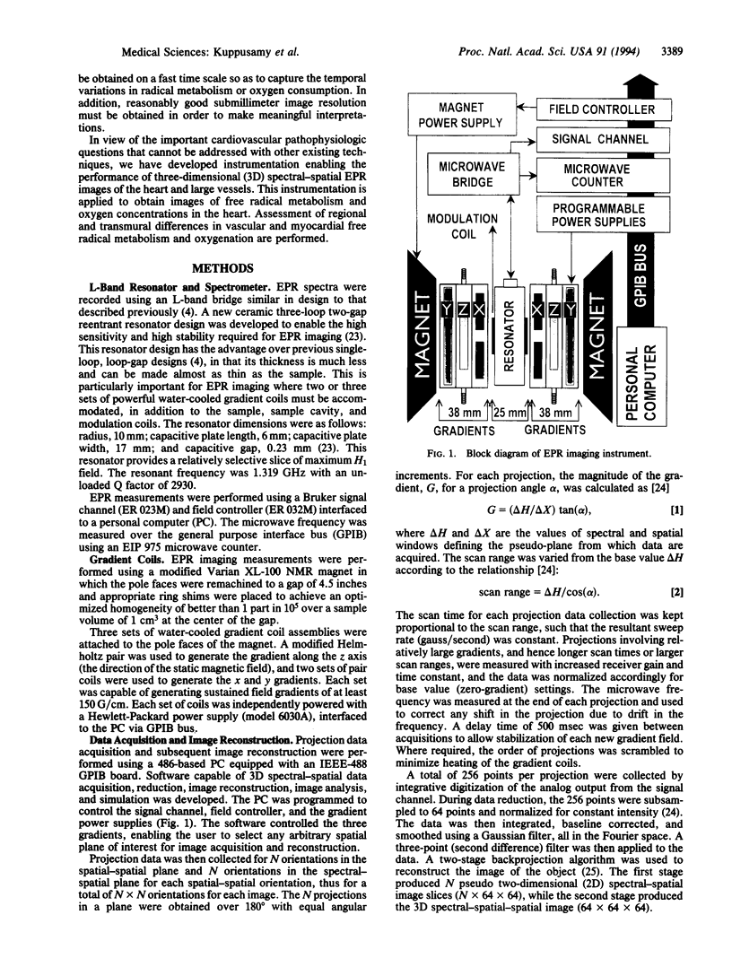
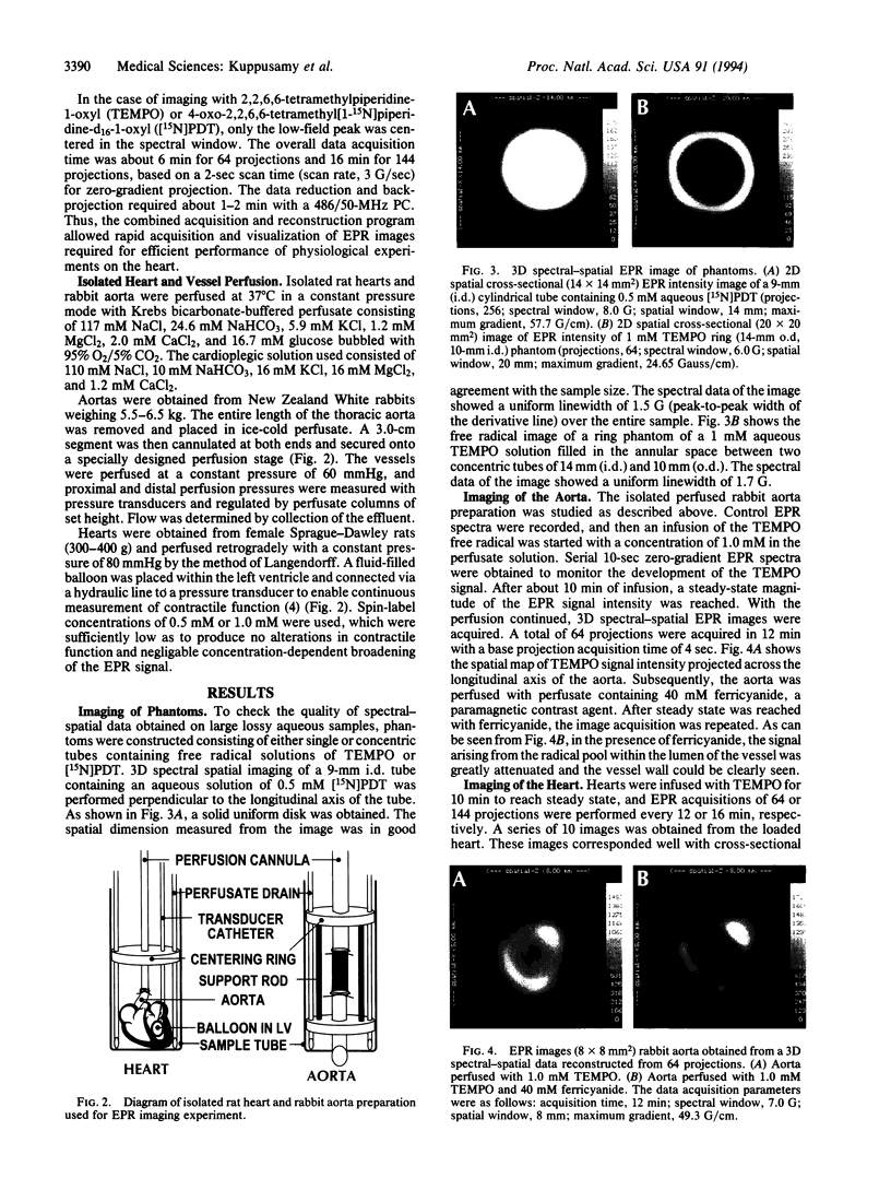
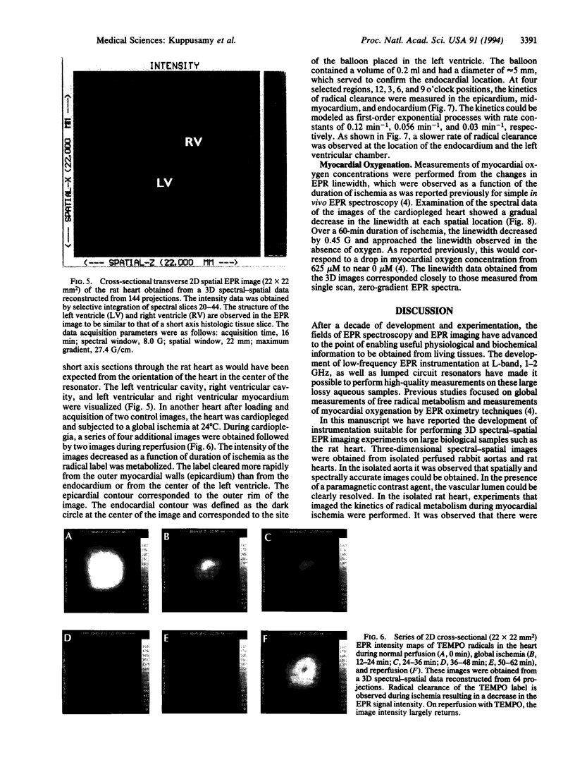
Abstract
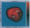
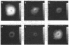
It has been hypothesized that free radical metabolism and oxygenation in living organs and tissues such as the heart may vary over the spatially defined tissue structure. In an effort to study these spatially defined differences, we have developed electron paramagnetic resonance imaging instrumentation enabling the performance of three-dimensional spectral-spatial images of free radicals infused into the heart and large vessels. Using this instrumentation, high-quality three-dimensional spectral-spatial images of isolated perfused rat hearts and rabbit aortas are obtained. In the isolated aorta, it is shown that spatially and spectrally accurate images of the vessel lumen and wall could be obtained in this living vascular tissue. In the isolated rat heart, imaging experiments were performed to determine the kinetics of radical clearance at different spatial locations within the heart during myocardial ischemia. The kinetic data show the existence of regional and transmural differences in myocardial free radical clearance. It is further demonstrated that EPR imaging can be used to noninvasively measure spatially localized oxygen concentrations in the heart. Thus, the technique of spectral-spatial EPR imaging is shown to be a powerful tool in providing spatial information regarding the free radical distribution, metabolism, and tissue oxygenation in living biological organs and tissues.
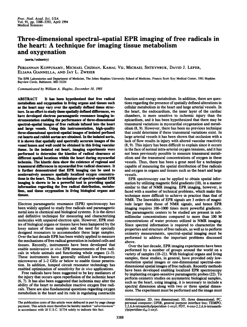
Full text
PDF
Images in this article
Selected References
These references are in PubMed. This may not be the complete list of references from this article.
- Alecci M., Colacicchi S., Indovina P. L., Momo F., Pavone P., Sotgiu A. Three-dimensional in vivo ESR imaging in rats. Magn Reson Imaging. 1990;8(1):59–63. doi: 10.1016/0730-725x(90)90213-l. [DOI] [PubMed] [Google Scholar]
- Berliner J. L., Fujii H. Magnetic resonance imaging of biological specimens by electron paramagnetic resonance of nitroxide spin labels. Science. 1985 Feb 1;227(4686):517–519. doi: 10.1126/science.2981437. [DOI] [PubMed] [Google Scholar]
- Berliner L. J., Fujii H., Wan X. M., Lukiewicz S. J. Feasibility study of imaging a living murine tumor by electron paramagnetic resonance. Magn Reson Med. 1987 Apr;4(4):380–384. doi: 10.1002/mrm.1910040410. [DOI] [PubMed] [Google Scholar]
- Dobrucki J. W., Sutherland R. M., Swartz H. M. Nonperturbing test for cytotoxicity in isolated cells and spheroids, using electron paramagnetic resonance. Magn Reson Med. 1991 May;19(1):42–55. doi: 10.1002/mrm.1910190105. [DOI] [PubMed] [Google Scholar]
- Quaresima V., Alecci M., Ferrari M., Sotgiu A. Whole rat electron paramagnetic resonance imaging of a nitroxide free radical by a radio frequency (280 MHz) spectrometer. Biochem Biophys Res Commun. 1992 Mar 16;183(2):829–835. doi: 10.1016/0006-291x(92)90558-3. [DOI] [PubMed] [Google Scholar]
- Reimer K. A., Jennings R. B. The "wavefront phenomenon" of myocardial ischemic cell death. II. Transmural progression of necrosis within the framework of ischemic bed size (myocardium at risk) and collateral flow. Lab Invest. 1979 Jun;40(6):633–644. [PubMed] [Google Scholar]
- Reimer K. A., Lowe J. E., Rasmussen M. M., Jennings R. B. The wavefront phenomenon of ischemic cell death. 1. Myocardial infarct size vs duration of coronary occlusion in dogs. Circulation. 1977 Nov;56(5):786–794. doi: 10.1161/01.cir.56.5.786. [DOI] [PubMed] [Google Scholar]
- Takeshita K., Utsumi H., Hamada A. ESR measurement of radical clearance in lung of whole mouse. Biochem Biophys Res Commun. 1991 Jun 14;177(2):874–880. doi: 10.1016/0006-291x(91)91871-9. [DOI] [PubMed] [Google Scholar]
- Zweier J. L., Kuppusamy P. Electron paramagnetic resonance measurements of free radicals in the intact beating heart: a technique for detection and characterization of free radicals in whole biological tissues. Proc Natl Acad Sci U S A. 1988 Aug;85(15):5703–5707. doi: 10.1073/pnas.85.15.5703. [DOI] [PMC free article] [PubMed] [Google Scholar]
- Zweier J. L., Kuppusamy P., Lutty G. A. Measurement of endothelial cell free radical generation: evidence for a central mechanism of free radical injury in postischemic tissues. Proc Natl Acad Sci U S A. 1988 Jun;85(11):4046–4050. doi: 10.1073/pnas.85.11.4046. [DOI] [PMC free article] [PubMed] [Google Scholar]
- Zweier J. L., Kuppusamy P., Williams R., Rayburn B. K., Smith D., Weisfeldt M. L., Flaherty J. T. Measurement and characterization of postischemic free radical generation in the isolated perfused heart. J Biol Chem. 1989 Nov 15;264(32):18890–18895. [PubMed] [Google Scholar]