

Background: miR-21 is overexpressed in many human cancers.
Results: FBXO11 (a member of the F-box subfamily lacking a distinct unifying domain) is a novel miR-21 target gene, and inhibits tumorigenesis.
Conclusion: miR-21 down-regulates FBXO11 which acts as a tumor suppressor in melanoma, prostate cancer and glioblastoma.
Significance: FBXO11 may have promise as a therapeutic target, and as a diagnostic and prognostic marker in cancer.
Keywords: cancer, glioblastoma, melanoma, microRNA (miRNA), prostate cancer, tumor suppressor gene, FBXO11, miR-21
Abstract
The microRNA miR-21 is overexpressed in most human cancers and accumulating evidence indicates that it functions as an oncogene. Since miRNAs suppress the expression of their target genes, we hypothesized that some miR-21 targets may act as tumor suppressors, and thus their expression would be anticipated to be reduced by the high miR-21 levels observed in various human cancers. By microarray analysis and quantitative PCR we identified and validated FBXO11 (a member of the F-box subfamily lacking a distinct unifying domain) as a miR-21 target gene. FBXO11 is a component of the SKP1-CUL1-F-box ubiquitin ligase complex that targets proteins for ubiquitination and proteosomal degradation. By loss of function and gain of function studies, we show that FBXO11 acts as a tumor suppressor, promotes apoptosis and mediates the degradation of the oncogenic protein BCL6. The critical role that FBXO11 plays in miR-21-mediated tumorigenesis was demonstrated by a rescue experiment, in which silencing FBXO11 in miR-21KD cancer cells restored their high tumorigenicity. Expression of miR-21 and FBXO11 are inversely correlated in tumor tissue, and their expression correlates with patient survival and tumor grade. High FBXO11 expression correlates with better patient survival and lower tumor grade consistent with its tumor suppressor activity. In contrast high miR-21 expression, which correlates with poor patient survival and higher tumor grade, is consistent with its oncogenic activity. Our results identify FBXO11 as a novel miR-21 target gene, and demonstrate that the oncogenic miRNA miR-21 decreases the expression of FBXO11, which normally acts as a tumor suppressor, and thereby promotes tumorigenesis.
Introduction
MicroRNAs (miRNAs)2 are a family of abundant, evolutionarily conserved, endogenous, non-protein coding, 20–23 nucleotide single-stranded RNAs that negatively regulate the expression of genes implicated in fundamental biological processes such as differentiation, proliferation, and apoptosis (1, 2). The human genome is predicted to encode approximately one thousand miRNAs, which regulate the expression of multiple targets by binding to the 3′-untranslated regions (UTR) of target mRNAs to promote mRNA degradation at a post-transcriptional level or by inhibiting the initiation of translation (1, 3). Each miRNA may simultaneously regulate the expression of hundreds of genes with the expression of ∼30% of human proteins apparently regulated by miRNAs (4).
miRNAs play an important role in the oncogenic process, and are predictive of tumor classification, prognosis and response (2). Expression profiling of miRNA reveals dysregulation of miRNAs in many tumor types (5). miRNAs can act as either tumor suppressors or oncogenes, and thus actively participate in the oncogenic process (6). miR-21 is frequently overexpressed in various human tumors and in cancer cell lines, including glioblastoma, head and neck cancer, ovarian cancer, B-cell lymphoma, hepatocellular carcinoma, cervical cancer, and lung cancer (7). Functional studies in cancer cell lines indicate that miR-21 plays an important role in the oncogenic process as indicated by its association with high proliferation, low apoptosis, high invasion, and metastatic potential (5, 8–13). Thus, miR-21 promotes oncogenesis and hence has been called an oncomiR.
A number of miR-21 target genes have been identified, including PTEN, PDCD4, and BTG2, which play important roles in the oncogenic process (14). We recently found that miR-21 regulates tumorigenesis in melanoma, glioblastoma and prostate cancer models (15, 16). Since miRNAs suppress the expression of their target genes, we hypothesized that some targets of miR-21 may act as tumor suppressors, and thus their expression would be anticipated to be inhibited by the high miR-21 levels observed in various human cancers. Using this approach we recently found that miR-21 suppressed the expression of IGFBP3, which appears to act as a tumor suppressor in human glioma (17). In the present study we identified and validated FBXO11 (a member of the F-box subfamily lacking a distinct unifying domain) by microarray analysis as a miR-21 target gene. FBXO11 is a component of the SKP1-CUL1-F-box ubiquitin ligase complex that targets proteins for ubiquitination and degradation (18, 19). FBXO11 targets a number of proteins, which play important roles in cell cycle control, differentiation and the metastatic process, for proteosomal degradation. By loss of function and gain of function studies we show that FBXO11 acts as a tumor suppressor in several cancer models, and acts by mediating the degradation of the oncogenic protein BCL6. Moreover, expression of miR-21 and FBXO11 is inversely correlated in tumor tissue, and their expression correlates with patient survival and tumor grade. High FBXO11 expression correlates with better patient survival and lower tumor grade in melanoma, prostate cancer, and glioblastoma consistent with its tumor suppressor activity.
MATERIALS AND METHODS
Biological Reagents and Cell Cultures
The biological activity of recombinant human interferon (IFNcon1, InterMune) was expressed in terms of international reference units/ml using the human NIH reference standard (20). Antibodies against the following proteins were used: PTEN, BCL6, and actin (Santa Cruz Biotechnology); PDCD4 and BTG2 (Abcam); CASP3 and cleaved poly (ADP-ribose) polymerase (PARP) (Cell Signaling Technology); STAT3 (BD Transduction Laboratories); and FBXO11 (Novus Biologicals). MT330 (UTHSC Department of Neurosurgery), SJ-G2 (St. Jude Children's Research Hospital), B16 (American Type Culture Collection, ATCC) and DU145 (ATCC) cell lines were grown in DMEM containing 10% Fetal Bovine serum (Atlanta Biologics) supplemented with penicillin (100 IU/ml) and streptomycin (100 μg/ml) at 37 °C with 5% CO2.
Gene Expression Analysis
Total RNA was isolated using RNeasy Mini kit (Qiagen) from empty vector and miR-21KD B16 cells, and submitted to the UTHSC Center of Genomics and Bioinformatics (Memphis, TN) for labeling and hybridization to Human-HT12 BeadChips (Illumina). Microarray data analysis was then carried out using GenomeStudio 3.4.0 and GeneSpring software 7.0 (Silicon Genetics), and expression values for each gene were normalized as described previously (21). The average fold-change in gene expression from three independent sets of GeneChip data were subjected to non-parametric t testing. In addition, 3–5 (1 μm) curls were cut from 36 glioblastoma patient biopsy specimens (UTHSC Tissue Services Core), RNA isolated using the RecoverAllTM Total Nucleic Acid Isolation Kit (Ambion), and gene expression was determined by quantitative real time PCR (qPCR) as previously described (15), using the following gene-specific primers: hFBXO11, 5′-GCCGAAAAGAACAGCGTGTC-3′ (forward) and 5′-GTTTTGCACGATGACCAAAGTT-3′ (reverse); mFBXO11, 5′-GGCGAGAGGGATGATGTTCC-3′ (forward) and 5′-TCTGCGAAGTTGGTATGGACTAT-3′ (reverse); mPTEN, 5′-TGCACAGTATCCTTTTGAAGACC-3′ (forward) and 5′-GAATTGCTGCAACATGATTGTCA-3′ (reverse); mPDCD4, 5′-CCACTGACCCTGACAATTTAAGC-3′ (forward) and 5′-TTTTCCGCAGTCGTCTTTTGG-3′ (reverse); mBTG2, 5′-ATGAGCCACGGGAAGAGAAC-3′ (forward) and 5′-GCCCTACTGAAAACCTTGAGTC-3′ (reverse); mACTB, 5′-AGTGTGACGTTGACATCCGTA-3′ (forward) and 5′-GCCAGAGCAGTAATCTCCTTCT-3′ (reverse); hACTB 5′-GGACTTCGAGCAAGAGATGG-3′ (forward) and 5′-AGCACTGTGTTGGCGTACAG-3′ (reverse). For miR-21 expression, total RNA (5 μg) was reverse-transcribed into first-strand cDNA and 40 ng of cDNA was used as a template for the PCR reaction with a forward primer specific to the mature miR-21 sequence as previously described (15). SYBR Green-based real-time PCR was performed on a Bio-Rad iCycler and gene expression normalized relative to U6 or β-actin expression for miRNA or mRNA, respectively.
Lentiviral Knockdown of miR-21 and FBXO11 Expression, and FBXO11 Overexpression
To knockdown miR-21 expression, oligonucleotides against the mature sequence of miR-21 gene were cloned into the lentiviral vector pLenti-U6-pgk-puro and antagomiR-21 lentivirus produced as previously described (15). To knockdown FBXO11 expression, miR-21 knockdown (miR-21KD) B16 and MT330 cells were transduced with the FBXO11 short hairpin RNA (shRNA)–miR lentiviral vector pGIPZ, which contains a miR-30 hairpin-based shRNA-miR structure against FBXO11 (OPEN Biosystems). To overexpress FBXO11, we purchased the FBXO11 expression clones, which contain the full open-reading frame of the FBXO11 gene (Genecopoeia, Product ID: Z3342 for human, and Product ID: Mm27237 for mouse). Transduced cells were selected with puromycin, and after selection stable pools with expression levels knocked down by >75% or overexpressed by >3-fold were maintained in growth medium without puromycin.
Immunoblot Analysis
Total cell lysates (25 μg) were separated by SDS-PAGE, transferred to polyvinylidene difluoride membranes (Millipore) and immunoblotted with the indicated antibodies, followed by IRDye800CW goat anti-mouse IgG or IRDye680 goat anti-rabbit IgG (LI-COR Biosciences). Blots were visualized on an Odyssey Infrared Imaging System (LI-COR Biosciences).
Cell Viability/Death
Cell viability and death was analyzed by the Live/Dead, viability/cytotoxicity assay (Molecular Probes), according to the manufacturer's protocol. Images were captured on a Zeiss LSM700 laser scanning confocal microscope.
Construction of Luciferase Reporter Gene Plasmids and Reporter Assays
The 3′-UTR of FBXO11 was amplified by PCR from genomic DNA of human 293T cells. After digestion with XhoI and BamHI, the PCR product was purified and cloned into pcDNA3.1-luc, resulting in the wild-type FBXO11 reporter plasmid, pcDNA3.1-Luc-wtUTR. The mutant FBXO11 reporter plasmid pcDNA3.1-Luc-muUTR was constructed by mutating the binding sites of miR-21 in the 3′-UTR of FBXO11 using PCR-based site-directed mutagenesis according to manufacturer's instructions (Stratagene). The primers for amplifying the wild-type 3′-UTR are 5′-GATACTCGAGAGTGAAGAAAATAAGCTGCAATTTTGTACAGATACC-3′ and 5′-GCGGATCCTCCTAGGAAGTCTCATTAAAACACT-3′, and the primers for mutant construct are 5′-GATACTCGAGAGTGAAGAAAGAATTCTGCAATTTTGTACAGATACCAACT-3′ and 5′-GCGGATCCTCCTAGGAAGTCTCATTAAAACACT-3′. Reporter gene binding assays were performed by co-transfection of 293T cells using wild-type and mutant reporter plasmids pcDNA3.1-Luc-wtUTR and pcDNA3.1-Luc-muUTR with miR-21 overexpressing (or knockdown) plasmid, respectively. pSV40-Renilla plasmid was co-transfected as an internal control. The ratio of luciferase and Renilla activities was determined at 24 h post-transfection using the dual luciferase reporter gene kit (Promega).
Tumor Formation in Mice
Animal experiments were performed in accordance with a study protocol approved by the Institutional Animal Care and Use Committee of the University of Tennessee Health Science Center. Xenografts were established in five-week-old male NOD.Cg-Prkdcscid Il2rgtm1Wjl/SzJ (NSG) mice (Jackson Laboratory) by injection of DU145 cells (1 × 10⁶) directly into the flanks (16). Tumors were measured weekly with a handheld caliper. For analysis of melanoma metastasis, C57/BL6 mice were injected via tail vein with B16 melanoma cells (1 × 10⁶) (16). For survival analysis mice were observed daily after injection and were sacrificed at the first sign of shortness of breath, decreased locomotion or reduced body weight (>20% of total body weight).
TCGA Data Query
To examine the relationship between miR-21 and FBXO11 expression in human cancer specimens from glioblastoma, prostate adenocarcinoma and cutaneous melanoma, we queried the TCGA data portal for all samples with Level 3 miRNA expression data available, as well as the accompanying clinical data. The data set was filtered for samples having expression data for miR-21, FBXO11 and clinical data, yielding a final set of 420 glioblastoma, 232 prostate cancer, and 216 melanoma independent patient samples. Statistical analyses were performed using Graphpad Prism.
Statistical Analyses
± S.D. ANOVA and post-hoc least significant difference analysis or Student's t-tests were performed. p values < 0.05 (*), 0.01 (**), and 0.001 (***) were considered statistically significant.
RESULTS
Identification of FBXO11 as a miR-21 Target Gene
To identify potential miR-21 targets, microarray analysis was performed on total RNA prepared from B16 melanoma cells in which miR-21 expression was knocked down by antagomiR-21 lentiviral transduction by ∼75% (miR-21KD B16 cells) (16), as well as from lung tumors from mice injected via tail-vein with miR-21KD B16 cells. Controls included B16 cells transduced with empty-vector (EV) and lung tumors from mice injected with EV-transduced cells. As shown in the heat map displayed in Fig. 1A, classical miR-21 targets such as PDCD4, BTG2, and PTEN were confirmed by this approach, as well as FBXO11. To determine whether the expression of these genes was indeed enhanced by miR-21KD, RNA was prepared from cells and tumor tissue and gene expression determined by qPCR. As shown in Fig. 1B, the expression of FBXO11, PTEN, PDCD4, BTG2 was enhanced in miR-21KD cells and in tumor tissue. Similar results were obtained when we performed immunoblotting for these proteins (Fig. 1C). We then examined the effect of miR-21KD on FBXO11 protein and gene expression in DU145 human prostate cancer cells, and SJ-G2 and MT330 human glioma cells, and found that miR-21KD consistently enhanced FBXO11 expression at the protein and gene level (Fig. 1, D and E, respectively).
FIGURE 1.

The effects of miR-21KD on target gene expression. A, total RNA was prepared from EV and miR-21KD murine B16 melanoma cells, and microarray analysis performed. B, PCR validation of the genes up-regulated by miR-21KD in both cells and tumor tissue (n = 3), and expressed relative to actin expression. C, lysates were prepared from miR-21KD B16 cells and immunoblotted as indicated. D, lysates were prepared from EV and miR-21KD human DU145 prostate cancer, and SJ-G2 and MT330 glioblastoma cells, and immunoblotted for FBXO11 and actin. E, RNA was extracted from miR-21KD DU145, SJ-G2 and MT330 cells, and gene expression determined by PCR, and normalized to actin expression (n = 3).
By binding to the 3′-UTR of target genes, miRNAs target mRNAs for degradation. Examining the 3′-UTR of FBXO11 we identified the 5–8-nucleotide seed sequence for miR-21 binding (Fig. 2A). To determine whether FBXO11 was a direct miR-21 target, the 3′-UTR of FBXO11 mRNA containing the predicted miR-21 target sequence as well as a corresponding mutated sequence were linked to luciferase, and a dual-luciferase (pcDNA3.1-Luc) reporter system was employed to evaluate miRNA:mRNA interactions. Overexpression of miR-21 in HEK293T cells down-regulated luciferase activity of the FBXO11 wild-type reporter constructs, while miR-21KD enhanced reporter activity (Fig. 2B). In contrast, luciferase constructs with mutated miR-21 seed target sequences in FBXO11 were unaffected by miR-21KD or overexpression. These results show that FBXO11 is a bona fide miR-21 target gene.
FIGURE 2.
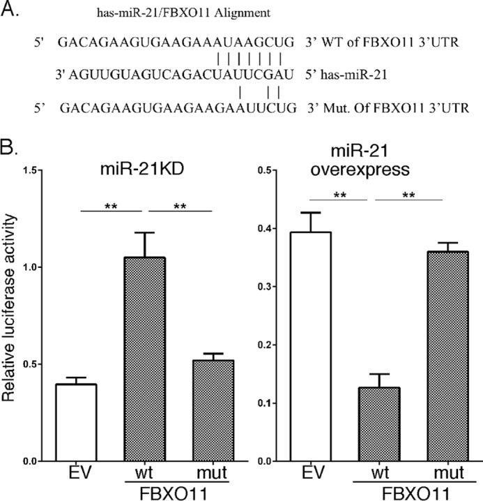
Identification of FBXO11 as a miR-21 target gene. A, sequence alignment of the miR-21 binding sequence with the 3′-UTR of FBXO11 gene (wild-type) and a mutant FBXO11 construct used for experiments. B, 293T cells were transiently cotransfected with empty vector (pcDNA3.1-Luc), or wild-type (pcDNA3.1-Luc-wtUTR) or mutant (pcDNA3.1-Luc-muUTR) FBXO11 reporter plasmids, and with antagomiR-21 plasmid to knockdown expression, or with miR-21 plasmid to overexpress miR-21. pSV40-Renilla plasmid was cotransfected as an internal control. The ratio of luciferase and Renilla activities was determined at 24 h post-transfection using the dual luciferase reporter gene kit (Promega).
FBXO11 Targets BCL6 for Degradation
FBXO11 is a component of the SCF ubiquitin ligase complex that targets proteins for degradation. BCL6 is the product of a proto-oncogene that is implicated in B-cell lymphoma, and is degraded by an FBXO11-mediated pathway (18). We first examined whether there was a relationship between the expression of FBXO11 and BCL6 by immunoblotting protein extracts from various human cell lines. In cells with high FBXO11 expression, such as PC3 and LNCaP prostate cancer cells, there were relatively low BCL6 levels (Fig. 3A). PC3 cells are noteworthy as they have extremely low miR-21 levels (15), and thus we anticipated that its target gene FBXO11 would be expressed at relatively high levels, while BCL6 levels would be low. In contrast, SJ-G2 and MT330 glioblastoma cells have low levels of FBXO11 consistent with their having high miR-21 expression (17), while their BCL6 levels are relatively high. To directly study the relationship between FBXO11 expression and BCL6 levels, we selected SJ-G2 and MT330 cells in which to overexpress FBXO11 by lentiviral-mediated transduction. FBXO11 overexpression resulted in markedly lower levels of BCL6 (Fig. 3B). In addition, miR-21KD in these cells, which leads to increased FBXO11 expression, also resulted in decreased protein levels of BCL6 (Fig. 3C).
FIGURE 3.

FBXO11 targets BCL6 for degradation. A, lysates of human prostate cancer cells (PC-3, LNCap, and DU145) and human glioma cells (SJ-G2 and MT330) were immunoblotted for FBXO11 and BCL6 protein expression. B, SJ-G2 and MT330 glioma cells were transduced with FBXO11-encoding lentivirus and lysates were prepared and immunoblotted for FBXO11 and BCL6 protein expression. C, lysates were prepared from miR-21KD SJ-G2 and MT330 glioma cells and immunoblotted for FBXO11 and BCL6 protein expression. D, cell lysates were prepared from EV and FBXO11 overexpressing MT330 cells treated with human IFN (1000 units/ml for 30 min), and STAT3 immunoprecipitates were immunoblotted for STAT3 and BCL6. To determine the role of STAT3 activation, IFN-treated cells were pretreated with WP-1066 (50 μm for 2 h), an inhibitor of STAT3 tyrosine phosphorylation. E, cell lysates were prepared from miR-21KD B16 cells transfected with FBXO11 shRNA or scrambled shRNA at various times after cycloheximide (CHX, 30 μg/ml) addition, and immunoblotted for BCL6 and FBXO11 protein expression. F, cell lysates were prepared from miR-21KD B16 cells transfected with FBXO11 shRNA or scrambled shRNA after MG132 (10 μm for 6 h) addition, and BCL6 immunoprecipitates were immunoblotted for BCL6 and FBXO11.
Previous studies have shown that BCL6 up-regulation is dependent on STAT3 and that these two proteins directly interact (22). Since we previously showed that the cytokine interferon induces miR-21 expression via a STAT3-dependent pathway (15), we examined whether IFN induced an interaction between STAT3 and BCL6, which was dependent on STAT3 activation. As shown in Fig. 3D, IFN induced the interaction of BCL6 with STAT3, which was blocked by an inhibitor of STAT3 activation WP1066 or by FBXO11 overexpression. To determine whether the interaction between FBXO11 and BCL6 was proteasome-dependent, miR-21KD B16 cells having high FBXO11 levels were treated with cycloheximide to inhibit protein synthesis or with MG-132, a proteasome inhibitor. Pretreatment with cycloheximide to inhibit protein synthesis in miR-21KD B16 cells with FBXO11 silenced rescued BCL6 protein levels (Fig. 3E). Furthermore, MG-132 treatment markedly enhanced the interaction between FBXO11 and BCL6, and shRNA knockdown of FBXO11 ablated this interaction (Fig. 3F). Thus, these results confirm that FBXO11 targets BCL6 for proteosomal degradation.
FBXO11 Promotes Apoptosis
Since miR-21 down-regulates the expression of FBXO11 and promotes cell survival and tumorigenesis, we next examined the role of FBXO11 in these processes. DU145 prostate cancer cells are relatively resistant to apoptosis induced by various agents (15). As shown in Fig. 4A, DU145 cells were relatively resistant to apoptosis induced by interferon-a cytokine, EF24-a curcumin analog that blocks the NF-κB signaling pathway (23), or staurosporine - a protein kinase inhibitor. In contrast, FBXO11 overexpression markedly sensitized DU145 cells to apoptosis induced by each of these agents with the greatest degree of apoptosis induced by EF24 and staurosporine. Similarly, FBXO11 overexpression in B16 melanoma cells sensitized cells to apoptosis (Fig. 4B). We next examined the effect of FBXO11 overexpression on the induction of cell death by sublethal amounts of EF24 using a live/dead cell fluorescent assay. While exposure of DU145 to 1 or 1.5 μm EF24 did not induce significant cell death in EV-transduced DU145 cells, a dose-dependent induction of cell death was observed in FBXO11-overexpressing cells (Fig. 4C). It is important to note that in FBXO11-overexpressing cells basal apoptosis is increased, and only a few live cells are observed upon treatment with 1.5 μm EF24. Thus, FBXO11 promotes apoptosis.
FIGURE 4.
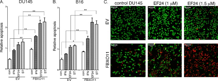
FBXO11 sensitizes cells to the induction of apoptosis. DU145 cells and FBXO11-overexpressing DU145 cells (A), or B16 cells and FBXO11 overexpressing B16 cells (B) were treated for 24 h with IFN (1000 IU/ml), EF24 (1 μm), or staurosporine (ST, 1 μm) and analyzed for apoptosis by cell death detection ELISA assays. Data represent the mean ± S.D. of at least three experiments performed in duplicate. C, DU145 cells and FBXO11 overexpressing DU145 cells were treated for 6 h with EF24 (1 or 1.5 μm) and analyzed by LIVE/DEAD Viability assay. Live cells fluoresce bright green, whereas dead cells fluoresce red-orange.
FBXO11 Inhibits Tumorigenesis
We next examined the effect of FBXO11 on the tumorigenicity of B16 melanoma cells in a well-studied mouse model for metastatic melanoma. Consistent with previous studies, injection of B16 cells into syngeneic C57BL6 mice via tail vein induces large highly pigmented lung tumors, and eventually results in death. As shown in Fig. 5A, animal survival is significantly prolonged (median survival of 26.2 ± 2.2 days as compared with 17.2 ± 1.8 days for EV-transduced cells) in mice injected with B16 cells that overexpress FBXO11, and only small pigmented lung tumors were found at necropsy in these mice. The effect of FBXO11 overexpression is analogous to our findings with miR-21KD B16 cells, in which animal survival was prolonged and only micrometastatic lesions were observed in the lungs of mice injected with miR-21KD B16 cells (16). In addition, FBXO11 overexpression in DU145 prostate cancer cells also inhibited their tumorigenesis. While subcutaneous injection of DU145 cells into immunocompromised mice resulted in the formation of rapidly growing, large tumor masses, FBXO11 overexpressing DU145 cells formed slower growing tumors (Fig. 5B). Taken together these findings indicate that FBXO11 sensitizes diverse cancer cell lines to apoptosis and inhibits their tumorigenicity, indicating that FBXO11 has tumor suppressive activity.
FIGURE 5.

The effects of FBXO11 expression on tumor formation. A, Kaplan-Meier analysis of survival data (n = 8) from C57BL6 mice that were injected with 106 EV or FBXO11 overexpressing B16 cells into the tail vein. B, NSG mice were injected subcutaneously with 106 EV or FBXO11 overexpressing D145 cells, and tumor growth was determined by caliper measurements. C, D, and E, NSG mice were injected subcutaneously with 106 miR-21KD B16 cells transduced with scrambled or shFBXO11, and tumor growth was determined twice per week by caliper measurements, and (C) Kaplan-Meier analysis of survival data (n = 8) was performed, and (D) H & E staining and (E) immunohistochemistry for caspase 3 activity and PARP cleavage from lung sections of injected mice.
Silencing FBXO11 Enhances the Tumorigenicity of miR-21KD B16 Cells
We previously found that miR-21KD inhibited tumorigenicity of prostate cancer, melanoma and glioblastoma cells (15, 16). Since FBXO11 expression is up-regulated by miR-21KD, we next addressed whether FBXO11 plays a direct role in the action of miR-21 on tumorigenesis. We tested several shRNA constructs against the FBXO11 gene, and found one that reduced FBXO11 expression in B16 miR-21KD cells by ∼80%. As shown in Fig. 5C, tail vein injection of miR-21KD cells transduced with FBXO11 shRNA markedly enhanced the tumorigenicity of these cells with animal survival reduced to 21.8 ± 1.9 days, as compared with mice injected with cells transduced with scrambled shRNA which survived 33.4 ± 2.8 days. At necropsy, H & E staining showed the lungs of mice injected with miR-21KD cells with FBXO11 silenced were filled with large highly pigmented lung tumors, while mice injected with miR-21KD cells with scrambled shRNA had only small metastatic lesions similar to the lesions in mice injected with miR-21KD B16 cells (Fig. 5D). Most interestingly, silencing FBXO11 resulted in marked reduction in apoptosis in tumor tissue as evidenced by PARP and caspase 3 staining (Fig. 5E). These results demonstrate that, while FBXO11 overexpression inhibits tumorigenesis, knockdown of FBXO11 restores tumorigenesis. Taken together these results demonstrate that FBXO11 is an important miR-21 target, which acts a tumor suppressor.
FBXO11 Expression Correlates with Better Survival of Cancer Patients and Inversely Correlates with miR-21 Expression
Since we found that FBXO11 suppressed tumor formation in several cancer cell lines we next examined whether there was a relationship between FBXO11 expression and tumor grade and cancer patient survival. We first examined the TCGA database for skin cancer specimens, which were classified according to Clark level (grade I/II is minimally invasive cancer and grade V is the most highly invasive form). As shown in Fig. 6A, high FBXO11 expression was found in the least invasive grade of skin cancer (Grade I/II) and decreased with increasing Clark level. In contrast, the inverse was observed with miR-21 expression, with low miR-21 levels found in low-grade skin cancer, and high levels in high-grade skin cancer. We then examined the relationship between FBXO11 and miR-21 expression in prostate cancer and glioblastoma specimens in the TCGA database, and found that the expression of miR-21was again inversely related to FBXO11 expression (Fig. 6B). To directly determine the relationship between miR-21 and FBXO11 expression, brain biopsy specimens from 36 glioblastoma patients were obtained, RNA was isolated, and expression of miR-21 and FBXO11 was determined by qPCR. As shown in Fig. 6C there was a statistically significant inverse correlation between miR-21 and FBXO11 expression (p = 0.0041). Moreover, there was a statistically significant linear relationship between FBXO11 expression and patient survival in glioblastoma, while there was an inverse relationship between miR-21 expression and patient survival. Taken together the results from the TCGA database and our own analysis of patient specimens showed that FBXO11 is a positive predictor of cancer patient survival, and that miR-21 and FBXO11 expression are negatively correlated. These findings bolster the hypothesis that miR-21 is an oncogene, while FBXO11 functions as a tumor suppressor.
FIGURE 6.

FBXO11 expression in vivo is inversely related to miR-21 expression, and high expression is associated with lower tumor grade and better patient survival. A, FBXO11 and miR-21 expression in the TCGA database for skin cancer according to Clark level (Grade 1 is the least aggressive and Grade V is the most aggressive). B, FBXO11 expression in the TCGA database of prostate cancer and glioblastoma patient samples was related to miR-21 expression. C, RNA was extracted from 36 glioblastoma patient biopsies, representing long-term (> 2 yr) and short-term (< 1 yr) survival after diagnosis. The expression of miR-21 and FBXO11 was determined by qPCR (n = 3), and normalized to the expression of U6A and actin, respectively.
DISCUSSION
Previous work in various cancer cell lines and animal models indicates that miR-21 is oncogenic. For example, miR-21 was the only consistently up-regulated miRNA in a study that profiled 540 clinical samples from cancer patients (5). Increased miR-21 expression has been substantiated in glioblastoma, head and neck carcinoma, ovarian cancer, B-cell lymphoma, and hepatocellular and cervical carcinoma (24). Induction of miR-21 expression in an inducible transgenic knock-in mouse model was found to result in spontaneous development of B-cell leukemia/lymphoma (25). Moreover, miR-21 expression modulates tumor number, incidence and size in a K-ras-dependent mouse lung cancer model (7). Thus, identification of new molecular targets of miR-21 may lead to improved therapeutic approaches to cancer.
A number of miR-21 target genes have been previously described, including PDCD4, BTG2 and PTEN. We found that the expression of these miR-21 target genes was up-regulated by miR-21KD in mouse melanoma cells, and in human brain and prostate cancer cells lines, demonstrating that these genes are true miR-21 targets. By gene expression analysis of miR-21KD B16 cell lines and tumor tissue, we identified FBXO11 as a novel miR-21 target gene. We found that FBXO11 expression was also up-regulated by miR-21KD in various human cancer cell lines. By luciferase reporter assays driven by wild-type and mutant 3′-UTR of FBXO11, we showed that FBXO11 expression was up-regulated by miR-21KD, and down-regulated by miR-21 overexpression. Our results identify FBXO11 as an important miR-21 target gene, which is the first evidence that FBXO11 expression is regulated through a miRNA-dependent pathway. FBXO11 is the mammalian homologue of the Caenorhabditis elegans DRE-1, which functions in the SKP1-CUL1-F Box (SCF) E3-ubiquitin ligase complex, and is the only F-box protein that is highly conserved across all major taxa (26). F-box proteins function as the substrate-recognition components of the SCF complexes, thus conferring substrate specificity to the ubiquitin-proteasome system (19). These E3 ubiquitin ligases control the levels of proteins involved in regulating cell cycle progression, DNA replication, gene transcription and apoptosis, and thus may play important roles in various biological processes including cancer.
FBXO11 has recently been identified as a tumor suppressor gene that is mutated or deleted in a subset of human diffuse large B-cell lymphomas (18). FBXO11 targets BCL6 for ubiquitination and proteosomal degradation by the SCF ubiquitin ligase complex. We found that there was an inverse relationship between FBXO11and BCL6 expression in various human cancer cell lines, and that FBXO11 overexpression resulted in markedly lower BCL6 levels. Consistent with the finding that FBXO11 directly mediates BCL6 degradation (18), we demonstrated that FBXO11 and BCL6 directly interact, and this interaction was not only dependent on the proteasome, as treatment with the proteasome inhibitor MG-132 enhanced their interaction, and inhibiting protein synthesis by cycloheximide also rescued BCL6 protein levels. In addition, the cytokine interferon induced the interaction of BCL6 with STAT3 transcription factor, and this interaction was blocked by a STAT3 inhibitor (WP1066) or by FBXO11 overexpression. These results are consistent with the report that BCL6 up-regulation is STAT3-dependent and that STAT3 and BCL6 directly interact with each other (22). It is of interest that the role of BCL6 in lymphoma is fairly well defined (27), but our study suggests that BCL6 may also play an important role in non-lymphoid cancers as well.
Most interestingly we found that, while miR-21 functions as a tumor promoter in various cancer cell line models, FBXO11 acts as a tumor suppressor. The antagonistic action of miR-21 on FBXO11 is not entirely surprising, since miRNAs act by suppressing the expression of their target proteins. We found that FBXO11 overexpression inhibits tumorigenesis and prolongs the survival of tumor-bearing mice, which is very similar to our findings with miR-21 knockdown. In addition, overexpression of another miR-21 target in B16 cells, IGFBP3, did not prolong the survival of tumor-bearing mice (data not shown). Moreover, using the B16 melanoma model to establish lung metastases by tail vein injection, we found that, while B16 cells normally establish large metastatic lung tumors in mice, overexpression of FBXO11 in the cells as well as miR-21 knockdown resulted in only small lung tumors. In addition, silencing FBXO11 in miR-21 knockdown B16 melanoma cells reversed this effect, so that animal survival was markedly reduced and the mice once again succumbed to large metastatic lung lesions. Our previous studies in various cancer cell lines indicated that miR-21 is a suppressor of apoptosis in human glioblastoma and prostate cancer cell lines, and in mouse melanoma cells (15–17). In the present study we show that FBXO11 overexpression promotes not only basal apoptosis but also the induction of apoptosis by a variety of agents in cancer cell lines from diverse origins. This result is not entirely surprising since miR-21, which inhibits apoptosis, suppresses FBXO11 expression, which normally promotes apoptosis. This is also borne out by our findings of reduced apoptosis as evidenced by increased PARP cleavage and caspase activation in tumor tissue derived from B16 cells, in which FBXO11 and miR-21 expression was concurrently knocked down. These results provide strong evidence that FBXO11 acts a tumor suppressor.
FBXO11 has been identified as a tumor suppressor in diffuse large B-cell leukemia (18). However, our findings with cell lines grown in vitro and in animal models in vivo suggest that FBXO11 may play a broader role in oncogenesis. Thus, we examined the relationship between FBXO11 expression with tumor grade and patient survival in clinical samples. We found that high FBXO11 expression was found in the least invasive skin cancer (Clark level I/II) and decreased with increasing Clark level. In contrast, the inverse was observed with miR-21 expression, with low miR-21 levels found in low-grade skin cancer, and high levels in high-grade skin cancer. We also found in the TCGA database that miR-21 expression was inversely related to FBXO11 expression in prostate cancer and glioblastoma specimens (Fig. 6B). Most interestingly, there was an inverse correlation between miR-21 and FBXO11 expression in brain biopsy specimens from glioblastoma patients who were long-term survivors as opposed to those who were only short-term survivors after diagnosis. Moreover, there was a linear relationship between FBXO11 expression and glioblastoma patient survival. Taken together the results from the TCGA database and our own analysis of patient specimens showed that FBXO11 is a positive predictor of cancer patient survival, and that miR-21 and FBXO11 expression is inversely correlated. These findings bolster the hypothesis that while miR-21 is an oncogene, FBXO11 functions as a tumor suppressor. Thus, FBXO11 may have diagnostic and prognostic utility in cancer. In summary, we identified FBXO11 as a novel miR-21 target gene in cancer, and characterized the role of the opposing actions of miR-21 and FBXO11 in various cancer models as well as in biopsy specimens from cancer patients, which suggests that miR-21 promotes tumorigenesis through suppressing the expression of the tumor suppressor FBXO11.
This work was supported in part by grants from the National Institutes of Health CA133322 (to L. M. P. and A. M. D.), the Department of Defense W81XWH-11-1-0533 (to L. M. P.), the Cancer Center Support Grant 21766 from the National Cancer Institute and the Assisi Foundation of Memphis (to A. M. D.), by the Muirhead Chair Endowment (to L. M. P.) of the UTHSC and by the American Lebanese Syrian Associated Charities (to A. M. D.).
- miRNA
- microRNA
- UTR
- untranslated region.
REFERENCES
- 1. Bartel D. P. (2004) MicroRNAs: genomics, biogenesis, mechanism, and function. Cell 116, 281–297 [DOI] [PubMed] [Google Scholar]
- 2. Calin G. A., Croce C. M. (2006) MicroRNA signatures in human cancers. Nat. Rev. Cancer 6, 857–866 [DOI] [PubMed] [Google Scholar]
- 3. Lim L. P., Lau N. C., Garrett-Engele P., Grimson A., Schelter J. M., Castle J., Bartel D. P., Linsley P. S., Johnson J. M. (2005) Microarray analysis shows that some microRNAs downregulate large numbers of target mRNAs. Nature 433, 769–773 [DOI] [PubMed] [Google Scholar]
- 4. Lewis B. P., Burge C. B., Bartel D. P. (2005) Conserved seed pairing, often flanked by adenosines, indicates that thousands of human genes are microRNA targets. Cell 120, 15–20 [DOI] [PubMed] [Google Scholar]
- 5. Volinia S., Calin G. A., Liu C. G., Ambs S., Cimmino A., Petrocca F., Visone R., Iorio M., Roldo C., Ferracin M., Prueitt R. L., Yanaihara N., Lanza G., Scarpa A., Vecchione A., Negrini M., Harris C. C., Croce C. M. (2006) A microRNA expression signature of human solid tumors defines cancer gene targets. Proc. Natl. Acad. Sci. U.S.A. 103, 2257–2261 [DOI] [PMC free article] [PubMed] [Google Scholar]
- 6. Ventura A., Jacks T. (2009) MicroRNAs and cancer: short RNAs go a long way. Cell 136, 586–591 [DOI] [PMC free article] [PubMed] [Google Scholar]
- 7. Hatley M. E., Patrick D. M., Garcia M. R., Richardson J. A., Bassel-Duby R., van Rooij E., Olson E. N. (2010) Modulation of K-Ras-dependent lung tumorigenesis by MicroRNA-21. Cancer Cell 18, 282–293 [DOI] [PMC free article] [PubMed] [Google Scholar]
- 8. Si M. L., Zhu S., Wu H., Lu Z., Wu F., Mo Y. Y. (2007) miR-21-mediated tumor growth. Oncogene 26, 2799–2803 [DOI] [PubMed] [Google Scholar]
- 9. Folini M., Gandellini P., Longoni N., Profumo V., Callari M., Pennati M., Colecchia M., Supino R., Veneroni S., Salvioni R., Valdagni R., Daidone M. G., Zaffaroni N. (2010) miR-21: an oncomir on strike in prostate cancer. Mol. Cancer 9, 12. [DOI] [PMC free article] [PubMed] [Google Scholar]
- 10. Löffler D., Brocke-Heidrich K., Pfeifer G., Stocsits C., Hackermüller J., Kretzschmar A. K., Burger R., Gramatzki M., Blumert C., Bauer K., Cvijic H., Ullmann A. K., Stadler P. F., Horn F. (2007) Interleukin-6 dependent survival of multiple myeloma cells involves the Stat3-mediated induction of microRNA-21 through a highly conserved enhancer. Blood 110, 1330–1333 [DOI] [PubMed] [Google Scholar]
- 11. Chan J. A., Krichevsky A. M., Kosik K. S. (2005) MicroRNA-21 is an antiapoptotic factor in human glioblastoma cells. Cancer Res. 65, 6029–6033 [DOI] [PubMed] [Google Scholar]
- 12. Zhu S., Wu H., Wu F., Nie D., Sheng S., Mo Y. Y. (2008) MicroRNA-21 targets tumor suppressor genes in invasion and metastasis. Cell Res. 18, 350–359 [DOI] [PubMed] [Google Scholar]
- 13. Wang P., Zou F., Zhang X., Li H., Dulak A., Tomko R. J., Jr., Lazo J. S., Wang Z., Zhang L., Yu J. (2009) microRNA-21 negatively regulates Cdc25A and cell cycle progression in colon cancer cells. Cancer Res. 69, 8157–8165 [DOI] [PMC free article] [PubMed] [Google Scholar]
- 14. Buscaglia L. E., Li Y. (2011) Apoptosis and the target genes of microRNA-21. Chin. J. Cancer 30, 371–380 [DOI] [PMC free article] [PubMed] [Google Scholar]
- 15. Yang C. H., Yue J., Fan M., Pfeffer L. M. (2010) IFN induces miR-21 through a signal transducer and activator of transcription 3-dependent pathway as a suppressive negative feedback on IFN-induced apoptosis. Cancer Res. 70, 8108–8116 [DOI] [PMC free article] [PubMed] [Google Scholar]
- 16. Yang C. H., Yue J., Pfeffer S. R., Handorf C. R., Pfeffer L. M. (2011) MicroRNA miR-21 Regulates the Metastatic Behavior of B16 Melanoma Cells. J. Biol. Chem. 286, 39172–39178 [DOI] [PMC free article] [PubMed] [Google Scholar]
- 17. Yang C. H., Yue J., Pfeffer S. R., Fan M., Paulus E., Hosni-Ahmed A., Sims M., Qayyum S., Davidoff A. M., Handorf C. R., Pfeffer L. M. (2014) MicroRNA-21 promotes glioblastoma tumorigenesis by downregulating IGFBP3. J. Biol. Chem. 289, 25079–25087 [DOI] [PMC free article] [PubMed] [Google Scholar]
- 18. Duan S., Cermak L., Pagan J. K., Rossi M., Martinengo C., di Celle P. F., Chapuy B., Shipp M., Chiarle R., Pagano M. (2012) FBXO11 targets BCL6 for degradation and is inactivated in diffuse large B-cell lymphomas. Nature 481, 90–93 [DOI] [PMC free article] [PubMed] [Google Scholar]
- 19. Skaar J. R., Pagan J. K., Pagano M. (2013) Mechanisms and function of substrate recruitment by F-box proteins. Nat. Rev. Mol. Cell Biol. 14, 369–381 [DOI] [PMC free article] [PubMed] [Google Scholar]
- 20. Pfeffer L. M., Mullersman J. E., Pfeffer S. R., Murti A., Shi W., Yang C. H. (1997) STAT3 as an adapter to couple phosphatidylinositol-3 kinase to the IFNAR-1 chain of the type I IFN receptor. Science 276, 1418–1420 [DOI] [PubMed] [Google Scholar]
- 21. Wei L., Sandbulte M. R., Thomas P. G., Webby R. J., Homayouni R., Pfeffer L. M. (2006) NFκB negatively regulates interferon-induced gene expression and anti-influenza activity. J. Biol. Chem. 281, 11678–11684 [DOI] [PMC free article] [PubMed] [Google Scholar]
- 22. Hideshima T., Mitsiades C., Ikeda H., Chauhan D., Raje N., Gorgun G., Hideshima H., Munshi N. C., Richardson P. G., Carrasco D. R., Anderson K. C. (2010) A proto-oncogene BCL6 is up-regulated in the bone marrow microenvironment in multiple myeloma cells. Blood 115, 3772–3775 [DOI] [PMC free article] [PubMed] [Google Scholar]
- 23. Yang C. H., Yue J., Sims M., Pfeffer L. M. (2013) The curcumin analog EF24 targets NF-κB and miRNA-21, and has potent anticancer activity in vitro and in vivo. PLoS One 8, e71130. [DOI] [PMC free article] [PubMed] [Google Scholar]
- 24. Krichevsky A. M., Gabriely G. (2009) miR-21: a small multi-faceted RNA. J. Cell Mol. Med. 13, 39–53 [DOI] [PMC free article] [PubMed] [Google Scholar]
- 25. Medina P. P., Nolde M., Slack F. J. (2010) OncomiR addiction in an in vivo model of microRNA-21-induced pre-B-cell lymphoma. Nature 467, 86–90 [DOI] [PubMed] [Google Scholar]
- 26. Shaye D. D., Greenwald I. (2011) OrthoList: a compendium of C. elegans genes with human orthologs. PLoS One 6, e20085. [DOI] [PMC free article] [PubMed] [Google Scholar]
- 27. Parekh S., Polo J. M., Shaknovich R., Juszczynski P., Lev P., Ranuncolo S. M., Yin Y., Klein U., Cattoretti G., Dalla Favera R., Shipp M. A., Melnick A. (2007) BCL6 programs lymphoma cells for survival and differentiation through distinct biochemical mechanisms. Blood 110, 2067–2074 [DOI] [PMC free article] [PubMed] [Google Scholar]