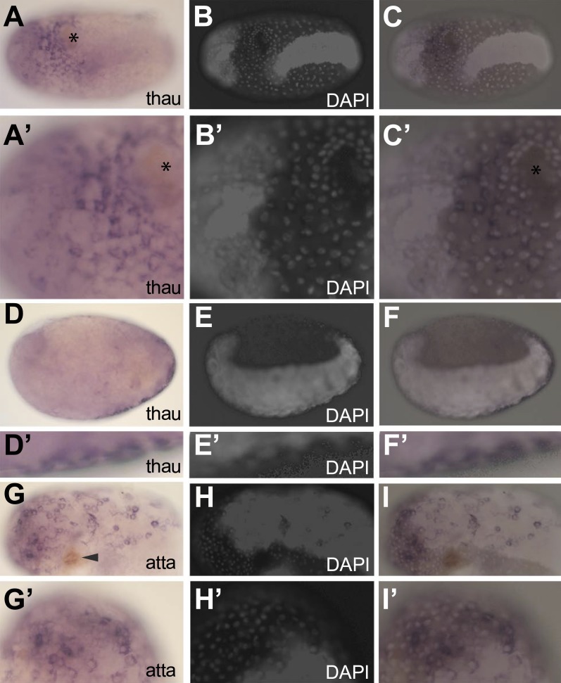
Figure 6. In situ hybridization showing expression of AMP genes in the serosa upon septic injury.
(A–F) Thaumatin1 in situ hybridization. (A) Superficial view. Thaumatin1 is expressed around the site of injury (asterix). Brown melanisation is observed around the site of injury. (A′) Magnification of the expression area shown in (A). Asterix marks the site of injury. (B) DAPI counterstaining of the same egg as in (A). The large polyploid serosal nuclei can be distinguished from the oversaturated DAPI signal from the germ-band. Head lobes to the left. (B′) magnification of (B). (C) Overlay of the in situ hybridization shown in A and the DAPI staining shown in (B). The thaumatin1 expression associates with the large polyploid serosal nuclei and is not found in the embryo proper. (C′) Magnification of the expression area shown in (C). (D) Focal plane through the egg. Thaumatin1 is expressed in a thin outer layer at the surface of the egg. (D′) Magnification of the expression area shown in (D). (E) DAPI staining of the same egg shown in (D). The embryo is brightly visible. Head to the left. (E′) Magnification of E. (F) Overlay of the in situ hybridization shown in D and the DAPI staining shown in (E). (F′) Magnification of the expression area. (G) Attacin1 in situ hybridization. Brown melanisation is visible around the site of injury (arrowhead). (G′) Magnification of the anterior region of the egg shown in (G). (H) DAPI staining of the same egg shown in G. The germ-band is brightly stained (head to the left) and the separate large serosal nuclei are visible. (H′) Magnification of the anterior of the egg shown in (H). (I) Overlay of the in situ hybridization shown in G and the DAPI staining shown in (H). Attacin1 is expressed in the large serosal cells covering the germ-band and is not expressed in the dense cells of the germ band. (I′) Magnification of the anterior of the egg shown in (I). The attacin1 staining associates with the large serosal nuclei.

