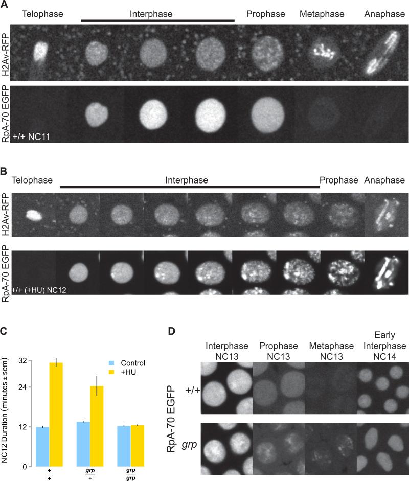
Figure 4. RpA-70 EGFP Marks Sites of Stalled Replication.
(A) RpA-70 EGFP uniformly localizes to interphase nuclei before NC13. An RpA-70 EGFP; H2Av RFP embryo was imaged by confocal microscopy. Successive representative images of a single NC11 stage nucleus are shown at the cell cycle stages indicated on top.
(B) RpA-70 EGFP and H2Av RFP as visualized in a HU-treated embryo by time-lapse confocal microscopy. Successive representative images of a single NC12 nucleus are shown at the cell cycle stages indicated on top.
(C) Wild type (+/+), grp/+, and grp mutant embryos were treated with HU and total NC12 duration was measured by time lapse confocal microscopy.
(D) Wild type (+/+) and grp mutant embryos expressing RpA-70 EGFP were visualized by time-lapse confocal microscopy. Successive representative images of two nuclei per genotype are shown at the cell cycle stages indicated.

