Abstract
Major functions of the hippocampus are to generate, organize and store memory. This is a complex process, which is orchestrated by a group of molecules, called plasticity-related molecules. To control these various plasticity-related molecules at the transcriptional level, we have been endowed with cAMP response element-binding protein (CREB), also known as a master regulator of memory. Interestingly, we have seen that this master regulator is regulated at the transcriptional level in the hippocampus by peroxisome proliferator-activated receptor α (PPARα), a nuclear hormone receptor family transcription factor that is known to control the metabolism of fatty acids in the liver, underlying a possible crosstalk between fat and memory. Although liver PPARα does not directly control hippocampal CREB, this opens up an important possibility to improve hippocampal functions and to be resistant to memory loss by PPARα ligands and maintaining normal levels of PPARα in the hippocampus.
Keywords: Fatty acid metabolism, Liver, PPARα, Hippocampus, CREB, Plasticity
Introduction
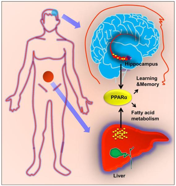
Although memory loss is a gerontology issue and problems with memory become increasingly common as people age, in some persons, memories last long time, even a life time. On the other hand, some people experience milder to substantial memory problems even at an earlier age. Although there are several risk factors of dementia, abnormal fat metabolism always poses a risk for memory and learning. For example, a diet rich in saturated long-chain fatty acids decreases memory and learning in mice (Granholm et al. 2008). It is known that people with high amounts of abdominal fat in their middle age are 3.6 times as likely to develop memory loss and dementia later in their life (2008). It has been also reported that brain volume is less in people with greater waistline (Debette et al. 2010). However, mechanisms by which abnormal lipid metabolism interacts with memory remain unknown. Therefore, understanding how fat is connected to memory and learning (Fig. 1) is important to developing effective approach to protect memory and learning.
Fig. 1.

A cartoon showing the possible crosstalk between hepatic fatty acid metabolism and hippocampal learning and memory. PPARα is present in both the liver and the hippocampus. While hepatic PPARα is involved in fatty acid oxidation, hippocampal PPARα participates in synaptic plasticity
Peroxisome Proliferator-Activated Receptors (PPARs)
PPARs are a group of three transcription factors (PPARα, PPARβ/δ and PPARγ) that consist of a DNA binding domain (DBD) in the N-terminus and a ligand binding domain (LBD) in the C-terminus (Marcus et al. 1993; Schoonjans et al. 1996). However, three isotypes of PPAR differ from each other in terms of their tissue distributions and physiological roles (Marcus et al. 1993; Schoonjans et al. 1996). For example, PPARγ is mainly expressed in white and brown adipose tissue, the large intestine and spleen. Several studies demonstrate an important role of PPARγ in the regulation of adipogenesis, energy balance, lipid biosynthesis, and inflammation (Murphy and Holder 2000). Although PPARβ/δ is expressed ubiquitously in virtually all tissues, it is particularly abundant in the liver, kidney, adipose tissue, and skeletal muscle. It is known to participate in fatty acid oxidation, mainly in skeletal and cardiac muscles. On the other hand, PPARα is highly expressed in the liver (Fig. 1), heart and kidney, tissues that use fat as an energy source (Keller et al. 1993; Marcus et al. 1993; Kersten et al. 2000).
PPARα Signaling Pathways
Being a member of the nuclear hormone receptor superfamily, activation of PPARα depends on ligands. However, PPARα is known to be associated with HSP72 and molecular reason for this association is poorly understood. After interaction with ligands, PPARα is translocated to the nucleus and it is suggested that the PPARα-HSP72 complex may be involved in translocating the ligand to the nucleus (Reddy and Mannaerts 1994). In the absence of ligand, the unliganded PPARα remains bound to the nuclear receptor corepressor (NCoR) and silencing mediator of retinoid and thyroid hormone receptor (SMRT) (Nagy et al. 1997). SMRT functions as a platform protein that facilitates the recruitment of histone deacetylases (HDACs) to the DNA promoters. However, in the presence of a ligand, PPARα forms a heterodimeric complex with 9-cis retinoic acid receptor RXRα. This PPARα:RXRα complex then binds to peroxisome proliferator response element (PPRE) present in the promoter of different genes. Different co-activators like PPARγ coactivator-1α (PGC-1α) and PRIP (peroxisome proliferator-activated receptor-interacting protein)/ASC2/AIB3/ RAP250/NCoA6 and histone acetylase (HAT) p300/CBP also play crucial role in the formation of active transcriptional complex (Nagy et al. 1997; Spiegelman and Heinrich 2004).
PPARα and Lipid Metabolism
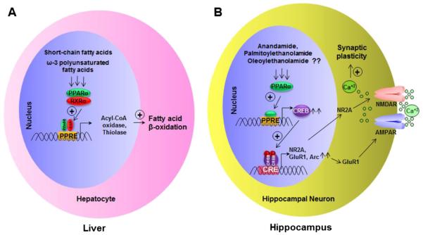
While mitochondria catalyze the β-oxidation of the bulk of short-, medium-, and long-chain fatty acids derived from diet, it is peroxisomes that take care of the β-oxidation of very long-chain fatty acids (VLCFA) (Reddy and Mannaerts 1994). After chain shortening of VLCFAs via β-oxidation, these fatty acids are transported to mitochondria for complete metabolism. Although activation of PPARα stimulates the β-oxidation pathway in both mitochondria and peroxisomes, it is more specific for peroxisomal β-oxidation than the mitochondrial one. The PPREs are found in the promoters of PPAR responsive genes, such as acyl-CoA oxidase, L-bifunctional protein, thiolase etc. (Fig. 2). It has been shown that upon activation of PPARα by different ligands (fibrate drugs, short-chain fatty acids, eicosanoids, etc.), the PPARα:RXRα heterodimer is recruited to the promoters of genes encoding the classical β-oxidation pathway (Reddy and Mannaerts 1994). Accordingly, the fatty acid β-oxidation pathway is decreased in PPARα null mice (Gonzalez 1997).
Fig. 2.
PPARα signaling pathways leading to fatty acid oxidation in the liver (a) and synaptic plasticity in the hippocampus (b). Short-chain fatty acids and ω-3 polyunsaturated fatty acids are known to activate the PPARα:RXRα heterodimeric complex in hepatocytes, which in turn binds to promoters of different genes encoding fatty acid β-oxidation. On the other hand, anandamides, palmitoylethanolamide and oleoylethanolamide may activate PPARα, which is recruited to the promoter of CREB in hippocampal neurons. Then CREB turns on the transcription of different plasticity-associated molecules
PPARα in Hippocampus
PPARα is known to be present in metabolically active organs and the hippocampus does not produce energy from fat metabolism, however, we (Roy et al. 2013) and other (Rivera et al. 2014) have shown the presence of PPARα in different subfields of the hippocampus of rodents. Although humans and other primates have been reported to contain considerably lower levels of PPARα in liver than rodents (Tugwood et al. 1998), PPARα is also present in the hippocampus of rhesus monkeys (Roy et al. 2013). While PPARβ and PPARγ are present in both nucleus and cytoplasm, PPARα is exclusively present in nucleus of hippocampal neurons (Roy et al. 2013).
Regulation of Hippocampal Plasticity by PPARα
While metabotropic receptors (e.g. NR2A, GluR1) play a crucial role in hippocampal plasticity, voltage-gated ion channel molecules like Kv1.1 and Scn1a contribute to neuronal excitability and discharge behavior. Interestingly, knockdown of PPARα decreases the expression of various plasticity-associated molecules (NR2A, NR2B, GluR1, and Arc), but not voltage-gated ion channel molecules, in hippocampal neurons (Roy et al. 2013), indicating selective role of PPARα in plasticity. Accordingly, PPARα null hippocampal neurons exhibit a weaker calcium influx and a smaller amplitude oscillation than wild type neurons in response to both AMPA and NMDA (Roy et al. 2013), suggesting that PPARα plays a role in controlling the synaptic plasticity in hippocampal neurons (Fig. 2).
Transcriptional regulation of CREB by PPARα
CREB has a well-documented role in synaptic plasticity in the brain (Ghosh et al. 1994). Interestingly, the Creb promoter harbors one consensus PPRE in the distal region (−1164 to −1152) and PPARα agonists induce the activation of Creb promoter in hippocampal neurons isolated from wild type, but not PPARα null, mice (Roy et al. 2013). Furthermore, PPARα agonists remain unable to activate a Creb promoter in which the PPRE is mutated, suggesting the involvement of PPARα in the transcription of Creb (Fig. 2). Accordingly, the recruitment of PPARα to the Creb promoter is seen in the hippocampus of wild type, but not PPARα null, mice (Roy et al. 2013). However, currently, it is not known whether PPARα alone, the classical PPARα:RXRα heterodimer or a new heterodimeric complex is recruited to the Creb promoter.
Control of Learning and Memory by PPARα
Although there are no significant differences in body weight, general motor behavior and average food consumption between wild type and PPARα null mice, PPARα null mice are also seen to be deficient in spatial learning and memory as compared to wild type mice (Roy et al. 2013). This is consistent to the involvement of CREB in the generation of long term memory and spatial learning (Ghosh et al. 1994; Stevens 1994). Upregulation of CREB and restoration of memory and learning in PPARα null mice by lentiviral delivery of PPARα into the hippocampus suggests an important role of PPARα in memory and learning (Roy et al. 2013).
Hippocampal Memory is not Directly Controlled by Hepatic PPARα
Although PPARα is present in the periphery (e, g, liver) as well as in the hippocampus, according to Campolongo et al. (Campolongo et al. 2009), PPARα in the gut generates a noradrenergic transmission in the basolateral amygdala in order to facilitate the retention of spatial memory. They have also suggested that this particular autonomic neurotransmission is absent in mice lacking expression of PPARα thus rendering them poor consolidators of spatial memory. In contrast, in order to dissect peripheral PPARα from CNS PPARα, bone marrow chimeric mice were generated and analyses of these mice delineate that the hippocampal memory apparatus is not regulated by peripheral PPARα (Roy et al. 2013). Interestingly, animals with peripherally-ablated PPARα have intact hippocampal NR2A and the ability of generating spatial memory, whereas, the ablation of PPARα in the CNS reduces hippocampal NR2A expression and makes animals markedly poor in consolidating spatial memory (Roy et al. 2013). Therefore, the peripheral PPARα may regulate noradrenergic neurotransmission to amygdala via vagal innervations in response to N-oleoylethanolamide (Campolongo et al. 2009), this pathway, however, is not the only mechanism involved in PPARα-mediated regulation of learning and memory. This could be an indirect mechanism as peripheral PPARα should not regulate the hippocampal master regulator CREB at the transcriptional level.
Pondering on Therapeutic Opportunities: Concluding Thoughts
What Does This Mean for Overweight People?
The same protein that controls fat metabolism in the liver is also present in the hippocampus in order to regulate memory and learning via transcriptional control of CREB (Fig. 1). However, the major and direct effect on spatial memory comes from hippocampal PPARα and the absence of this transcription factor in the hippocampus completely abrogates the learning and memory acquisition process via inhibition of Creb transcription and subsequent suppression of different plasticity-associated molecules. Because PPARα directly controls the expression of genes involved in lipid metabolism, overweight people may have abnormal lipid metabolism and depleted PPARα in the liver. However, all overweight people do not suffer from memory loss. Although lipid is an important risk factor for memory loss, many overweight people have normal memory probably because of having normal PPARα in the hippocampus. On the other hand, those having memory loss may be deficient in expression either due to a genetic polymorphism in PPARα or in altered expression of the gene due to hormonal imbalances or epigenetic mechanisms. Therefore, this study indicates that people may suffer from memory-related problems only when they lose PPARα in the hippocampus. However, the genetic or physiological basis for this hypothesis needs to be investigated.
Would PPARα be Beneficial for Dementia And Memory Loss?
Alzheimer’s disease (AD), the most common human neurodegenerative disorder, accounts for 60 to 70% cases of dementia. At present, nothing is known about the level of PPARα in hippocampus of patients with mild cognitive impairment and AD. Although according to Brune et al. (Brune et al. 2003), polymorphism in the PPARα gene influences the risk for AD, a later study (Sjolander et al. 2009) did not find any significant differences in genotype or allele distributions between AD patients and controls. However, PPARα level goes down in different organs during aging (Iemitsu et al. 2002; Gelinas and McLaurin 2005), suggesting that it may also go down in the hippocampus during age-related disorders. Under that condition, maintaining PPARα in the hippocampus may preserve our precious memory and learning as lentiviral delivery of PPARα in the hippocampus of PPARα null mice improves spatial learning and memory.
Possible Beneficial Effects of Ligands
We also must remember that being a nuclear hormone receptor, the function of PPARα is dependent on ligands. Therefore, ligands must be present for proper functioning of PPARα in the hippocampus. Various endocannabinoids like anandamide, palmitoylethanolamide and oleoylethanolamide are known to be present in the brain. Accordingly, type 1 cannabinoid receptors (CB1) are abundant in cortex and hippocampus (Monory et al. 2006). Several studies have also demonstrated that these endocannabinoids are capable of activating PPARα (Kozak et al. 2002; Fu et al. 2003; Sun et al. 2006). Therefore, it is possible that these endocannabinoids may serve as ligands of PPARα in hippocampus to support memory and learning. It has been shown that omega-3 fatty acids such as eicosapentaenoic acid (EPA) and docosahexaenoic acid (DHA) present in fatty fish are essential for brain health. Studies have shown that people having more of these fatty acids in their blood or a long-term habit of eating fish every week are less likely to develop Alzheimer’s disease (Sinn et al. 2012; Cederholm et al. 2013). Here, it is important to remember that both DHA and EPA are ligands of PPARα. Therefore, it is possible that DHA and EPA support hippocampal memory and learning via PPARα. Since ligands of PPARα are also capable of upregulating the level of PPARα via PPARα, supplementation of BBB-permeable PPARα ligands may be helpful in protecting memory and learning.
Is There Any Health Concern From PPARα Ligands?
Although gemfibrozil (FDA-approved since 1981) and fenofibrate (FDA-approved since 1998) are currently being used for patients with hyperlipidemia, long-term administration of some of the PPARα ligands like clofibrate and ciprofibrate to the rodents is known to cause hepatomegaly and hepatic tumor (Reddy and Mannaerts 1994). However, induction of hepatic tumor promotion by fibrate drugs has not been demonstrated in human, other primates, and guinea pig (Braun et al. 1999; Yeldandi et al. 2000), species that have lost their ability to synthesize vitamin C (ascorbic acid) due to inherent loss of the gulonolactone oxidase gene. According to Braun et al. (Braun et al. 1999), the evolutionary loss of the gulonolactone oxidase gene may contribute to the missing carcinogenic effect of peroxisome proliferators in humans, since ascorbate synthesis is accompanied by H2O2 production, ultimately causing harmful effects. Furthermore, recent studies have also revealed that humans have considerably lower levels of PPARα in liver than rodents, and this difference may, in part, explain the species differences in the carcinogenic response to fibrate drugs (Gonzalez et al. 1998). However, no such tumorigenic effects have been reported so far for anandamide and other endocannabinoids. Therefore, ligands of PPARα may not cause human health problems, while supporting hippocampal plasticity signaling.
Acknowledgments
This study was supported by grants from NIH (AT6681 and NS83054) and Alzheimer’s Association (IIRG-12-241179).
Contributor Information
Avik Roy, Department of Neurological Sciences, Rush University Medical Center, 1735 West Harrison St, Suite 320, Chicago, IL 60612, USA.
Kalipada Pahan, Department of Neurological Sciences, Rush University Medical Center, 1735 West Harrison St, Suite 320, Chicago, IL 60612, USA; Division of Research and Development, Jesse Brown Veterans Affairs Medical Center, 820 South Damen Avenue, Chicago, IL, USA.
References
- Abdominal fat boosts later dementia risk. Harv Ment Health Lett. 2008;25:7. [PubMed] [Google Scholar]
- Braun L, Mile V, Schaff Z, Csala M, Kardon T, Mandl J, Banhegyi G. Induction and peroxisomal appearance of gulonolactone oxidase upon clofibrate treatment in mouse liver. FEBS Lett. 1999;458:359–362. doi: 10.1016/s0014-5793(99)01184-9. [DOI] [PubMed] [Google Scholar]
- Brune S, Kolsch H, Ptok U, Majores M, Schulz A, Schlosser R, Rao ML, Maier W, Heun R. Polymorphism in the peroxisome proliferator-activated receptor alpha gene influences the risk for Alzheimer’s disease. J Neural Transm. 2003;110:1041–1050. doi: 10.1007/s00702-003-0018-6. [DOI] [PubMed] [Google Scholar]
- Campolongo P, Roozendaal B, Trezza V, Cuomo V, Astarita G, Fu J, McGaugh JL, Piomelli D. Fat-induced satiety factor oleoylethanolamide enhances memory consolidation. Proc Natl Acad Sci U S A. 2009;106:8027–8031. doi: 10.1073/pnas.0903038106. [DOI] [PMC free article] [PubMed] [Google Scholar]
- Cederholm T, Salem N, Jr, Palmblad J. omega-3 fatty acids in the prevention of cognitive decline in humans. Adv Nutr. 2013;4:672–676. doi: 10.3945/an.113.004556. [DOI] [PMC free article] [PubMed] [Google Scholar]
- Debette S, Beiser A, Hoffmann U, Decarli C, O’Donnell CJ, Massaro JM, Au R, Himali JJ, Wolf PA, Fox CS, Seshadri S. Visceral fat is associated with lower brain volume in healthy middle-aged adults. Ann Neurol. 2010;68:136–144. doi: 10.1002/ana.22062. [DOI] [PMC free article] [PubMed] [Google Scholar]
- Fu J, Gaetani S, Oveisi F, Lo Verme J, Serrano A, Rodriguez De Fonseca F, Rosengarth A, Luecke H, Di Giacomo B, Tarzia G, Piomelli D. Oleylethanolamide regulates feeding and body weight through activation of the nuclear receptor PPAR-alpha. Nature. 2003;425:90–93. doi: 10.1038/nature01921. [DOI] [PubMed] [Google Scholar]
- Gelinas DS, McLaurin J. PPAR-alpha expression inversely correlates with inflammatory cytokines IL-1beta and TNF-alpha in aging rats. Neurochem Res. 2005;30:1369–1375. doi: 10.1007/s11064-005-8341-y. [DOI] [PubMed] [Google Scholar]
- Ghosh A, Ginty DD, Bading H, Greenberg ME. Calcium regulation of gene expression in neuronal cells. J Neurobiol. 1994;25:294–303. doi: 10.1002/neu.480250309. [DOI] [PubMed] [Google Scholar]
- Gonzalez FJ. Recent update on the PPAR alpha-null mouse. Biochimie. 1997;79:139–144. doi: 10.1016/s0300-9084(97)81506-4. [DOI] [PubMed] [Google Scholar]
- Gonzalez FJ, Peters JM, Cattley RC. Mechanism of action of the nongenotoxic peroxisome proliferators: role of the peroxisome proliferator-activator receptor alpha. J Natl Cancer Inst. 1998;90:1702–1709. doi: 10.1093/jnci/90.22.1702. [DOI] [PubMed] [Google Scholar]
- Granholm AC, Bimonte-Nelson HA, Moore AB, Nelson ME, Freeman LR, Sambamurti K. Effects of a saturated fat and high cholesterol diet on memory and hippocampal morphology in the middle-aged rat. J Alzheimers Dis. 2008;14:133–145. doi: 10.3233/jad-2008-14202. [DOI] [PMC free article] [PubMed] [Google Scholar]
- Iemitsu M, Miyauchi T, Maeda S, Tanabe T, Takanashi M, Irukayama-Tomobe Y, Sakai S, Ohmori H, Matsuda M, Yamaguchi I. Aging-induced decrease in the PPAR-alpha level in hearts is improved by exercise training. Am J Physiol Heart Circ Physiol. 2002;283:H1750–H1760. doi: 10.1152/ajpheart.01051.2001. [DOI] [PubMed] [Google Scholar]
- Keller H, Dreyer C, Medin J, Mahfoudi A, Ozato K, Wahli W. Fatty acids and retinoids control lipid metabolism through activation of peroxisome proliferator-activated receptor-retinoid X receptor heterodimers. Proc Natl Acad Sci U S A. 1993;90:2160–2164. doi: 10.1073/pnas.90.6.2160. [DOI] [PMC free article] [PubMed] [Google Scholar]
- Kersten S, Desvergne B, Wahli W. Roles of PPARs in health and disease. Nature. 2000;405:421–424. doi: 10.1038/35013000. [DOI] [PubMed] [Google Scholar]
- Kozak KR, Gupta RA, Moody JS, Ji C, Boeglin WE, DuBois RN, Brash AR, Marnett LJ. 15-Lipoxygenase metabolism of 2-arachidonylglycerol. Generation of a peroxisome proliferator-activated receptor alpha agonist. J Biol Chem. 2002;277:23278–23286. doi: 10.1074/jbc.M201084200. [DOI] [PubMed] [Google Scholar]
- Marcus SL, Miyata KS, Zhang B, Subramani S, Rachubinski RA, Capone JP. Diverse peroxisome proliferator-activated receptors bind to the peroxisome proliferator-responsive elements of the rat hydratase/dehydrogenase and fatty acyl-CoA oxidase genes but differentially induce expression. Proc Natl Acad Sci U S A. 1993;90:5723–5727. doi: 10.1073/pnas.90.12.5723. [DOI] [PMC free article] [PubMed] [Google Scholar]
- Monory K, et al. The endocannabinoid system controls key epileptogenic circuits in the hippocampus. Neuron. 2006;51:455–466. doi: 10.1016/j.neuron.2006.07.006. [DOI] [PMC free article] [PubMed] [Google Scholar]
- Murphy GJ, Holder JC. PPAR-gamma agonists: therapeutic role in diabetes, inflammation and cancer. Trends Pharmacol Sci. 2000;21:469–474. doi: 10.1016/s0165-6147(00)01559-5. [DOI] [PubMed] [Google Scholar]
- Nagy L, Kao HY, Chakravarti D, Lin RJ, Hassig CA, Ayer DE, Schreiber SL, Evans RM. Nuclear receptor repression mediated by a complex containing SMRT, mSin3A, and histone deacetylase. Cell. 1997;89:373–380. doi: 10.1016/s0092-8674(00)80218-4. [DOI] [PubMed] [Google Scholar]
- Reddy JK, Mannaerts GP. Peroxisomal lipid metabolism. Annu Rev Nutr. 1994;14:343–370. doi: 10.1146/annurev.nu.14.070194.002015. [DOI] [PubMed] [Google Scholar]
- Rivera P, Arrabal S, Vargas A, Blanco E, Serrano A, Pavon FJ, Rodriguez de Fonseca F, Suarez J. Localization of peroxisome proliferator-activated receptor alpha (PPARalpha) and N-acyl phosphatidylethanolamine phospholipase D (NAPE-PLD) in cells expressing the Ca(2+)-binding proteins calbindin, calretinin, and parvalbumin in the adult rat hippocampus. Front Neuroanat. 2014;8:12. doi: 10.3389/fnana.2014.00012. [DOI] [PMC free article] [PubMed] [Google Scholar]
- Roy A, Jana M, Corbett GT, Ramaswamy S, Kordower JH, Gonzalez FJ, Pahan K. Regulation of cyclic AMP response element binding and hippocampal plasticity-related genes by peroxisome proliferator-activated receptor alpha. Cell Rep. 2013;4:724–737. doi: 10.1016/j.celrep.2013.07.028. [DOI] [PMC free article] [PubMed] [Google Scholar]
- Schoonjans K, Peinado-Onsurbe J, Lefebvre AM, Heyman RA, Briggs M, Deeb S, Staels B, Auwerx J. PPARalpha and PPARgamma activators direct a distinct tissue-specific transcriptional response via a PPRE in the lipoprotein lipase gene. EMBO J. 1996;15:5336–5348. [PMC free article] [PubMed] [Google Scholar]
- Sinn N, Milte CM, Street SJ, Buckley JD, Coates AM, Petkov J, Howe PR. Effects of n-3 fatty acids, EPA v. DHA, on depressive symptoms, quality of life, memory and executive function in older adults with mild cognitive impairment: a 6-month randomised controlled trial. Br J Nutr. 2012;107:1682–1693. doi: 10.1017/S0007114511004788. [DOI] [PubMed] [Google Scholar]
- Sjolander A, Minthon L, Bogdanovic N, Wallin A, Zetterberg H, Blennow K. The PPAR-alpha gene in Alzheimer’s disease: lack of replication of earlier association. Neurobiol Aging. 2009;30:666–668. doi: 10.1016/j.neurobiolaging.2007.07.018. [DOI] [PubMed] [Google Scholar]
- Spiegelman BM, Heinrich R. Biological control through regulated transcriptional coactivators. Cell. 2004;119:157–167. doi: 10.1016/j.cell.2004.09.037. [DOI] [PubMed] [Google Scholar]
- Stevens CF. CREB and memory consolidation. Neuron. 1994;13:769–770. doi: 10.1016/0896-6273(94)90244-5. [DOI] [PubMed] [Google Scholar]
- Sun Y, Alexander SP, Kendall DA, Bennett AJ. Cannabinoids and PPARalpha signalling. Biochem Soc Trans. 2006;34:1095–1097. doi: 10.1042/BST0341095. [DOI] [PubMed] [Google Scholar]
- Tugwood JD, Holden PR, James NH, Prince RA, Roberts RA. A peroxisome proliferator-activated receptor-alpha (PPARalpha) cDNA cloned from guinea-pig liver encodes a protein with similar properties to the mouse PPARalpha: implications for species differences in responses to peroxisome proliferators. Arch Toxicol. 1998;72:169–177. doi: 10.1007/s002040050483. [DOI] [PubMed] [Google Scholar]
- Yeldandi AV, Rao MS, Reddy JK. Hydrogen peroxide generation in peroxisome proliferator-induced oncogenesis. Mutat Res. 2000;448:159–177. doi: 10.1016/s0027-5107(99)00234-1. [DOI] [PubMed] [Google Scholar]


