ABSTRACT
A knee brace for medial knee osteoarthritis (OA) is required to restrict knee adduction moment (KAM), but must not restrict knee flexion during swing phase. There is no report of a knee brace with both functions. The purpose of this study is to investigate the effect of the custom-made hinged knee brace for patients with knee OA compared to the hinged knee brace generally used, and to assess the KAM and knee flexion angle during swing phase. Fifteen patients (average age: 71.6 ± 7.8 years old) with medial knee OA participated. Gait analysis was performed using a 3-D motion analysis system to measure two conditions: hinged knee brace (KB), and a custom-made hinged knee brace with knee-flexion support- equipped rubber tubes at the posterior of the lateral and medial side poles (KBF). The peak KAM with KBF was significantly smaller than those with the KB (P=0.004, the difference between these conditions of KAM: 0.06 Nm/kg). The peak knee flexion angles during swing phase with KBF were also significantly larger than those with the KB (P=0.004, the difference between these conditions of knee flexion angle: 1.5 degrees). The custom-made brace with one type of tube actuator in the present study could decrease KAM and make for a small increase of knee flexion angle as opposed to the hinged knee brace.
Key Words: Knee osteoarthritis, Hinged knee brace, Knee adduction moment, Knee flexion angle
INTRODUCTION
Knee osteoarthritis (OA) is a common disease highly prevalent among the middle-aged and the elderly.1) Patients with medial knee OA are reported around 10 times more often than patients with lateral knee OA.2) The external knee adduction moment (KAM) is the frontal plane moment at which the tibia tends to rotate into adduction (varus). Increased KAM results in medial knee joint compression.3) Therefore, decrease of the knee adduction moment (KAM) is considered important for the treatment and prevention of medial knee OA.
Several rigid valgus knee braces have been reported to be effective for decreasing KAM and medial compression force.4-7) However, Richard et al.4) have reported that the valgus brace caused reduction of flexion during swing phase. The restriction of knee flexion brings about reduced foot clearance which can cause a fall. Raja and Dewan8) described in a systematic review that further work is required to design a better valgus brace that could relieve the restriction on knee flexion during swing phase.
Hinged knee braces are widely used for the treatment of patients with knee OA in clinical situations. However, few studies have reported the effect of the brace. Richards et al.4) reported that the simple hinged brace had no restriction on knee flexion during swing phase, but it also had no restriction of the KAM. They have indicated that the simple hinged knee brace is supportive, but not as supportive in the frontal plane as a rigid valgus brace.
Furthermore, for knee OA patients without knee braces, the knee flexion angle during swing phase is decreased compared to the knee flexion angle of their healthy counterparts.9) Therefore, if the knee brace can increase the knee flexion angle during swing phase and decrease KAM, it will be a more adequate brace for patients with knee OA.
In light of the above-mentioned, a custom-made hinged knee brace with knee flexion support was designed to serve two necessary functions. The purpose of this study was to investigate the effect of the custom-made brace compared to the hinged knee brace generally used by assessment of KAM and knee flexion angle during swing phase.
SUBJECTS AND METHODS
Subjects
Fifteen patients (4 men and 11 women) with medial knee OA participated in the current study, all of whom were recruited from a local orthopedic clinic. Knee OA was diagnosed by an orthopedic surgeon on the basis of knee pain, age (over 50 years old), stiffness within 30 minutes, crepitus and osteophytes using the criteria for the classification of OA of the knee by the American College of Rheumatology10). The average ± SD age, height, body weight, and body mass index of the participants were 71.6 ± 7.8 years, 155.1 ± 9.7 cm, 59.6 ± 7.9 kg, and 24.7 ± 2.1 kg/m2, respectively. The OA grade number of each knee using the Kellegren-Lawrence (KL) severity level was as follows: grade I, 2 knees; grade II, 9 knees; grade III, 4 knees. The average femoro-tibial angle imaged on radiography during standing was 182.3 ± 3.5 degrees. The average (SD) of knee flexion and extension was 135.2 ± 12.0 degrees (ranging from 105 to 152 degrees) and –5.1 ± 3.3 degrees (ranging from 0 to –11 degrees), respectively. Exclusion criteria were: (1) being unable to walk 10 m without any assistive devices; (2) neurological disorder (e.g., stroke); (3) diagnosis of arthritis at another lower extremity joint (ankle or hip joint); and (4) total knee displacement of either knee joint. All participants were informed as to the nature of the study, and informed consent was obtained in writing, as required by the Ethics Committee of the School of Health Sciences, Toyohashi SOZO University.
Knee brace with flexion support
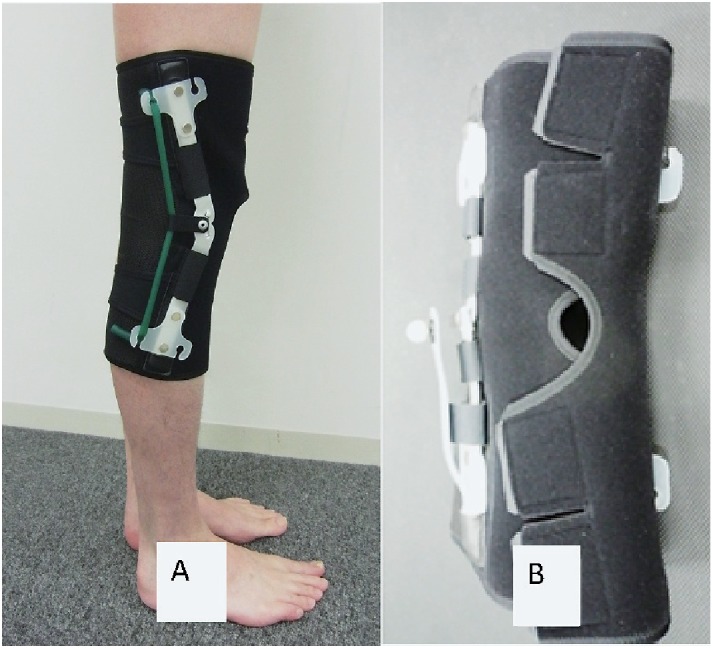
The custom-made knee brace with flexion support (KBF: Figure 1A) was modified based on the design of the general hinged knee brace (Geltex Light Sports, Nippon Sigmax Co., Ltd, Japan). Exercise Tubing (green tube, TheraBandTM, Hygenic Corporation, USA) was the elastic tube used for the actuator. The tubes were equipped at the posterior of the lateral and medial side poles. To determine the tube and the strength that applied to KBF, we tried three types of tube strength (1, 2 and 3 kg forces with these tubes extended and the hook attachable) for three healthy volunteers before this study. As a result, the tube strength (length) with 1 kg force could give the knee flexion support during the swing phase most effectively. Therefore, we used the tube strength (length) with 1 kg force for KBF in this study. The characteristics of the assistive knee flexion moment using the lateral and medial tubes were 0.44, 0.38, 0.32, 0.26, and 0.12 Nm, respectively, at 0, 15, 30, 45 and 60 degrees of knee flexion.
Fig. 1.

(A) A custom-made hinged knee brace with flexion support (KBF).
(B) Attachment for the knee joint marker.
Instrumentation
Three-dimensional trajectory data were obtained using an 8-camera motion analysis system (Vicon Nexus; Vicon Motion System, Oxford, UK). Trajectory data were sampled at 120 Hz and digitally recorded. Ground reaction forces were collected at a rate of 120 Hz using custom-made two-piece force plates (Kistler Z11942, 2×0.8 m, Z12091, 2×0.4 m; Kistler Instrument, Winterthur, Switzerland) embedded in the floor, and the force plate and 3D motion analysis system were synchronized. Thirty-five reflective sphere markers (14 mm in diameter) were attached to anatomical locations according to the VICON Plug-in Gait (full body model) marker placement protocol, as described in our previous study.11) Figure 1B shows the attachment of the knee joint marker using a custom-offset-arm. The knee joint marker was located at the same place as the lateral epicondyle on the sagittal plane and the attached marker plate in KB and KBF. Data collection was performed at the gait analysis laboratory of Toyohashi SOZO University.
Procedures
All subjects participated in two tasks: free gait with KB or KBF (Figure 1A). They all wore the same type of shirts and pants, and performed the tasks barefooted. Subjects were asked to traverse a 10-m walkway, and to then perform two trials by walking with KB and KBF, one time each.
Data analysis
Kinematic and kinetic data from the three trials were averaged for analysis. All data were normalized to 100% of a gait cycle which is from the foot contact on the floor to the next foot contact in the same measured limb. Peak KAM during the stance phase at which the foot is on the ground (approximately 0–60% of the gait cycle) and peak knee flexion angle during swing phase at which the foot was not in contact with the ground (approximately 60–100% of the gait cycle) were obtained as the main outcomes using Plug-in Gait model processing with force plate data. The kinematic data were filtered using the Woltring filter with a predicted mean squared error value of 20. Knee moments were calculated using inverse dynamics. The knee joint moments were normalized to body mass.
To determine differences existing between the two conditions, paired t-test was performed for each peak KAM during stance phase and peak knee flexion angles during swing phase. The difference was set for the significance level of p<0.05. All statistical analyses were performed with SPSS, Version 16.0 (IBM Japan, Tokyo, Japan).
RESULTS
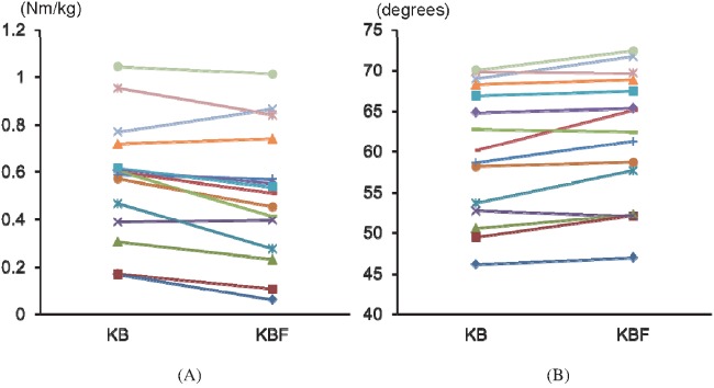
Table 1 shows the KAM during stance phase and knee flexion angle during swing phase under KB and KBF conditions. The peak KAM in KBF was significantly smaller than that of KB (p=0.004). The peak knee flexion angles of KBF during swing phase were significantly larger than those of KB conditions (p=0.003). The changes of each subject in two conditions were presented in Figure 2 (A) and (B).
Table 1.
Comparison of the peak knee adduction moment and knee flexion angle during swing phase between KB and KBF with t-test
| mean ± SD | KB | KBF | p |
|---|---|---|---|
| Peak knee adduction moment (Nm/kg) | 0.57 ± 0.25 | 0.51 ± 0.28 | 0.004 |
| Peak knee flexion angle (degrees) | 60.1 ± 8.1 | 61.6 ± 8.0 | 0.003 |
KB: hinged knee brace; KBF: hinged knee brace with knee flexion support.
Fig. 2.
Change of KB and KBF during swing phase. (A) The peak knee adduction moment, (B) the knee flexion angle.
DISCUSSION
KAM with KBF was decreased significantly comparing to that with KB, and the average change was 0.06 Nm/kg. The knee flexion angle with KBF during swing phase was significantly larger than that with KB, and the average change was 1.5 degrees. Because both of the average changes were small, we conducted the additional analysis below.
As the sub-analysis, the smallest real differences (SRD), used to indicate the magnitude of change (or differences between two brace conditions) that would exceed the expected trial-to-trial variability, were described in our previous study with the same measurement setting as in the present study.11) SRD was calculated using the following equation: 1.96×√–2×standard error of measurement.12) The SRDs of the peak KAM and knee flexion angles were 0.05 Nm/kg and 3.5 degrees, respectively.11) The difference of KAM between KB and KBF, 0.06 Nm/kg, was larger than the SRD of KAM. However, the difference of knee flexion angle between these conditions, 1.5 degrees, was smaller than the SRD of knee flexion angle.
It was considered that the adequate effects of KBF would be only the decrease of KAM at this tube strength. Previous reports have indicated that KAMs during gait are associated with medial compartment osteoarthritis.13-15) Therefore, KBF is considered to be a more effective brace than KB for patients with medial knee OA, because it decreases KAM. The KBF was attached to two of the same tubes of the same length on the lateral and medial posterior side of the brace poles. All subjects with medial knee OA had knee varus alignment, and the lateral tube of KBF was assumed to be more extended than the medial tube due to the knee varus deformity. Then the extended lateral tube was thought to give the varus deformity a straight alignment. This mechanism might have caused a decrease in KAM. However, the real strength of the correction and the different lengths of the lateral and medial tubes were not measured. Thus, we could not conclude the mechanism of the effect. Figure 2(A) shows that the effect might be obtained at around 0.6 Nm/kg, assuming therefore that the correction force for knee varus alignment would be small.
For the improvement with smaller than the SRD of the knee flexion angle, the small restriction of the knee flexion angle during the swing phase among the study participants could be attributed to the result. Typical knee flexion during swing phase of gait ranges from 60 to 70 degrees.16, 17) The subjects in the current study attained on average 60 degrees of knee flexion in these brace conditions suggesting that the brace conditions did not adversely affect the knee flexion angle during the swing. It is also possible that the knee flexion support force was not adequate to further assist in knee flexion at the larger angles. At 60 degrees of knee flexion, the knee flexion support force of KBF was only 0.12 Nm.
So far, a knee brace with two functions, which restricts KAM during stance phase and increases knee flexion angle during swing phase, has not been reported in the literature. An adequate effect was not obtained from the knee flexion angle using KBF in the present study. In future study, we must investigate whether the KBF has two functions due to changes in the tube length (tube strength).
There are limitations to this study. First, the sample size was small, and the participants whose improvement in knee flexion was investigated during swing phase should have included patients with decreased knee flexion angle. Second, there are no data of a no-brace condition as the control, because the knee marker of skin and the marker on the brace could not be attached in exactly the same place. Third, this study used one type of tube all of the same length. Therefore, the appropriate type and length of tube for each patient could not be found. The knee flexion angle during swing phase in the participants was close to the normal range. However, because the primary feature of KBF is the ability to change the strength of knee flexion assistance by using tubes of different lengths and strengths, adequate conditions for flexion support using a variety of tube strengths and lengths for patients with knee OA should be established in the future.
CONCLUSION
The effect of the custom-made hinged knee brace with knee flexion support for patients with knee OA was investigated to assess KAM during the stance phase and knee flexion angle during swing phase. The custom-made brace could significantly decrease KAM during the stance phase and increased the knee flexion angle during swing phase as opposed to the hinged knee brace. However, the knee flexion angle was only a small change within the smallest real difference in one type of tube actuator in the present study.
ACKNOWLEDGEMENTS
We are grateful to the participants and staff (students and faculty) of Toyohashi SOZO University.
Conflict of Interest
None
REFERENCES
- 1).Yoshimura N, Muraki S, Oka H, Mabuchi A, En-Yo Y, Yoshida M, Saika A, Yoshida H, Suzuki T, Yamamoto S, Ishibashi H, Kawaguchi H, Nakamura K, Akune T. Prevalence of knee osteoarthritis, lumbar spondylosis, and osteoporosis in Japanese men and women: the research on osteoarthritis/osteoporosis against disability study. J Bone Miner Metab, 2009; 27: 620–628. [DOI] [PubMed]
- 2).Ahlback S. Osteoarthrosis of the knee. A radiographic investigation. Acta Radiol Diagn (Stockh), 1968; Suppl 277: 7–72. [PubMed]
- 3).Walter JP, D’Lima DD, Colwell CW, Fregly BJ. Decreased knee adduction moment does not guarantee decreased medial contact force during gait. J Orthop Res, 2010; 28: 1348–1354. [DOI] [PMC free article] [PubMed]
- 4).Richards JD, Sanchez-Ballester J, Jones RK, Darke N, Livingstone BN. A comparison of knee braces during walking for the treatment of osteoarthritis of the medial compartment of the knee. J Bone Joint Surg Br, 2005; 87: 937–939. [DOI] [PubMed]
- 5).Self BP, Greenwald RM, Pflaster DS. A biomechanical analysis of a medial unloading brace for osteoarthritis in the knee. Arthritis Care Res, 2000; 13: 191–197. [DOI] [PubMed]
- 6).Pollo FE, Otis JC, Backus SI, Warren RF, Wickiewicz TL. Reduction of medial compartment loads with valgus bracing of the osteoarthritic knee. Am J Sports Med, 2002; 30: 414–421. [DOI] [PubMed]
- 7).Draganich L, Reider B, Rimington T, Piotrowski G, Mallik K, Nasson S. The effectiveness of self-adjustable custom and off-the-shelf bracing in the treatment of varus gonarthrosis. J Bone Joint Surg Am, 2006; 88: 2645–2652. [DOI] [PubMed]
- 8).Raja K, Dewan N. Efficacy of knee braces and foot orthoses in conservative management of knee osteoarthritis: a systematic review. Am J Phys Med Rehabil, 2011; 90: 247–262. [DOI] [PubMed]
- 9).Gok H, Ergin S, Yavuzer G. Kinetic and kinematic characteristics of gait in patients with medial knee arthrosis. Acta Orthop Scand, 2002; 73: 647–652. [DOI] [PubMed]
- 10).Altman R, Asch E, Bloch D, Bole G, Borenstein D, Brandt K, Christy W, Cooke TD, Greenwald R, Hochberg M, et al. Development of criteria for the classification and reporting of osteoarthritis. Classification of osteoarthritis of the knee. Diagnostic and Therapeutic Criteria Committee of the American Rheumatism Association. Arthritis Rheum, 1986; 29: 1039–1049. [DOI] [PubMed]
- 11).Ota S, Ueda M, Aimoto K, Suzuki Y, Sigward SM. Acute influence of restricted ankle dorsiflexion angle on knee joint mechanics during gait. Knee, 2014. [DOI] [PubMed]
- 12).Beckerman H, Roebroeck ME, Lankhorst GJ, Becher JG, Bezemer PD, Verbeek ALM. Smallest real difference, a link between reproducibility and responsiveness. Qual Life Res, 2001; 10: 571–578. [DOI] [PubMed]
- 13).Miyazaki T, Wada M, Kawahara H, Sato M, Baba H, Shimada S. Dynamic load at baseline can predict radiographic disease progression in medial compartment knee osteoarthritis. Ann Rheum Dis, 2002; 61: 617–622. [DOI] [PMC free article] [PubMed]
- 14).Landry SC, McKean KA, Hubley-Kozey CL, Stanish WD, Deluzio KJ. Knee biomechanics of moderate OA patients measured during gait at a self-selected and fast walking speed. J Biomech, 2007; 40: 1754–1761. [DOI] [PubMed]
- 15).Amin S, Luepongsak N, McGibbon CA, LaValley MP, Krebs DE, Felson DT. Knee adduction moment and development of chronic knee pain in elders. Arthritis Rheum, 2004; 51: 371–376. [DOI] [PubMed]
- 16).Perry J, ed. Gait analysis. 1992, Slack Incorporated: Thoroface.
- 17).Murray MP, Drought AB, Kory RC. Walking patterns of normal men. J Bone Joint Surg Am, 1964; 46: 335–360. [PubMed]


