

Original citation: J Clin Invest. 2014;124(4):1853–1867. doi:10.1172/JCI73531.
Citation for this corrigendum: J Clin Invest. 2015;125(3):1362. doi:10.1172/JCI81340.
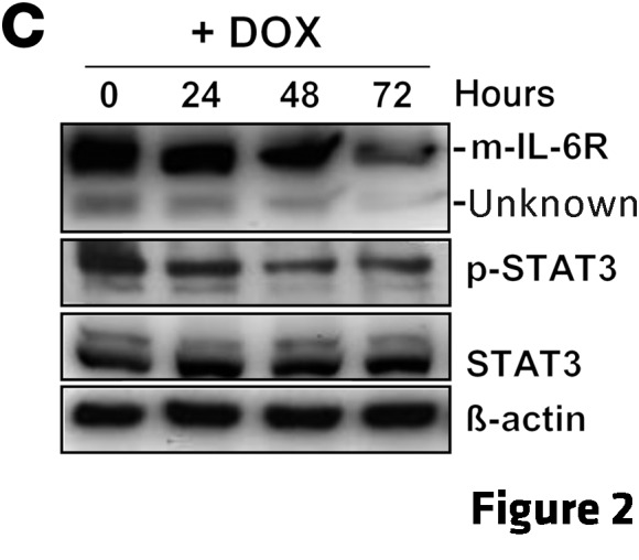
Following the publication of this article, the authors became aware that the epitope for the IL-6R antibody (SC-661, Santa Cruz Biotechnology Inc.) is not present in the soluble variant of IL-6R (s–IL-6R), which lacks the C-terminal domain. The authors originally designated the bands detected by SC-661 as membrane-bound IL-6R (m–IL-6R; ~70 kDa) and s–IL-6R (~50 kDa); however, the identity of the 50-kDa band is not known. Thus, no conclusions regarding s–IL-6R can be made from the data presented. Accordingly, the labels on Figures 2C, 3A, 3B, 3E, and 8D have been corrected to indicate that the 50-kDa band detected by SC-661 has not been characterized. The corrected figures appear at right.


In addition, this error affected portions of the text in the Results and Discussion. The corrected sentences appear below:
Results, 4th paragraph:
Besides the membrane-bound IL-6R (m–IL-6R), we also observed a signal at 50 kDa, which represents an uncharacterized band. Densitometric quantification of IL-6R signals with normalization to β-actin showed a statistically significant decrease in m–IL-6R after DOX treatment (Supplemental Figure 3A).
Results, 5th paragraph:
Interestingly, the mesenchymal-like CRC lines SW480 and SW620 showed a more pronounced expression of m–IL-6R when compared with 5 CRC lines with epithelial traits. Furthermore, the mesenchymal lines displayed STAT3 phosphorylation, which was not detectable in the epithelial lines.
Discussion, final paragraph:
For activation of STAT3, the IL-6R requires the ligand IL-6, which allows binding to gp130. In vivo, IL-6 can be generated by tumor cells or by tumor stromal cells, such as macrophages or fibroblasts (12). The RNA used for the expression analysis of human CRC samples was isolated from nonmicrodissected tumors. Therefore, several stromal cells could have contributed to IL-6 expression. The analysis of human CRC samples suggests that the IL-6R/STAT3/miR-34a loop is also manifest in primary human colorectal tumors with mesenchymal characteristics and might represent a useful prognostic marker for cancer progression. In line with these findings, we recently showed that the loss of miR-34a expression by epigenetic silencing in primary tumors is associated with increased lymph node infiltration and metastasis in colon cancer patients (57). Besides STAT3 and IL-6R, which are already established targets for cancer treatment, our results suggest that restoring miR-34a function using mimetics may have therapeutic potential for the treatment of invasive CRCs. Furthermore, recombinant, soluble gp130 may be suitable for the treatment of invasive CRC displaying MIR34A inactivation and therefore upregulation of IL-6R expression.
Finally, the following information in the supplemental data file has been corrected: (a) findings related to the quantification of soluble IL-6R protein expression have been removed, (b) the β-actin antibody utilized has been specified as Sigma-Aldrich (A2066), and (c) the unedited image for the β-actin blot corresponding to the p53 blot in Supplemental Figure 5A has been provided.
The authors regret the errors.
Footnotes
See the related article beginning on page 1853.