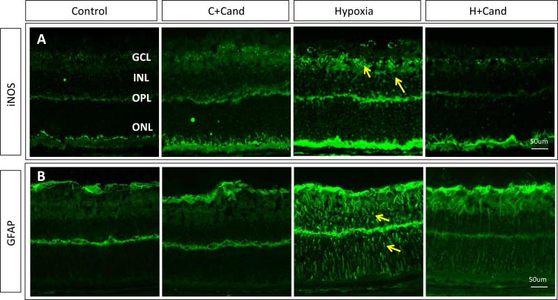
Figure- 4. Candesartan ameliorated hypoxia-induced Muller cell activation and suppressed iNOS expression.
(A) Representative images of immunostained retinas with iNOS (green), showing higher expression in hypoxia than normoxia or hypoxia-candesartan. (B) Representative images of retinas from different treatment groups stained with GFAP (green), a marker for stressed Muller cells. In hypoxia, Muller cell activation is remarkably higher compared to normoxia or normoxia-candesartan and the hypoxic retinas treated with candesartan. GCL- Ganglion Cell Layer; IPL- Inner Plexiform Layer; INL- Inner Nuclear Layer; ONL- Outer Nuclear Layer; DAPI-Diamidino-2-phenylindole; GFAP- Glial fibrillary acidic protein. Similar findings were observed in 3-additional retinas.

