Abstract
The array of diagnostic workup for pyrexia of unknown origin (PUO) generally revolves in searching for infections, inflammatory/autoimmune, and endocrine etiologies. A differential diagnosis of fever, hemolytic anemia, and thrombocytopenia can have etiologies varying from infections like malaria, dengue, cytomegalovirus, Ebstein barr virus, Parvovirus, infective endocarditis, to autoimmune disorder (systemic lupus erythromatosis), vasculitis, hemolytic uremic syndrome, thrombotic thrombocytopenic purpura (TTP), autoimmune hemolytic anemia/Evan's syndrome, paroxysmal nocturnal hemoglobinuri (PNH), or drugs. Nutritional deficiencies (especially vitamin B12 deficiency) as a cause of fever, hemolytic anemia, and thrombocytopenia are very rare and therefore rarely thought of. Severe vitamin B12 deficiency may cause fever and if accompanied by concurrent hyper-homocysteinemia and hypophosphatemia can sometimes lead to severe hemolysis mimicking the above-mentioned conditions. We present a case that highlights vitamin B12 and vitamin D deficiency as an easily treatable cause of PUO, hemolytic anemia, and thrombocytopenia, which should be actively looked for and treated before proceeding with more complicated and expensive investigation or starting empiric treatments.
Keywords: Fever, hemolytic anemia, homocysteine, hypophosphatemia, vitamin B12, vitamin D
Introduction
Nutritional deficiencies are the most common cause of anemia in the tropical countries.[1] Deficiencies of vitamin B12 and folate can cause severe anemia and cytopenias due to ineffective hematopoiesis and can sometimes mirror hemolytic anemia. Also megaloblastic anemia, presenting solely as pyrexia, can be found in only a small proportion of cases and is poorly characterized. This etiology can often be missed and delay the diagnosis if not actively looked for in cases pyrexia of unknown origin (PUO).
Case Report
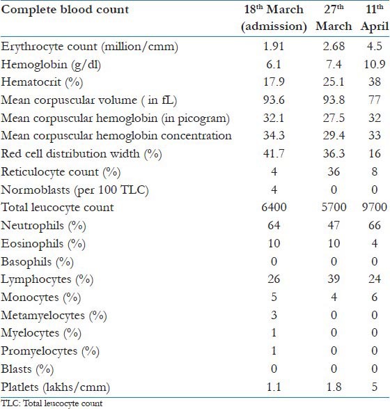
A 17 years old girl presented with complains of low-grade intermittent fever, fatigue, anorexia, and generalized weakness since 1 month. She had no other localizing symptom like cough, abdominal pain, diarrhea, headache, or burning micturition. She had been treated with oral antibiotics for fever by her local practitioner 15 days back, but symptoms did not resolve. Apart from this, she had normal menstruation, and there was no history of addictions or any other drug ingestion. Dietary history revealed that she was strict vegetarian since birth. She was admitted to our center for further workup. On examination, she was febrile with temperature of 38.80C, pulse rate was 104 beats/min, respiratory rate was 16/min, blood pressure was 110/70 mm of mercury. Her body mass index was around 20 kg/m2. Severe pallor, mild icterus, hyperpigmentation of knuckles was noted on general examination. There was no lymphadenopathy, clubbing, or pedal edema. Abdominal, respiratory, neurological clinical examinations were normal. Investigations revealed severe anemia and thrombocytopenia as shown in [Table 1].
Table 1.
Progression of complete blood count with treatment of vitamin B12 started on 23rd March and disappearance of fever on 26th March

Workup for fever did not reveal any abnormality. There was no evidence of malarial parasite on blood smear examinations on two occasions. Even antigen test for malaria and dengue (dengue NS1) were negative. Repeated blood cultures and urine culture were all negative. Widal test was negative. Thyroid and cortisol hormones were in normal range. Workup for anemia revealed peripheral blood smear showing anisocytosis, mild poikilocytosis, polychromasia, microcytosis, macrocytosis, occasional Cabbot rings, and Howel Jolley bodies. Presence of a schistocytes and hypersegmented neutrophils were the other abnormalities that were noted. Erythrocyte sedimentation rate (ESR) was 36 mm/h. Reticulocyte count was 4% with absolute reticulocyte count of 1.7% and reticulocyte index of 0.7 (reticulocyte index >2 constitutes a normal response). Serum iron studies showed serum iron-128 micro/l, serum iron saturation -34.6%, total iron binding capacity 370 micro/dl and serum ferritin was 68.62 ng/ml. Vitamin B12 level (by automated ELISA) was found to be very low at 77 pmol/L (normal-240-900 pmol/L). RBC folate and serum folate levels were 608 ng/ml and 5.85 ng/ml, respectively, i. e. within normal range. Anti-intrinsic factor and anti-parietal cell antibodies were absent. There was no evidence of active blood loss from any body site. Repeatedly done stool routine examination was normal. Urine examination was normal with RBC-0-1/high power field (hpf), pus cells-1-2/hpf. Urine testing for hemosiderin granules and hemoglobinuria was negative. Human immunodeficiency virus (HIV), hepatitis-B surface antigen (HBsAg), anti-hepatitis C virus antibodies, and veneral disease research lab (VDRL) tests were all negative. Glucose6 phosphatase (G6PD) activity in RBCs was normal. Serum haptoglobin measures (by immune turbidimetric method) were very low with 1 mg/100 ml (normal 90-200 mg/100 ml) suggestive of severe hemolysis. Direct and indirect COOMBS test were negative. Prothrombin time and activated prothrombin time were within normal range. Testing for antiphospholipid antibody was negative. Screening tests for PNH with acid HAM and sucrose lysis test were negative and complements levels (C3 and C4) were within normal limits. Hemoglobin electrophoresis was normal with HbA-97.1%, HbF-0%, and HbA2-2.9%. Tests for sickling and osmotic fragility were negative. Autoimmune diseases were ruled out as anti-nuculear antibodies (ANA), and rheumatoid factor (RA) were negative. Protein electrophoresis showed a normal albumin/globulin ratio and did not show presence of any M-band or cryoglobulins. Chest radiography showed normal lung fields and a normal mediastinum. Ultrasonography (USG) showed normal liver and spleen and kidneys. 2D echocardiography ruled out endocarditis. Bone marrow aspiration and biopsy were performed on the 3rd day revealed marked marrow hypercellularity, erythroid hyperplasia with increased normoblasts an increased erythroid to myeloid ratio. Also giant metamyelocytes and megakaryocyte were seen with adequate marrow iron stores. There was no evidence granulomas, hemoparasites, malignancy. IgM ELISA with for Ebstein Barr virus (EBV), cytomegalovirus (CMV), and parvo-virus were all negative. Other biochemical parameters showed serum albumin 4 g/dl, serum calcium-7.3 mg/dl, phosphorus-2.5mg/dl, vitamin D-3 ng/ml, total cholesterol-137 mg/dl, uric acid-5.1 mg/dl, creatinine-0.6 mg/dl, Creatine phosphokinase (CPK)-62mU/ml, total bilirubin-2.2 mg/dl with indirect bilirubin of 1.8 mg/dl, serum glutamate pyruvate transaminase (SGPT)-46 mu/ml, serum glutamate oxaloacetate transaminase (SGOT)-181 mu/l and a very high level of lactate dehydrogenase (LDH)-8051mu/ml. Serum homocystein levels were high 45 micromoles/l. After common infections, autoimmune conditions and other hemalotogical conditions had been excluded, a provisional diagnosis of vitamin B12 deficiency induced pyrexia, hemolytic anemia and thrombocytopenia was postulated and treatment was started with treatment of injection cyanocobalamin 1000 mcg OD supplemented, vitamin D 60,000 IU/week and calcium. Following treatment, there was improvement in blood parameters and patient became totally afebrile by the 4th day of starting treatment. After a week, intravenous therapy, patient was discharged on oral preparation of combination of vitamin B12 and pyridoxine for hyperhomocysteinemia. Patient had refused gastrointestinal endoscopy for further evaluation of vitamin B12 deficiency.
After 2 weeks, on follow-up, patient was afebrile and repeat levels of vitamin B12 levels were 2000 pg/l, LDH had decreased to-341 mU/ml, total bilirubin to 0.5 mg/dl (with indirect bilirubin-0.1mg/dl) and homocsyteine had decreased to12 micromoles/l. The repeat CBC is as shown in [Table 1].
Discussion
In our patient's, the dramatic response in fever and blood parameters to cobalamin supplements supports our theory that the pyrexia was attributable directly to nutritional vitamin B12 deficiency as other etiologies were adequately ruled out by suitable available tests.
Although the incidence of low-grade fever in nutritional megaloblastic anemia varies from 28% to 60%,[1] literature search suggests that fever as a presenting symptom of vitamin B12 deficiency is rare. There are only few case reports where vitamin B12 deficiency was solely attributed as the cause of pyrexia.[2,3,4] The exact cause of pyrexia in megaloblastic anemia is not known but a proposed mechanism is that megaloblastic anemia leads to hyperplasia and thus increased activity within the bone marrow leading to systemic pyrexia. In majority of patients with pyrexia due to vitamin B12 deficiency, patients have a minimal rise of temperature (≤38.5°C) as in our case. Rarely fever is greater than 38.5 °C as was seen in our case. Fever is more common is patients with severe anemia and in patients having high MCV, low hematocrit (<20%), thrombocytopenia (<100 × 109/ L), high LDH (>1000 IU/L), and unconjugated hyperbilirubinemia (>1.5 mg/dL),[4] which correlates with our case. In majority of the patients, this fever subsides 24-72 h after supplementation of vitamin B12 and/or folate, suggesting the rapid correction of ineffective hematopoiesis as was also seen in our case. The level of pyrexia usually correlates with severity of anemia,[3,4] but the levels of vitamin B12 do not exactly correlate with degree of pyrexia as seen in our case and few other cases.[2,3,4] The absence of neurological features at such a low vitamin B12 levels was notable.
Mechanisms of hemolysis in patients with vitamin B12 deficiency are not entirely understood, it is believed that the hemolysis results from intramedullary destruction as well as from pseudo-thrombotic microangiopathy (TMA) that accompanies vitamin B12 deficiency.[5,6] The patient reported here had severe microangiopathic hemolysis as evidenced by the very low levels of serum haptoglobin, with elevated LDH and indirect hyperbilirubinemia as well as the presence of schistocytes in the peripheral blood smears. Although normally vitamin B12 deficiency presents with high MCV, in our case, the near normal MCV could be explained by average size of macrocytes and schistocytes.
The role of homocysteine in increasing the risk of hemolysis in vitamin B12 and folate deficiency has been demonstrated in vitro.[7] We postulate that the high homocysteine levels in our case may have played a role in ensuing hemolysis in addition to intramedullary destruction of RBCs. With the additional impact of hyperhomocysteinemia on vitamin B12 deficiency, the hemolysis is more severe and occur in both intravascular and intramedullary settings, resulting in a drastic reduction of circulating red blood cell mass as was seen in our case. Our claim is supported by another recent study.[8]
The low levels of calcium and phosphorus at presentation prompted us to check for vitamin D levels, which were found to be very low. Vitamin D deficiency prevails in epidemic proportions all over the Indian subcontinent, with a prevalence of 70% -100% in the general population.[9] In India, widely consumed food items such as dairy products are rarely fortified with vitamin D. Indian socio-religious and cultural practices do not facilitate adequate sun exposure, thereby negating potential benefits of plentiful sunshine. Consequently, subclinical vitamin D deficiency is highly prevalent in both urban and rural settings, and across all socioeconomic and geographic strata as was seen in our patient.[9] The role of this hypovitaminosis D-induced hypophosphtemia may also have contributed to severity of hemolysis as effect of hypophosphatemia causing hemolysis is well known.[10] However, the levels of serum phosphorus may sometimes be normal or even spuriously increased in cases of severe hemolysis due to leakage of phosphorus from the RBCs[10] So concurrent presence of such deficiencies should be looked for and corrected for timely recovery.
A differential diagnosis of fever, hemolytic anemia, and thrombocytopenia can range from infections like malaria, dengue, cytomegalovirus, Ebstein barr virus, parvovirus, infective endocarditis, autoimmune disorder (systemic lupus erythromatosis), vasculitis, hemolytic uremic syndrome, thrombotic thrombocytopenic purpura (TTP), autoimmune hemolytic anemia/Evan's syndrome, and so on. All these etiologies were excluded by suitable tests in our case. TTP and HUS were ruled out due to absence of precipitating cause (though not always necessary), renal dysfunction and mental changes. The presence of very high LDH levels and a low reticulocyte count and a relatively milder thrombocytopenia in our patient also favors the cause as pseudo-thrombotic microangiopathy (TMA) caused by cobalamin deficiency over TTP.[5] Although testing for ADAMTS-13 levels would have been ideal if it is available to rule out TTP.
Conclusion
Vitamin B12 deficiency is an important and easily treatable cause of PUO. All patients presenting with pyrexia and cytopenia with hemolytic picture should be carefully evaluated for possible vitamin B12 and folate deficiency in order to prevent the unnecessary burden of investigations and treatments. More studies evaluating the possible roles of cytokine signaling and bone marrow stromal microenvironment might help in understanding the pathophysiological mechanism of pyrexia in megaloblastic anemia. Presence of hyperhomocysteinemia and hypovitaminosis D-induced hypophosphatemia in vitamin B12 deficiency are additional risk factor for severe hemolysis in megaloblastic anemias.
Footnotes
Source of Support: Nil.
Conflict of Interest: None declared.
References
- 1.Tahlan A, Bansal C, Palta A, Chauhan S. Spectrum and analysis of bone marrow findings in anemic cases. Indian J Med Sci. 2008;62:336–9. [PubMed] [Google Scholar]
- 2.Negi RC, Kumar J, Kumar V, Singh K, Bharti V, Gupta D, et al. Vitamin B12 deficiency presenting as pyrexia. J Assoc Physicians India. 2011;59:379–80. [PubMed] [Google Scholar]
- 3.Manuel K, Padhi S, G’ boy Varghese RG. Pyrexia in a patient with megaloblastic anemia: A case report and literature review. Iran J Med Sci. 2013;38:198–201. [PMC free article] [PubMed] [Google Scholar]
- 4.McKee LC., Jr Fever in megaloblastic anemia. South Med J. 1979;72:1423–4. doi: 10.1097/00007611-197911000-00023. 1428. [DOI] [PubMed] [Google Scholar]
- 5.Noël N, Maigné G, Tertian G, Anguel N, Monnet X, Michot JM, et al. Hemolysis and schistocytosis in the emergency department: Consider pseudothrombotic microangiopathy related to vitamin B12 deficiency. QJM. 2013;106:1017–22. doi: 10.1093/qjmed/hct142. [DOI] [PubMed] [Google Scholar]
- 6.Antony AC. Megaloblastic anemias. In: Hoffman R, Benz EJ Jr, Shattil SJ, Furie B, Cohen HJ, Silberstein LE, McGlave P, editors. Hematology, Basic Principles and Practices. 4th ed. Philadelphia, PA: Elsevier Churchill Livingstone; 2005. pp. 519–56. [Google Scholar]
- 7.Ventura P, Panini R, Tremosini S, Salvioli G. A role for homocysteine increase in haemolysis of megaloblastic anemias due to vitamin B12 and folate deficiency: Results from an in vitro experience. Biochim Biophys Acta. 2004;1739:33–42. doi: 10.1016/j.bbadis.2004.08.005. [DOI] [PubMed] [Google Scholar]
- 8.Acharya U, Gau JT, Horvath W, Ventura P, Hsueh CT, Carlsen W. Hemolysis and hyperhomocysteinemia caused by cobalamin deficiency: Three case reports and review of the literature. J Hematol Oncol. 2008;1:26. doi: 10.1186/1756-8722-1-26. [DOI] [PMC free article] [PubMed] [Google Scholar]
- 9.Ritu G, Gupta A. Vitamin D deficiency in India: Prevalence, causalities and interventions. Nutrients. 2014;6:729–75. doi: 10.3390/nu6020729. [DOI] [PMC free article] [PubMed] [Google Scholar]
- 10.FR, Demay MB, Krane SM, Kronenberg HM. Bone and mineral metabolism in health and diseases. In: Longo DL, Fauci AS, Kasper DL, Hauser SL, Jameson JL, editors. Harrison's Principles of Internal Medicine. 18th ed. New York: McGraw-Hill; 2012. pp. 3082–95. [Google Scholar]


