Abstract
Sirenomelia (mermaid syndrome) is a rare congenital fetal anomaly with characteristic feature of complete or partial fusion of lower limbs. Although, this syndrome is incompatible with life due to the association of several congenital visceral abnormalities; however, there are few reports of surviving infants. Our first case was a live born, normally delivered at term by a 27-year-old third gravida of lower socioeconomic status with history of tobacco use. Examination of the baby revealed caudal dysgenesis having fusion of lower limbs, single leg with 1 foot and 5 toes. There was no identifiable external genitalia and anus. The second case was a 34 week, 1.6 kg preterm infant of unidentified sex born to a 28-year-old primigravida mother with overt diabetes mellitus. Incidentally, both the infants died few hours after birth and we report these cases due to their rarity and term live birth.
Keywords: Caudal regression syndrome, mermaid syndrome, Potter's facies, sirenomelia
INTRODUCTION
Sirenomelia is a rare and fatal congenital defect characterized by varying degrees of lower limb fusion, thoracolumbar spinal anomalies, sacrococcygeal agenesis, genitourinary, and anorectal atresia.[1] The incidence of sirenomelia is 0.8-1 case/100,000 births with male to female ratio being 3:1.[2] The rarity of the case is obvious from the fact that many a gynecologist might not have come across a case of sirenomelia in their whole professional carrier. There is a strong association with maternal diabetes where relative risk is 1:200-250 and up to 22% of fetuses with this anomaly will have mothers with diabetes.[3,4] We report two cases of sirenomelia where maternal drug abuse and overt diabetes may have been the cause of this rare anomaly.
CASE REPORTS
Case 1
A 27-year-old unbooked G3P1L1A1 at 39 weeks 5 days of gestational age with previous one live vaginal birth and one first trimester spontaneous abortion was admitted in the labor room with pain in the abdomen. She had no history of prior antenatal care and belonged to a tribal community with lower socioeconomic status. There was history of tobacco use both before and during pregnancy. She was otherwise healthy with no known history of genetic or congenital anomaly in her family.
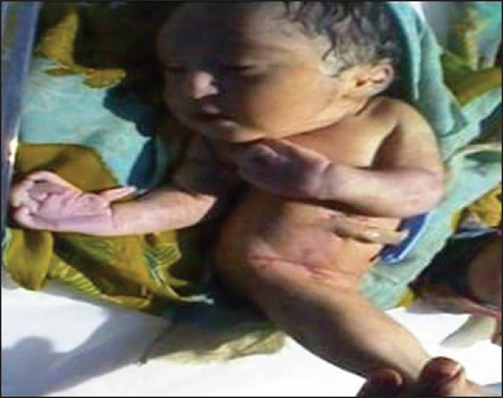
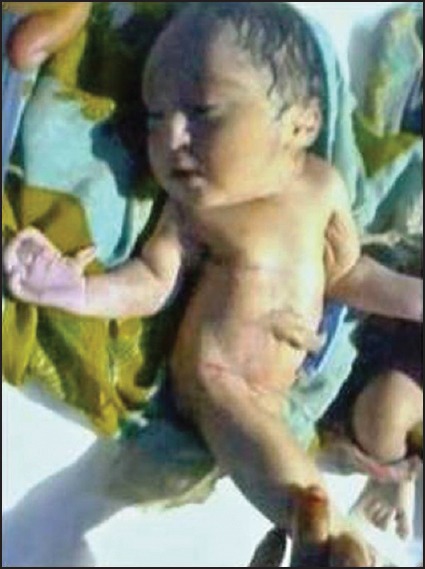
On examination, she was observed to be in the second stage of labor with cephalic presentation and regular fetal heart rate. She delivered a term 2.5 kg baby with multiple congenital anomalies. The Apgar score was 3 at 1’ and 0 at 5 min. The baby died within 30 min postbirth in spite of resuscitation attempts by neonatologist. On physical examination, the infant showed narrow chest, bilateral hypoplastic thumb, fused lower limbs with a single foot and 5 toes, absent external genitalia, imperforate anus and umbilical cord with single umbilical artery [Figure 1]. There were also prominent epicanthal folds, hypertelorism, downward curved nose, receding chin, low-set soft dysplastic ears and small slit-like mouth suggestive of Potter's facies [Figure 2]. Autopsy was declined by the parents. Intrapartum and the postpartum period of mother was uneventful.
Figure 1.

Photograph of the baby showing fusion of lower limbs, hypoplastic thumb, absent external genitalia and features of Potter's facies
Figure 2.

Sirenomeliac baby with narrow chest and Potter's facies (prominent infraorbital folds, small slit-like mouth, receding chin, downward curved nose, and low-set soft dysplastic ears)
Case 2
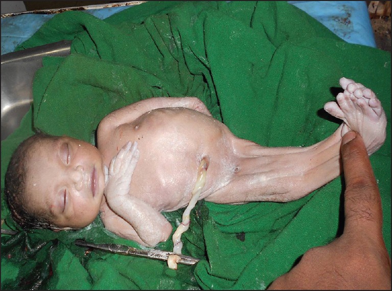
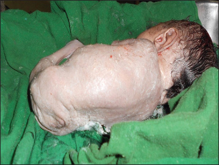
A preterm baby weighing 1.6 kg was delivered vaginally at 34 weeks gestation by a 23-year-old primigravida with an unsupervised pregnancy. Postpartum investigation revealed the presence of diabetes mellitus. There was no history of drug intake and radiation exposure. The Apgar score was 3 at 1’ and same at 5 min following which the baby was shifted to neonatal intensive care unit, but died 12 h postbirth due to severe respiratory distress. There was very scanty amniotic fluid drained at the time of delivery. The new born baby had gross anomalies like narrow chest indicating lung hypoplasia, fused both lower limbs and feet with 10 toes, absence of external genitalia, imperforate anus and single umbilical artery [Figures 3 and 4]. Examination of the fused lower limbs showed the presence of all thigh and leg bones thus classifying our patient as Type I of Stocker and Heifetz classification. The infant also had features of Potter's facies including prominent infraorbital folds, small slit-like mouth, receding chin, downward curved nose, and low-set ears. Ultrasonography revealed bilateral renal agenesis. On autopsy, there was an absence of both kidneys, ureters, urinary bladder, seminal vesicle, and urethra. The gastrointestinal system ended in a blind loop at the rectosigmoid area and was filled with meconium. Two pea sized gonads suggestive of testes were seen bilaterally posterior to pubis. Right pneumothorax with collapsed right lung was evident. Examination of brain, heart, liver, adrenal glands, and pancreas revealed normal anatomy.
Figure 3.

Sirenomeliac baby with fused lower limbs containing 10 toes, Potter's facies, narrow chest, and absent external genitalia
Figure 4.

Photograph of baby showing imperforate anus
DISCUSSION
Anomalies observed in sirenomelia are described as the most severe form of caudal regression syndrome.[5] Fusion of the lower extremities, presence of single umbilical and persistent vitelline artery are major features of sirenomelia.[6]
Although the primary molecular defect resulting in sirenomelia remains unclear, two main pathogenic hypotheses namely the vascular steal hypothesis and the defective blastogenesis hypothesis are proposed. According to vascular steal hypothesis,[7] fusion of the limbs results from a deficient blood flow and nutrient supply to the caudal mesoderm, which in turn results in agenesis of midline structures and subsequent abnormal approximation of both lower limb fields. However in defective blastogenesis hypothesis,[8] the primary defect in development of caudal mesoderm is attributed to a teratogenic event during the gastrulation stage. Such defect interferes with the formation of notochord, resulting in abnormal development of caudal structures. Maternal diabetes, tobacco use, retinoic acid and heavy metal exposure are possible environmental factors.[9] In our first case, there was history of tobacco use before and during pregnancy, while in the second case the mother had overt diabetes.
Sirenomelia is usually fatal within a day or two of birth because of complications associated with abnormal kidney and urinary bladder development and function. In literature approximately 300 cases[5] are reported worldwide of which 14 are from India. In most of the cases the diagnosis was performed after birth. In antenatal period, sirenomelia can be diagnosed as early as 13 weeks by using high resolution or color Doppler sonography.[10,11] The condition is usually incompatible with life due visceral abnormalities especially that of renal system. Exceptional cases without renal agenesis have survived, the best example being Tiffany Yorks, a 13-year-old girl who was born with fused legs. Over the years, she has undergone numerous operations to separate her lower extremities.[12]
The facial abnormality usually found in sirenomeliac infants known as Potter's facies, which includes large, low-set ears, prominent epicanthic fold, hypertelorism, flat nose and receding chin. In both of our cases, features of Potter's facies were present. When features of Potter's facies are combined with oligamnios and pulmonary hypoplasia it is known as Potter's syndrome,[13] which was present in our second case. In our first case, the right thumb was hypoplastic, which was also previously reported.[14] Stocker and Heifetz classified Sirenomeliac infants from Type I to Type VII according to the presence or absence of bones within the lower limb.[15] Although we did not have radiographs to classify our case with certainty, nevertheless based on external examination, we suggest our first and second case belonged to Type IV (partially fused femurs and fused fibula) and Type I (all thigh and leg bones are present), respectively.
CONCLUSION
Sirenomelia is a rare and lethal congenital anomaly. When diagnosed antenatally, termination should be offered. However, prevention is possible and should be the goal. Regular antenatal checkup with optimum maternal blood glucose level in preconceptional period and in first trimester should be maintained to prevent this anomaly.
ACKNOWLEDGMENT
We would like to thank the Department of OBGYN, SCB Medical College, Cuttack for their valuable support and co-operation of patients and their families admitted to this hospital.
Footnotes
Source of Support: Nil.
Conflict of Interest: None declared.
REFERENCES
- 1.Valenzano M, Paoletti R, Rossi A, Farinini D, Garlaschi G, Fulcheri E. Sirenomelia. Pathological features, antenatal ultrasonographic clues, and a review of current embryogenic theories. Hum Reprod Update. 1999;5:82–6. doi: 10.1093/humupd/5.1.82. [DOI] [PubMed] [Google Scholar]
- 2.Reddy KR, Srinivas S, Kumar S, Reddy S, Hariprasad Irfan GM. Sirenomelia a rare presentation. J Neonatal Surg. 2012;1:7. [PMC free article] [PubMed] [Google Scholar]
- 3.Aslan H, Yanik H, Celikaslan N, Yildirim G, Ceylan Y. Prenatal diagnosis of Caudal regression syndrome: A case report. BMC Pregnancy Childbirth. 2001;1:8. doi: 10.1186/1471-2393-1-8. [DOI] [PMC free article] [PubMed] [Google Scholar]
- 4.González-Quintero VH, Tolaymat L, Martin D, Romaguera RL, Rodríguez MM, Izquierdo LA. Sonographic diagnosis of caudal regression in the first trimester of pregnancy. J Ultrasound Med. 2002;21:1175–8. doi: 10.7863/jum.2002.21.10.1175. [DOI] [PubMed] [Google Scholar]
- 5.Duhamel B. From the Mermaid to Anal Imperforation: The syndrome of Caudal regression. Arch Dis Child. 1961;36:152–5. doi: 10.1136/adc.36.186.152. [DOI] [PMC free article] [PubMed] [Google Scholar]
- 6.Twickler D, Budorick N, Pretorius D, Grafe M, Currarino G. Caudal regression versus sirenomelia: Sonographic clues. J Ultrasound Med. 1993;12:323–30. doi: 10.7863/jum.1993.12.6.323. [DOI] [PubMed] [Google Scholar]
- 7.Sadler TW, Rasmussen SA. Examining the evidence for vascular pathogenesis of selected birth defects. Am J Med Genet A. 2010;152A:2426–36. doi: 10.1002/ajmg.a.33636. [DOI] [PubMed] [Google Scholar]
- 8.Duesterhoeft SM, Ernst LM, Siebert JR, Kapur RP. Five cases of caudal regression with an aberrant abdominal umbilical artery: Further support for a caudal regression-sirenomelia spectrum. Am J Med Genet A. 2007;143A:3175–84. doi: 10.1002/ajmg.a.32028. [DOI] [PubMed] [Google Scholar]
- 9.Naveena S, Mrudula C. Sirenomelia — The mermaid syndrome: A case report. IOSR J Dent Med Sci. 2013;7:01–4. [Google Scholar]
- 10.Vijayaraghavan SB, Amudha AP. High-resolution sonographic diagnosis of sirenomelia. J Ultrasound Med. 2006;25:555–7. doi: 10.7863/jum.2006.25.4.555. [DOI] [PubMed] [Google Scholar]
- 11.Sahu L, Singh S, Gandhi G, Agarwal K. Sirenomelia: A case report with literature review. Int J Reprod Contracept Obstet Gynecol. 2013;2:430–2. [Google Scholar]
- 12.Shah DS, Tomar G, Preetkiran Sirenomelia. Indian J Radiol Imaging. 2006;16:203–4. [Google Scholar]
- 13.Dharmraj M, Gaur S. Sirenomelia: A rare case of foetal congenital anomaly. J Clin Neonatol. 2012;1:221–3. doi: 10.4103/2249-4847.106006. [DOI] [PMC free article] [PubMed] [Google Scholar]
- 14.Banerjee A, Faridi MM, Banerjee TK, Mandal RN, Aggarwal A. Sirenomelia. Indian J Pediatr. 2003;70:589–91. doi: 10.1007/BF02723165. [DOI] [PubMed] [Google Scholar]
- 15.Stocker JT, Heifetz SA. Sirenomelia. A morphological study of 33 cases and review of the literature. Perspect Pediatr Pathol. 1987;10:7–50. [PubMed] [Google Scholar]


