

Abstract
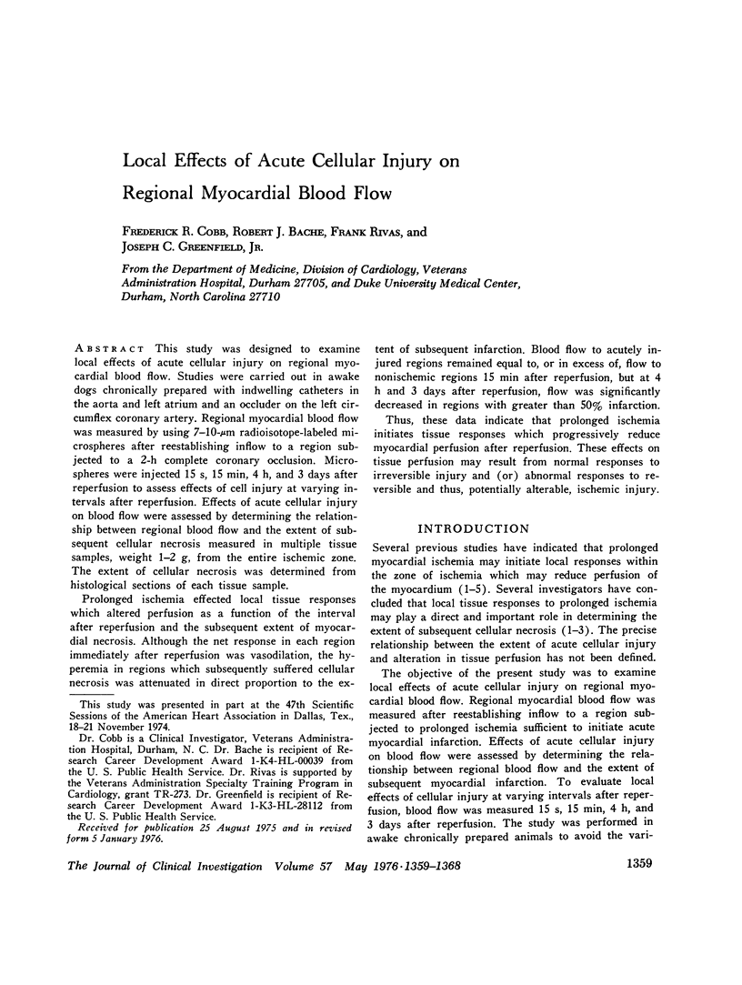
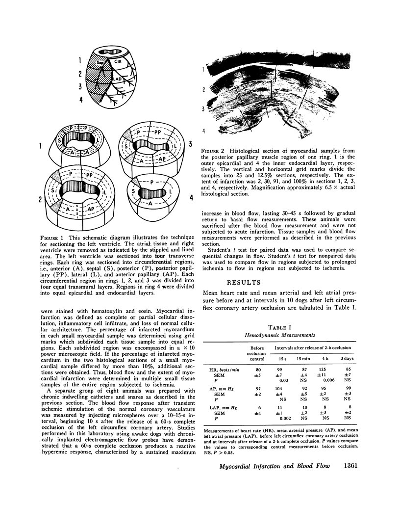
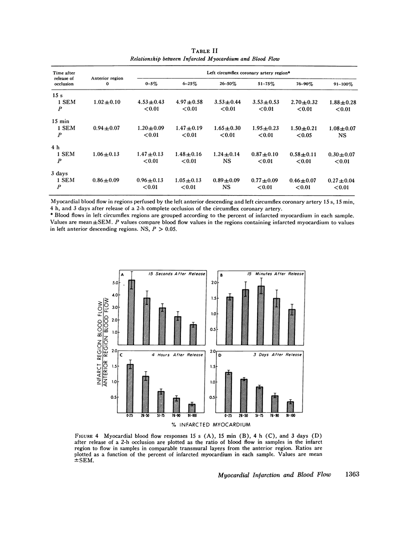
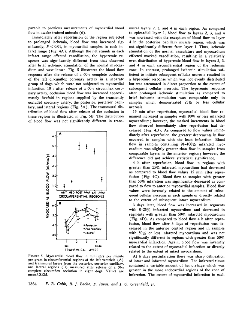
This study was designed to examine local effects of acute cellular injury on regional myocardial blood flow. Studies were carried out in awake dogs chronically prepared with indwelling catheters in the aorta and left atrium and an occluder on the left circumflex coronary artery. Regional myocardial blood flow was measured by using 7-10-mum radioisotope-labeled microspheres after reestablishing inflow to a region subjected to a 2-h complete coronary occlusion. Microspheres were injected 15 s, 15 min, 4 h, and 3 days after reperfusion to assess effects of cell injury at varying intervals after reperfusion. Effects of acute cellular injury on blood flow were assessed by determining the relationship between regional blood flow and the extent of subsequent cellular necrosis measured in multiple tissue samples, weight 1-2 g, from the entire ischemic zone. The extent of cellular necrosis was determined from histological sections of each tissue sample. Prolonged ischemia effected local tissue responses which altered perfusion as a function of the interval after reperfusion and the subsequent extent of myocardial necrosis. Although the net response in each region immediately after reperfusion was vasodilation, the hyperemia in regions which subsequently suffered cellular necrosis was attenuated in direct proportion to the extent of subsequent infarction. Blood flow to acutely injured regions remained equal to, or in excess of, flow to nonischemic regions 15 min after reperfusion, but at 4 h and 3 days after reperfusion, flow was significantly decreased in regions with greater than 50% infarction. Thus, these data indicate that prolonged ischemia initiates tissue responses which progressively reduce myocardial perfusion after reperfusion. These effects on tissue perfusion may result from normal responses to irreversible injury and (or) abnormal responses to reversible and thus, potentially alterable, ischemic injury.
Full text
PDF
Images in this article
Selected References
These references are in PubMed. This may not be the complete list of references from this article.
- Braunwald E., Maroko P. R. The reduction of infarct size--an idea whose time (for testing) has come. Circulation. 1974 Aug;50(2):206–209. doi: 10.1161/01.cir.50.2.206. [DOI] [PubMed] [Google Scholar]
- Cobb F. R., Bache R. J., Greenfield J. C., Jr Regional myocardial blood flow in awake dogs. J Clin Invest. 1974 Jun;53(6):1618–1625. doi: 10.1172/JCI107712. [DOI] [PMC free article] [PubMed] [Google Scholar]
- Cox J. L., McLaughlin V. W., Flowers N. C., Horan L. G. The ischemic zone surrounding acute myocardial infarction. Its morphology as detected by dehydrogenase staining. Am Heart J. 1968 Nov;76(5):650–659. doi: 10.1016/0002-8703(68)90164-6. [DOI] [PubMed] [Google Scholar]
- Flores J., DiBona D. R., Beck C. H., Leaf A. The role of cell swelling in ischemic renal damage and the protective effect of hypertonic solute. J Clin Invest. 1972 Jan;51(1):118–126. doi: 10.1172/JCI106781. [DOI] [PMC free article] [PubMed] [Google Scholar]
- Grayson J., Irvine M., Parratt J. R., Cunningham J. Vasospastic elements in myocardial infarction following coronary occlusion in the dog. Cardiovasc Res. 1968 Jan;2(1):54–62. doi: 10.1093/cvr/2.1.54. [DOI] [PubMed] [Google Scholar]
- Hellstrom H. R. Coronary artery stasis after induced myocardial infarction in the dog. Cardiovasc Res. 1971 Jul;5(3):371–375. doi: 10.1093/cvr/5.3.371. [DOI] [PubMed] [Google Scholar]
- JENNINGS R. B., SOMMERS H. M., SMYTH G. A., FLACK H. A., LINN H. Myocardial necrosis induced by temporary occlusion of a coronary artery in the dog. Arch Pathol. 1960 Jul;70:68–78. [PubMed] [Google Scholar]
- Kloner R. A., Ganote C. E., Jennings R. B. The "no-reflow" phenomenon after temporary coronary occlusion in the dog. J Clin Invest. 1974 Dec;54(6):1496–1508. doi: 10.1172/JCI107898. [DOI] [PMC free article] [PubMed] [Google Scholar]
- Kloner R. A., Ganote C. E., Whalen D. A., Jr, Jennings R. B. Effect of a transient period of ischemia on myocardial cells. II. Fine structure during the first few minutes of reflow. Am J Pathol. 1974 Mar;74(3):399–422. [PMC free article] [PubMed] [Google Scholar]
- Leaf A. Cell swelling. A factor in ischemic tissue injury. Circulation. 1973 Sep;48(3):455–458. doi: 10.1161/01.cir.48.3.455. [DOI] [PubMed] [Google Scholar]
- Maroko P. R., Kjekshus J. K., Sobel B. E., Watanabe T., Covell J. W., Ross J., Jr, Braunwald E. Factors influencing infarct size following experimental coronary artery occlusions. Circulation. 1971 Jan;43(1):67–82. doi: 10.1161/01.cir.43.1.67. [DOI] [PubMed] [Google Scholar]
- Redding V. J., Rees J. R. Early changes in collateral flow following coronary artery ligation: the role of the sympathetic nervous system. Cardiovasc Res. 1968 Jul;2(3):219–225. doi: 10.1093/cvr/2.3.219. [DOI] [PubMed] [Google Scholar]
- Rubio R., Berne R. M. Release of adenosine by the normal myocardium in dogs and its relationship to the regulation of coronary resistance. Circ Res. 1969 Oct;25(4):407–415. doi: 10.1161/01.res.25.4.407. [DOI] [PubMed] [Google Scholar]
- Willerson J. T., Watson J. T., Hutton I., Templeton G. H., Fixler D. E. Reduced myocardial reflow and increased coronary vascular resistance following prolonged myocardial ischemia in the dog. Circ Res. 1975 Jun;36(6):771–781. doi: 10.1161/01.res.36.6.771. [DOI] [PubMed] [Google Scholar]