Abstract
Purpose
The knowledge of the function of the collateral ligaments – i.e., superficial medial collateral ligament (sMCL), deep medial collateral ligament (dMCL) and lateral collateral ligament (LCL) – in the entire range of knee flexion is important for soft tissue balance during total knee arthroplasty. The objective of this study was to investigate the length changes of different portions (anterior, middle and posterior) of the sMCL, dMCL and LCL during in vivo weightbearing flexion from full extension to maximal knee flexion.
Methods
Using a dual fluoroscopic imaging system eight healthy knees were imaged while performing a lunge from full extension to maximal flexion. The length changes of each portion of the collateral ligaments were measured along the flexion path of the knee.
Results
All anterior portions of the collateral ligaments were shown to have increasing length with flexion except that of the sMCL which showed a reduction in length at high flexion. The middle portions showed minimal change in lengths except that of the sMCL which showed a consistent reduction in length with flexion. All posterior portions showed reduction in lengths with flexion.
Conclusions
These data indicated that every portion of the ligaments may play important roles in knee stability at different knee flexion range. The soft tissue releasing during TKA may need to consider the function of the ligament portions along the entire flexion path including maximum flexion.
Keywords: In vivo Length Change Pattern, Medial Collateral Ligament, Lateral Collateral Ligament, Lunge
Introduction
Soft tissue balance is a critical procedure during the total knee arthroplasty (TKA) surgery where the minimal tensions in the medial and lateral collateral ligaments (MCL and LCL) are evenly maintained intra-operatively at full extension and 90° of flexion [6,7,9,26,31,32]. Soft tissue balancing beyond 90° of flexion has not been indicated in previous TKA procedures [8,33]. Most contemporary TKAs could result in knee flexion, in average, between 100 to 130° [20,24,34]. High flexion TKAs have been pursued over decades that are aimed to accommodate knee flexion up to 150° [14,15,21]. However, how to balance the soft tissues in high flexion has not been clearly reported in literature.
Numerous investigations have been reported on the biomechanics of the knee ligaments [1,16,17,19,31,35]. In situ forces of the MCL and LCL during high flexion have been reported using cadaveric knees [35]. Park et al. [31] studied the in vivo elongation of the MCL and LCL from full extension to 90° flexion of the knee by dividing the MCL and LCL into three functional portions using MRI and dual fluoroscopic system. Bergamini et al. [2] described the relevant lengths of cruciate and collateral ligaments from 0° to 90° of flexion using skin markers and stereophotogrammetry in movement analysis of cadaver knees. However, the in vivo functions of the collateral ligaments during full range of weightbearing flexion and especially in high flexion are not reported in literature.
The objective of this study is to investigate the length changes of different portions of the superficial MCL (sMCL), deep MCL (dMCL) and LCL of the knee during weightbearing flexion from full extension to maximal knee flexion in living subjects. The length changes of the collateral ligaments at high flexion angles were specifically examined. It was hypothesized that during high flexion of the knee, different portions of the collateral ligaments have different patterns of length changes and may play a significantly different role in knee stability. This study emphasizes that every portion of the colateral ligaments may play different roles in knee stability in whole flexion range of the knee and may be useful for addressing biomechanical restrictions to the knee in high flexion.
Materials and Methods
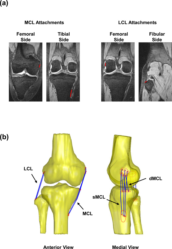
Eight right knees from eight healthy subjects (mean ± SD age: 32±11.8 years; mean ± SD body mass index: 24.2±3.5 Kg/m2; 6 males and 2 females; all right legged) with no history of knee injuries or chronic knee pain were recruited with institutional review board (IRB) approval. One knee of each subject was imaged using a 3.0T MR scanner (Siemens, Malvern, PA) with a fat suppressed 3D spoiled gradient recalled sequence. Sagittal and coronal images with a 1 mm thickness were captured in a 180 mm × 180 mm field of view and a resolution of 512 × 512 pixels. These images were used to create 3D anatomical models of the femur, tibia and fibula in a solid modeling software (Rhinoceros©, Robert McNeel & Assoc., Seattle, WA) [4], together with the femoral, tibial and fibular attachment areas of the sMCL, dMCL and LCL [22,23,31] (Fig. 1a). The collateral ligaments were each evenly divided into three portions: the anterior (AP), middle (MP) and posterior portions (PP) [3,31] (Fig. 1b) by dividing each attachment width in to 3 equal parts, and connecting the centroids of corresponding femoral and tibial (or fibular) segments with a line.
Figure 1.
(a) Coronal plane MR images of the knee were digitized and used to create the femoral and tibial attachment areas of the MCL and LCL. (b) Anterior view (left) and medial view (right) of a 3D knee model created using Sagittal plane MR images. The knee model includes the 3D surface of the femur, tibia and fibula (yellow), and the attachments of the medial and lateral collateral ligaments. Each Ligament was evenly divided into 3 portions (blue curves), by connecting the centroids of corresponding segments (red circles).
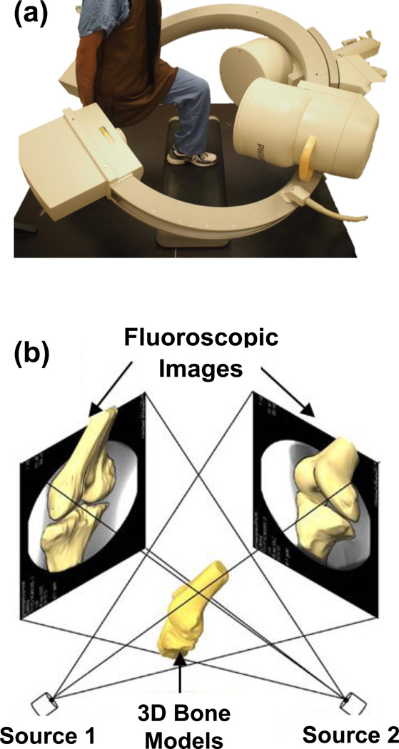
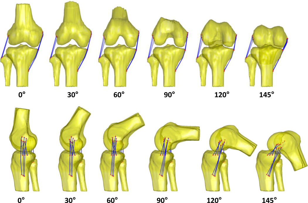
After MR scanning, each subject performed a quasi-static single-legged lunge from full extension to maximal flexion in the field of view of two orthogonally positioned fluoroscopes (BV Pulsera, Philips, Bothell, WA). The subjects were instructed to hold the knee position for one second at each selected flexion angle (~ every 30° from full extension to maximal flexion of 145°) and allowed to use the contralateral leg and a handrail to keep their body stable if necessary (Fig. 2a). Flexion angles of the knee were monitored using a goniometer. The orthogonal fluoroscopic images and the 3D model of the knee were imported into a virtual space that replicated the dual fluoroscopic imaging system in the solid modeling software (Fig. 2b) [4,17]. The outline of each bone was extracted from the fluoroscopic images. The projections of the 3D femur, tibia and fibula models were matched to their corresponding outlines on the fluoroscopic images to reproduce the 3D positions of the knee joint bones at each tested flexion angle (Fig. 3). The attachment areas of each ligament were therefore determined along the flexion path of the knee.
Figure 2.
(a) A subject performing a quasi-static deep lunge (right knee) in the dual fluoroscopic imaging system. (b) Reproduction of kinematics of the knee in a virtual DFIS. Each bony model was individually matched to the corresponding fluoroscopic images, viewed from two different directions.
Figure 3.
Anterior and medial view of the length changes of the collateral ligaments for a typical knee from full extension to maximal flexion. The lengths of each portion were measured along the path of flexion.
An optimization procedure was implemented to find the shortest 3D wrapping path of each ligament portion around the bones at each tested flexion angle of the knee and the lengths of each portion were determined (Fig. 3). This technique has been described in previous studies for measurements of ligament kinematics [4,13,17,29,31]. The initial length of each ligament portion was defined as the length measured at the full extension position. The length change of the portion was calculated as the length change relative to that of measured at full extension position (i.e., (l - l0) / (l0); where l0 is the initial length at full extension) and reported in percentage (%). Data Interpolation was used to determine exact values at each exact flexion angle during data analysis.
The accuracy of the above procedure for reproducing knee joint kinematics has been extensively evaluated [5,12,18]. Using standard geometries, the system has an accuracy of 0.1 mm in translation and 0.1° in rotation [18]. For human knee joint, the system has an accuracy of 0.1 mm in translation and a repeatability of 0.3° in rotation [5].
Statistical analysis
A two-way analysis of variance (ANOVA) with repeated measures and Newman-Keuls post hoc test were used to detect statistically significant differences in the change of the length of each ligament’s portion at each tested flexion angle. The dependent variables were defined as the length changes of the AP, MP and PP of the sMCL, dMCL and LCL. The flexion angle and the ligament portion were independent variables. The length changes of the ligament portions were analyzed along the flexion path. A statistically significant difference was obtained when p < 0.05.
Based on preliminary data on the length change patterns of the collateral ligaments along the knee flexion path from 0° to 90° of flexion [31], with an alpha of 5%, and power of 85%, 8 subjects were needed. The statistical analysis was performed using Statistica® (StatSoft, Inc, Tulsa, OK).
Results
sMCL
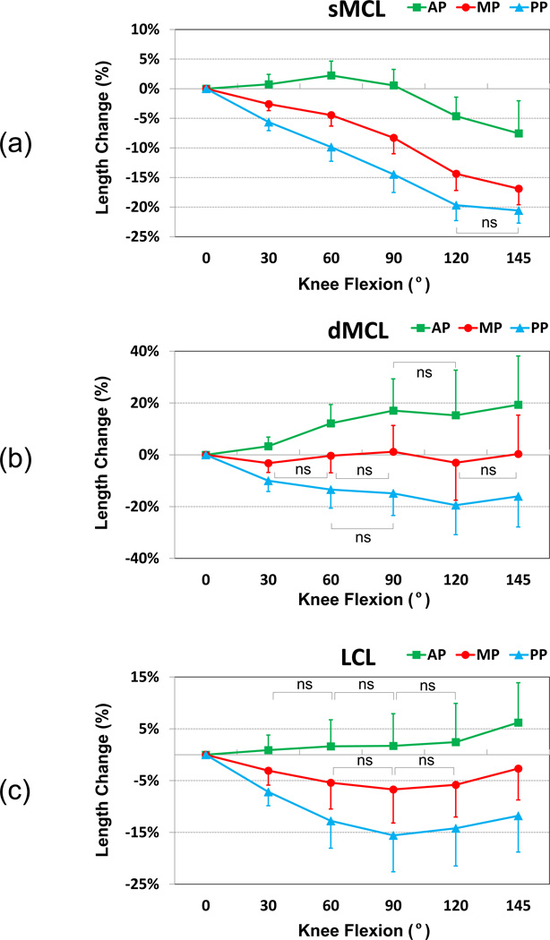
The AP length increased from full extension to 60° of flexion by about 2.2% (Fig. 4a). Beyond 90°, the AP length decreased consistently and reached −7.6% at maximal flexion (p<0.001). The MP length was slowly reduced with flexion to −8.3% at 90° of flexion (p<0.001). Beyond 90°, the MP length decreased sharply and reached −16.9% at maximal flexion (p<0.001). The PP length was reduced consistently along the flexion path and reached −20.6% at maximal flexion (p<0.001). The relative length changes of the sMCL portions (AP vs. MP vs. PP) were statistically significantly different at all flexion angles from full extension to maximum knee flexion- except for PP between 120° and maximum flexion, Fig. 4a. During the path of knee flexion, the greatest length reduction was observed in the posterior portion of the SMCL (−20.6%, Fig. 4a).
Figure 4.
The length changes of the (a) sMCL, (b) dMCL and (c) LCL normalized in to their initial lengths at full extension. Means and standard deviations are shown.
dMCL
The AP length increased from full extension to 90° of flexion by 17.1% (p<0.05) (Fig. 4b). Thereafter, the AP length was rather constant until 135°. At maximal flexion, the AP length slightly increased to 19.3% (p<0.01). The MP length was close to isometric along the flexion path with a variation within ±3.2%. The PP length decreased consistently to −19.4% from full extension to 120° of flexion (p<0.001). Thereafter, it slightly increased back to −16% at maximal flexion. The relative length changes of the dMCL portions (AP vs. MP vs. PP) were statistically significantly different at all knee flexion angles. The AP showed the greatest elongation along the knee flexion path, while the PP showed a significant length reduction.
LCL
The patterns of the relative length change of the LCL portions (AP vs. MP vs. PP) were statistically significantly different at all knee flexion angles with maximum elongation in the anterior portion and maximum length reduction in the posterior portion of the LCL (Fig. 4c). The AP length did not apparently change from full extension to 120° of flexion (Fig. 4c). At maximal flexion, the AP length slightly increased 6.2% (p<0.01). The MP length consistently decreased to −6.7% from full extension to 90° of flexion (p<0.01). Thereafter, the MP length slowly increased and reached −2.7% at maximal flexion. The PP length consistently decreased to −15.6% from full extension to 90° of flexion (p<0.001). Thereafter, it slightly increased back to −11.8% at maximal flexion.
Discussion
The most important finding of the present study was that different portions of the collateral ligaments had distinct functional roles along the in vivo knee flexion path from full extension to maximal knee flexion. The anterior portions were shown to have increasing length with flexion except that of the sMCL showing reduction in length at high flexion. The middle portions had minimal changes in lengths except that of the sMCL showing consistent reduction in length with flexion. The posterior portions showed reduction in lengths with flexion.
Few studies have studied the function of the collateral ligaments in knee joint stability [30,34,35]. In a cadaveric study, Yang et al. measured a peak force in the MCL at 60° of knee flexion and minimal forces at higher flexion angles. This is consistent with our observation on elongation patterns of the sMCL where the AP was elongated the most at 60° of flexion. Park et al. reported length changes of the collateral ligaments from full extension to 90° of flexion that were similar in trend with our data at low flexion range of the knee (i.e., in the MCL, the AP length increased from full extension to 90° of flexion, the MP length was close to isometric, and the PP length decreased consistently. In the LCL, the AP length did not considerably change, while the PP length consistently decreased).
The length change patterns of the collateral ligament may provide important implications to soft tissue balancing during TKA. The AP of the sMCL reached the longest at 60° and then consistently decreased till maximal flexion. Therefore, releasing the AP of the sMCL may affect the knee before 120° of flexion as the AP is rather evenly elongated in this range of flexion. However, as both MP and PP showed consistently decreasing in length with flexion, releasing the MP and PP may be more efficient to release the medial tightness of the knee in low flexion angles and may not affect the knee at high flexion angles.
The length of the AP of the dMCL peaked at 90° of flexion and the length reached the maximum at maximal flexion. Meanwhile, the length change pattern of the PP was opposite to the AP from full extension to 90° of flexion where the length of the AP peaked at maximal flexion and the PP peaked at full extension. The length of the MP of the dMCL retained consistently from full extension to maximal flexion of the knee. It is unclear in contemporary TKAs if the dMCL is completely resected due to joint opening and medial meniscus removal. Based on the data of normal knees, removing the AP could affect the knee in mid- and high-ranges of flexion. The PP can affect the knee at low flexion angles. The MP will affect the knee from full extension to maximal flexion of the knee. The data indicated that dMCL functions in the entire range of knee flexion. To retain the normal function of the knee after surgery, it may be important to maintain the dMCL function.
The length of the AP of the LCL consistently increased from full extension to maximal flexion and the lengths of MP and PP decreased from full extension to 90° of flexion and slightly increased from 90° to maximal flexion. Therefore, releasing of the AP during surgery could affect the knee at entire flexion range, especially at high flexion angles. Both the MP and the PP of the LCL can affect the lateral tightness at low flexion angles.
It should be noted that this study was based on the in vivo weightbearing quasi-static single-legged lunge. The reference length of each ligament is unknown. Therefore, the length change of each ligament with respect to its length at full extension was calculated. This could provide the ligament length change patterns along the flexion path, but it is technically challenging to determine in vivo ligament strains. As the knee kinematics is loading dependent, future studies should investigate the functions of the collateral ligaments during various dynamic flexion-extension activities of the knee, especially in high flexion activities. It is shown that body mass index (BMI) as well as body fat alter the kinematics of dynamic activities such as gait [28]. MBI can affect the muscular strength and static balance control [10]. BMI and body fat may affect knee kinematics in high knee flexion. Therefore, it is interesting to investigate the effect of body mass index on the high flexion of the knee. Gender and race may also have an influence on knee kinematics in high flexion due to different bone anatomy [11,25], different patterns of muscle recruitment and muscle activation [27], and different daily activities/habits (such as kneeling and squatting in Asians). It is warranted to recruit more subjects in future to analyze the gender and race effect on collateral ligaments of the knee in high flexion angles. Despite these limitations, this study provided quantitative data on the path dependent length change patterns of the collateral ligaments during a weightbearing high flexion of the knee joint. The data may have useful implications for development of knee replacement surgical procedures that are aimed at addressing the biomechanical restrictions to the knee in high flexion. Future investigations should validate these conclusions using intra observations or cadaveric experimental setups.
Conclusion
Understanding of the in vivo length change patterns of the knee ligaments is essential for optimizing soft tissue balancing during TKAs in order to achieve full range of flexion after the surgery. The data of this study indicated that different portions of the collateral ligaments function differently along the flexion path of the knee. Clinically, the soft tissue releasing during TKA may need to consider the effect of the ligament portions along the entire flexion path, not only at full extension and 90° of flexion.
Acknowledgement
The authors gratefully acknowledge the support of the National Institutes of Health (R01 AR055612), and the scholarship provided by the Chinese PLA General Hospital.
Footnotes
Conflict of Interest
There is no potential conflict of interest to declare.
Reference
- 1.Amiri S, Cooke TD, Wyss UP. A multiple-bundle model to characterize the mechanical behavior of the cruciate ligaments. Knee. 2011;18:34–41. doi: 10.1016/j.knee.2010.01.003. [DOI] [PubMed] [Google Scholar]
- 2.Bergamini E, Pillet H, Hausselle J, Thoreux P, Guerard S, Camomilla V, Cappozzo A, Skalli W. Tibio-femoral joint constraints for bone pose estimation during movement using multi-body optimization. Gait & posture. 2011;33:706–711. doi: 10.1016/j.gaitpost.2011.03.006. [DOI] [PubMed] [Google Scholar]
- 3.Blankevoort L, Huiskes R. Ligament-bone interaction in a three-dimensional model of the knee. J Biomech Eng. 1991;113:263–269. doi: 10.1115/1.2894883. [DOI] [PubMed] [Google Scholar]
- 4.DeFrate LE, Gill TJ, Li G. In vivo function of the posterior cruciate ligament during weightbearing knee flexion. Am J Sports Med. 2004;32:1923–1928. doi: 10.1177/0363546504264896. [DOI] [PubMed] [Google Scholar]
- 5.DeFrate LE, Papannagari R, Gill TJ, Moses JM, Pathare NP, Li G. The 6 Degrees of Freedom Kinematics of the Knee After Anterior Cruciate Ligament Deficiency: An In Vivo Imaging Analysis. Am J Sports Med. 2006;34:1240–1246. doi: 10.1177/0363546506287299. [DOI] [PubMed] [Google Scholar]
- 6.Delport H, Labey L, De Corte R, Innocenti B, Vander Sloten J, Bellemans J. Collateral ligament strains during knee joint laxity evaluation before and after TKA. Clin Biomech (Bristol, Avon) 2013;28:777–782. doi: 10.1016/j.clinbiomech.2013.06.006. [DOI] [PubMed] [Google Scholar]
- 7.Engh GA. The difficult knee: severe varus and valgus. Clin Orthop Relat Res. 2003:58–63. doi: 10.1097/01.blo.0000092987.12414.fc. [DOI] [PubMed] [Google Scholar]
- 8.Ghosh KM, Merican AM, Iranpour F, Deehan DJ, Amis AA. Length-change patterns of the collateral ligaments after total knee arthroplasty. Knee Surg Sports Traumatol Arthrosc. 2012;20:1349–1356. doi: 10.1007/s00167-011-1824-2. [DOI] [PubMed] [Google Scholar]
- 9.Halewood C, Risebury M, Thomas NP, Amis AA. Kinematic behaviour and soft tissue management in guided motion total knee replacement. Knee Surg Sports Traumatol Arthrosc. 2014 doi: 10.1007/s00167-014-2933-5. [DOI] [PubMed] [Google Scholar]
- 10.Handrigan G, Hue O, Simoneau M, Corbeil P, Marceau P, Marceau S, Tremblay A, Teasdale N. Weight loss and muscular strength affect static balance control. Int J Obes (Lond) 2010;34:936–942. doi: 10.1038/ijo.2009.300. [DOI] [PubMed] [Google Scholar]
- 11.Hoshino Y, Wang JH, Lorenz S, Fu FH, Tashman S. The effect of distal femur bony morphology on in vivo knee translational and rotational kinematics. Knee Surg Sports Traumatol Arthrosc. 2012;20:1331–1338. doi: 10.1007/s00167-011-1661-3. [DOI] [PMC free article] [PubMed] [Google Scholar]
- 12.Hosseini A, Gill TJ, Li G. In vivo anterior cruciate ligament elongation in response to axial tibial loads. J Orthop Sci. 2009;14:298–306. doi: 10.1007/s00776-009-1325-z. [DOI] [PMC free article] [PubMed] [Google Scholar]
- 13.Jordan SS, DeFrate LE, Nha KW, Papannagari R, Gill TJ, Li G. The in vivo kinematics of the anteromedial and posterolateral bundles of the anterior cruciate ligament during weightbearing knee flexion. Am J Sports Med. 2007;35:547–554. doi: 10.1177/0363546506295941. [DOI] [PubMed] [Google Scholar]
- 14.Kim YH, Choi Y, Kim JS. Range of motion of standard and high-flexion posterior cruciate-retaining total knee prostheses a prospective randomized study. J Bone Joint Surg Am. 2009;91:1874–1881. doi: 10.2106/JBJS.H.00769. [DOI] [PubMed] [Google Scholar]
- 15.Kim YH, Choi Y, Kwon OR, Kim JS. Functional outcome and range of motion of high-flexion posterior cruciate-retaining and high-flexion posterior cruciate-substituting total knee prostheses. A prospective, randomized study. J Bone Joint Surg Am. 2009;91:753–760. doi: 10.2106/JBJS.H.00805. [DOI] [PubMed] [Google Scholar]
- 16.Konig C, Matziolis G, Sharenkov A, Taylor WR, Perka C, Duda GN, Heller MO. Collateral ligament length change patterns after joint line elevation may not explain midflexion instability following TKA. Med Eng Phys. 2011;33:1303–1308. doi: 10.1016/j.medengphy.2011.06.008. [DOI] [PubMed] [Google Scholar]
- 17.Li G, DeFrate LE, Sun H, Gill TJ. In vivo elongation of the anterior cruciate ligament and posterior cruciate ligament during knee flexion. Am J Sports Med. 2004;32:1415–1420. doi: 10.1177/0363546503262175. [DOI] [PubMed] [Google Scholar]
- 18.Li G, Wuerz TH, DeFrate LE. Feasibility of using orthogonal fluoroscopic images to measure in vivo joint kinematics. J Biomech Eng. 2004;126:314–318. doi: 10.1115/1.1691448. [DOI] [PubMed] [Google Scholar]
- 19.Li G, Zayontz S, Most E, DeFrate LE, Suggs JF, Rubash HE. In situ forces of the anterior and posterior cruciate ligaments in high knee flexion: an in vitro investigation. J Orthop Res. 2004;22:293–297. doi: 10.1016/S0736-0266(03)00179-7. [DOI] [PubMed] [Google Scholar]
- 20.Li N, Tan Y, Deng Y, Chen L. Posterior cruciate-retaining versus posterior stabilized total knee arthroplasty: a meta-analysis of randomized controlled trials. Knee Surg Sports Traumatol Arthrosc. 2012 doi: 10.1007/s00167-012-2275-0. [DOI] [PubMed] [Google Scholar]
- 21.Lionberger DR, Eggers MD, Brewer KE, Fang L. Improved knee flexion following high-flexion total knee arthroplasty. J Orthop Surg Res. 2012;7:22. doi: 10.1186/1749-799X-7-22. [DOI] [PMC free article] [PubMed] [Google Scholar]
- 22.Liu F, Gadikota HR, Kozanek M, Hosseini A, Yue B, Gill TJ, Rubash HE, Li G. In vivo length patterns of the medial collateral ligament during the stance phase of gait. Knee Surg Sports Traumatol Arthrosc. 2011;19:719–727. doi: 10.1007/s00167-010-1336-5. [DOI] [PMC free article] [PubMed] [Google Scholar]
- 23.Liu F, Yue B, Gadikota HR, Kozanek M, Liu W, Gill TJ, Rubash HE, Li G. Morphology of the medial collateral ligament of the knee. J Orthop Surg Res. 2010;5:69. doi: 10.1186/1749-799X-5-69. [DOI] [PMC free article] [PubMed] [Google Scholar]
- 24.Luo SX, Zhao JM, Su W, Li XF, Dong GF. Posterior cruciate substituting versus posterior cruciate retaining total knee arthroplasty prostheses: a meta-analysis. Knee. 2012;19:246–252. doi: 10.1016/j.knee.2011.12.005. [DOI] [PubMed] [Google Scholar]
- 25.Mendiguchia J, Ford KR, Quatman CE, Alentorn-Geli E, Hewett TE. Sex differences in proximal control of the knee joint. Sports Med. 2011;41:541–557. doi: 10.2165/11589140-000000000-00000. [DOI] [PMC free article] [PubMed] [Google Scholar]
- 26.Mihalko WM, Whiteside LA. Bone resection and ligament treatment for flexion contracture in knee arthroplasty. Clin Orthop Relat Res. 2003:141–147. doi: 10.1097/01.blo.0000030512.43495.74. [DOI] [PubMed] [Google Scholar]
- 27.Padua DA, Carcia CR, Arnold BL, Granata KP. Gender differences in leg stiffness and stiffness recruitment strategy during two-legged hopping. J Mot Behav. 2005;37:111–125. doi: 10.3200/JMBR.37.2.111-126. [DOI] [PMC free article] [PubMed] [Google Scholar]
- 28.Page Glave A, Di Brezzo R, Applegate DK, Olson JM. The effects of obesity classification method on select kinematic gait variables in adult females. J Sports Med Phys Fitness. 2014;54:197–202. [PubMed] [Google Scholar]
- 29.Papannagari R, DeFrate LE, Nha KW, Moses JM, Moussa M, Gill TJ, Li G. Function of posterior cruciate ligament bundles during in vivo knee flexion. Am J Sports Med. 2007;35:1507–1512. doi: 10.1177/0363546507300061. [DOI] [PubMed] [Google Scholar]
- 30.Park SE, DeFrate LE, Suggs JF, Gill TJ, Rubash HE, Li G. The change in length of the medial and lateral collateral ligaments during in vivo knee flexion. Knee. 2005;12:377–382. doi: 10.1016/j.knee.2004.12.011. [DOI] [PubMed] [Google Scholar]
- 31.Park SE, DeFrate LE, Suggs JF, Gill TJ, Rubash HE, Li G. Erratum to "The change in length of the medial and lateral collateral ligaments during in vivo knee flexion". Knee. 2006;13:77–82. doi: 10.1016/j.knee.2004.12.012. [DOI] [PubMed] [Google Scholar]
- 32.Sasanuma H, Sekiya H, Takatoku K, Takada H, Sugimoto N. Evaluation of soft-tissue balance during total knee arthroplasty. J Orthop Surg (Hong Kong) 2010;18:26–30. doi: 10.1177/230949901001800106. [DOI] [PubMed] [Google Scholar]
- 33.Schirm AC, Jeffcote BO, Nicholls RL, Jakob H, Kuster MS. Sensitivity of knee soft-tissues to surgical technique in total knee arthroplasty. Knee. 2011;18:180–184. doi: 10.1016/j.knee.2010.05.005. [DOI] [PubMed] [Google Scholar]
- 34.Sumino T, Gadikota HR, Varadarajan KM, Kwon YM, Rubash HE, Li G. Do high flexion posterior stabilised total knee arthroplasty designs increase knee flexion? A meta analysis. Int Orthop. 2011;35:1309–1319. doi: 10.1007/s00264-011-1228-4. [DOI] [PMC free article] [PubMed] [Google Scholar]
- 35.Yang Z, Wickwire AC, Debski RE. Development of a subject-specific model to predict the forces in the knee ligaments at high flexion angles. Med Biol Eng Comput. 2010;48:1077–1085. doi: 10.1007/s11517-010-0653-7. [DOI] [PubMed] [Google Scholar]


