Abstract
Triceps tendon disruption is a rare orthopaedic injury that can lead to poor outcomes if misdiagnosed or managed inappropriately. This case report illustrates the importance of early, precise diagnosis of triceps rupture by clinical and radiological examination with appropriate management. A weightlifter who had fallen while riding his bike presented with pain, swelling around the posterior aspect of the left arm just above the elbow. Physical examination revealed ecchymosis and weakness in elbow extension. A radiograph of the elbow showed a small fleck of bone proximal to the tip of the olecranon. The patient was initially stabilised. Early intervention in the form of primary tendon repair was performed within 3 days and rehabilitation was started. The patient improved significantly to his best possible functional status with Mayo elbow score of 85. Early intervention was the key to better prognosis.
Background
Distal triceps tendon rupture is a very rare entity, representing less than 1% of all upper extremity tendon injuries.1 Acute rupture of the distal triceps tendon is usually missed because of associated swelling and pain resulting from fracture. Early accurate diagnosis is important because primary repair of the tendon is possible up to 3 weeks after injury.2 Reconstruction is a much more complex procedure and postoperative recovery is slower.2
Case presentation
A 40-year-old man, a weightlifter, presented to our casualty department after having had a fall while riding a two wheeler. He was in pain and had a swelling over the posterior aspect of the left elbow, which was increasing. He gave a history of anabolic steroid use over the past 6 months. There was no other significant medical history. On examination, there was ecchymosis and moderate swelling over the posterior aspect of the left arm just above the elbow, and there was weakness in extension of the elbow joint (figure 1). On motor examination, the grade of power was 1/5. Active extension of the elbow, keeping the arm by the side of the trunk, was possible probably due to controlled action of flexors. The patient did not have tingling or numbness in his hand. Active flexion of the elbow from 10° to 120° was possible. Owing to coexistent swelling, no appreciable tendon defect was palpated.
Figure 1.
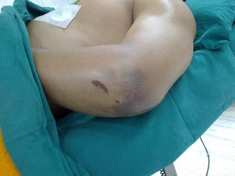
Image showing swelling and contusion around the elbow in a case of triceps rupture.
Investigations
Radiographs showed a small fleck of bone (arrow) proximal to the tip of the olecranon (figure 2).
Figure 2.
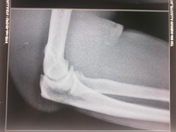
Avulsed osseous fragment from the olecranon.
MRI showed rupture of triceps with a large haematoma; the anconeus was intact (figure 3).
Figure 3.
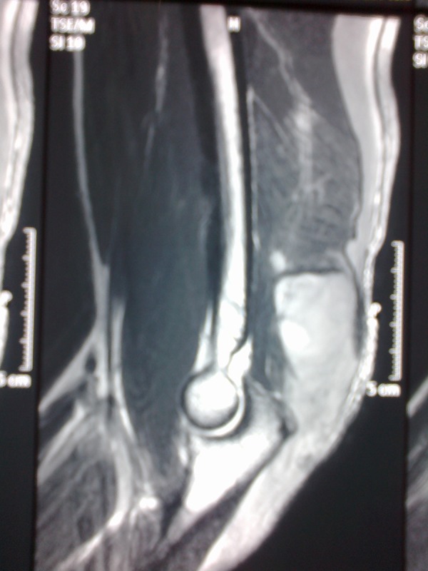
Sagittal view of T2-weighted MRI showing triceps tendon disruption with large haematoma.
Treatment
In this case, repair of the triceps tendon, which was completely torn, was performed on the third day after injury. A Krackow stitch was applied on the ruptured end of the triceps (figures 4 and 5). A four suture strand instead of the conventional two strand was used for better strength and more contact surface. Intraoperatively, the fleck of bone could not be identified. Intraoperative range of movement was 0–120°. The elbow was placed in a posterior splint at 20° flexion for approximately 5 days, after which gentle passive motion was allowed. Flexion was limited to 90°. At the end of 3 weeks, flexion past 90° was encouraged and managed with active assistance. At 4–6 weeks, active flexion and extension against gravity were allowed but no forced extension was permitted for an additional 4 weeks.3 At 12 weeks, the patient was allowed active triceps resistance exercises with a light weight of about 10 pounds. Gradually, resistance exercises were increased and at 1-year follow-up the patient is able to do tricep resistance exercises with heavier weights (figure 6). Mayo elbow scoring system at 1 year is 85 out of 100.
Figure 4.
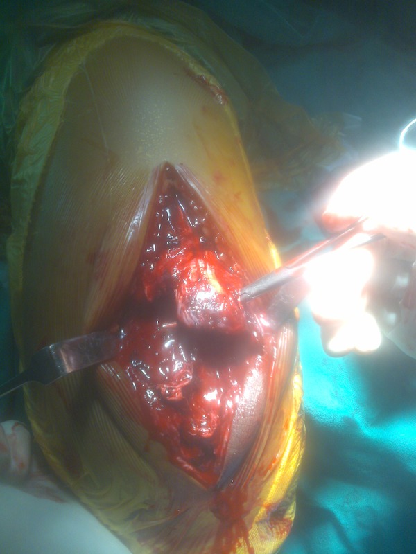
Intraoperative photograph displaying triceps tendon disruption.
Figure 5.
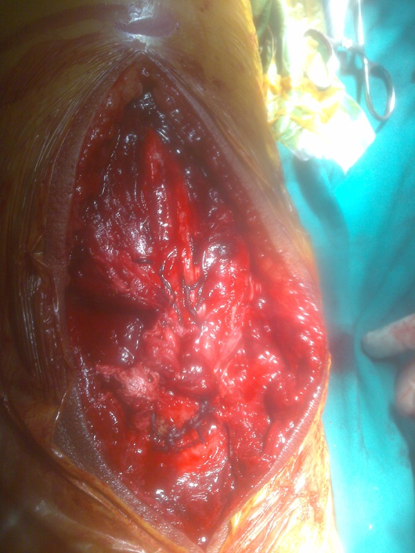
Triceps tendon after repair.
Figure 6.
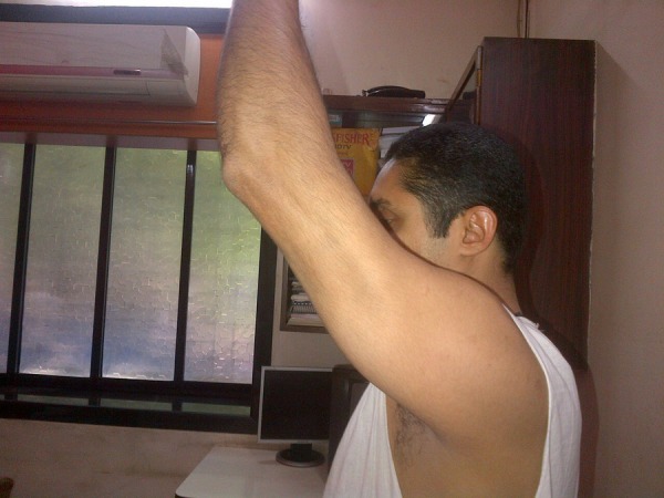
After 12 months, range of movement is full with adequate regain in muscle strength and movement against gravity and Mayo elbow scoring system score of 85.
Outcome and follow-up
The patient has been followed up for 1 year and is able to perform active resistance exercises with heavier weights and is kept under follow-up for further rehabilitation. On the latest examination, the patient's grade of power was 5/5 and Mayo elbow scoring system rate was 85 out of 100.
Discussion
Distal triceps tendon rupture is an infrequently seen condition in orthopaedics.4 It is the least common of all tendon injuries.1 Avulsion or rupture of the distal triceps tendon is usually the result of trauma and is seen more often in men aged 30–50 years who are involved in active sports such as football, boxing or weightlifting.5 Indirect trauma is the most common cause of injury and usually results from falling on an outstretched arm, resulting in pain and swelling at the elbow. Medical conditions that may increase the propensity to tendon disruption include hyperparathyroidism,6 chronic renal failure and haemodialysis, metabolic bone disease, olecranon bursitis,7 and systemic and anabolic steroid use.8 The mechanism of disruption involves a deceleration stress on the contracted triceps muscle, with or without a concomitant blow to the posterior aspect of the elbow, resulting in distal avulsion at the tendo-osseous insertion.9 The tendon usually retracts, and bone from the proximal olecranon becomes embedded in it, resulting in fracture.3
Diagnosis of distal triceps tendon rupture is usually prone to be missed when the injury is in the acute phase because of swelling and pain associated with the fracture. Although plain radiographs are helpful in ruling out other elbow pathology, MRI is used to confirm the diagnosis and guide management.10 In management of triceps avulsion it is important to differentiate between partial and complete tears. If a patient can perform active elbow extension against gravity (power of triceps >3/5), the injury is partial and can be treated non-operatively with splint protection for 4 weeks at 30° flexion. If a tear of more than 50% is shown on MRI together with significant loss of triceps extension (power <3/5), then operative repair of the torn tendon is recommended.9 In our case, the grade of power was 1/5 on examination hence extension against gravity was not possible. As per various studies, operative repair employing Krackow stitch was performed.
The most common form of repair reattaches the tendon through drill holes in the olecranon.9 Other techniques include reattachment to the olecranon periosteal flap, or to the intraosseous anchor. In cases where an avulsed flake of bone from the tendon's insertion on olecranon is of reasonable size, fixation with Kirschner wires and a tension band may be attempted.11 Graft reinforcement of the reattachment using the palmaris longus, plantaris, semitendinosus, Achilles tendon,2 7 9 12 anconeus muscle rotation,13 a flap from the proximal tricipital aponeurosis, or forearm fascia, may be necessary in patients with local or systemic risk factors for tendon rupture, those with chronic retracted tears and when tissue quality is in question. We think that the best way to obtain a solid insertion is to repair tendon insertion with a transosseous reinsertion.
Various suturing techniques for tendon repair include Bunnell, Kessler, Tajima, Tsuge, Pennington,14 Krackow,10 etc. We used the Krackow stitch technique on the ruptured end of the triceps. A four suture strand instead of the conventional two strand decreased the period of immobilisation.
Postoperative physiotherapy in the form of post splint at 20° flexion, increasing flexion by 10° every week with passive elbow extension up to 6 weeks, active elbow extension from 6 weeks and active elbow extension against resistance from 12 weeks was the usual protocol in various studies.15–18
Potential postoperative complications include olecranon bursitis secondary to wire suturing, flexion contracture, irritation from underlying internal fixation, rerupture, complex regional pain syndrome, infection, heterotopic ossification and compartment syndrome.2 19
Learning points.
For every injury involving the elbow, triceps rupture should be included in the differential diagnosis.
It is important to classify triceps ruptures as either complete or partial. This helps in guiding management.
Primary repair of the triceps rupture as early as possible gives good results.
Patients should be appropriately rehabilitated.
Footnotes
Contributors: SR wrote the primary manuscript. DS and AP critically analysed the manuscript and made corrections. JJB made final corrections before submission.
Competing interests: None.
Patient consent: Obtained.
Provenance and peer review: Not commissioned; externally peer reviewed.
References
- 1.Anzel SH, Covey KW, Weiner AD et al. Disruption of muscles and tendons: an analysis of 1,014 cases. Surgery 1959;45:406–14. [PubMed] [Google Scholar]
- 2.Van Riet RP, Morrey BF, Ho E et al. Surgical treatment of distal triceps rupture. J Bone Joint Surg Am 2003;85-A:1961–7. [DOI] [PubMed] [Google Scholar]
- 3.Bach BR Jr, Warren RF, Wickiewicz TL. Triceps rupture: a case report and literature review. Am J Sports Med 1987;15:285–9. 10.1177/036354658701500319 [DOI] [PubMed] [Google Scholar]
- 4.Waugh RL, Hatchcock TA, Elliot JL. Ruptures of muscle and tendons. Surgery 1949;25:370–92. [PubMed] [Google Scholar]
- 5.Pantazopoulos T, Exarchou E, Stavrou Z et al. Avulsion of the triceps tendon. J Trauma 1975;15:827–9. 10.1097/00005373-197509000-00013 [DOI] [PubMed] [Google Scholar]
- 6.Preston FS, Adicaff A. Hyperparathyroidism with avulsion of three major tendons: report of a case. N Engl J Med 1962;266:968–71. 10.1056/NEJM196205102661903 [DOI] [PubMed] [Google Scholar]
- 7.Clayton ML, Thirupathi RG. Rupture of triceps tendon with olecranon bursitis. Clin Orthop Relat Res 1984;184:183–5. [PubMed] [Google Scholar]
- 8.Liow RY, Tavares S. Bilateral rupture of quadriceps tendon associated with anabolic steroids. Br J Sports Med 1995;29:77–9. 10.1136/bjsm.29.2.77 [DOI] [PMC free article] [PubMed] [Google Scholar]
- 9.Farrar EL, Lippert FG. Avulsion of the triceps tendon. Clin Orthop Relat Res 1981;161:242–6. [PubMed] [Google Scholar]
- 10.Yeh PC, Dodds SD, Smart LR et al. Distal triceps rupture. Am Acad Orthop Surg 2010;18:31–40. [DOI] [PubMed] [Google Scholar]
- 11.Rajasekhar C, Kakarlapudi TK, Bhamra MS. Avulsion of the triceps tendon. Emerg Med J 2002;19:271–2. 10.1136/emj.19.3.271 [DOI] [PMC free article] [PubMed] [Google Scholar]
- 12.Stannard JP, Bucknell AL. Rupture of triceps tendon associated with steroid injections. Am J sports Med 1993;21:482–5. 10.1177/036354659302100327 [DOI] [PubMed] [Google Scholar]
- 13.Sanchez- sotelo J, Morrey BF. Surgical techniques for reconstruction of chronic insufficiency of the triceps. Rotation flap using anconeus and tendoachilles allograft. J Bone Joint Surg Br 2002;84:1116–20. 10.1302/0301-620X.84B8.12902 [DOI] [PubMed] [Google Scholar]
- 14.Rawson S, Cartmell S, Wong J. Suture techniques for tendon repair; a comparative review. Muscles Ligaments Tendons J 2013;3:220–8. [PMC free article] [PubMed] [Google Scholar]
- 15.Foulk DM, Galloway MT. Partial triceps disruption: a case report. Sports Health 2011;3:175–8. 10.1177/1941738111398613 [DOI] [PMC free article] [PubMed] [Google Scholar]
- 16.Khiami F, Tavassoli S, De Ridder Baeur L et al. Distal partial ruptures of triceps brachii tendon in an athlete. Orthop Traumatol Surg Res 2012;98:242–6. 10.1016/j.otsr.2011.09.022 [DOI] [PubMed] [Google Scholar]
- 17.Sharma P, Vijayargiya M, Tendon S et al. Triceps tendon avulsion: a rare injury. Ethiop J Health 2014;24:97–9. 10.4314/ejhs.v24i1.14 [DOI] [PMC free article] [PubMed] [Google Scholar]
- 18.Kokkalis ZT, Mavrogenis AF, Spyridonos S et al. Triceps brachii distal tendon reattachment with a double-row technique. Orthopedics 2013;36:110–16. 10.3928/01477447-20130624-22 [DOI] [PubMed] [Google Scholar]
- 19.Brumback RJ. Compartment syndrome causing avulsion of the origin of the triceps muscle. J Bone Joint Surg 1987;69A:1445. [PubMed] [Google Scholar]


