Fig. 1.
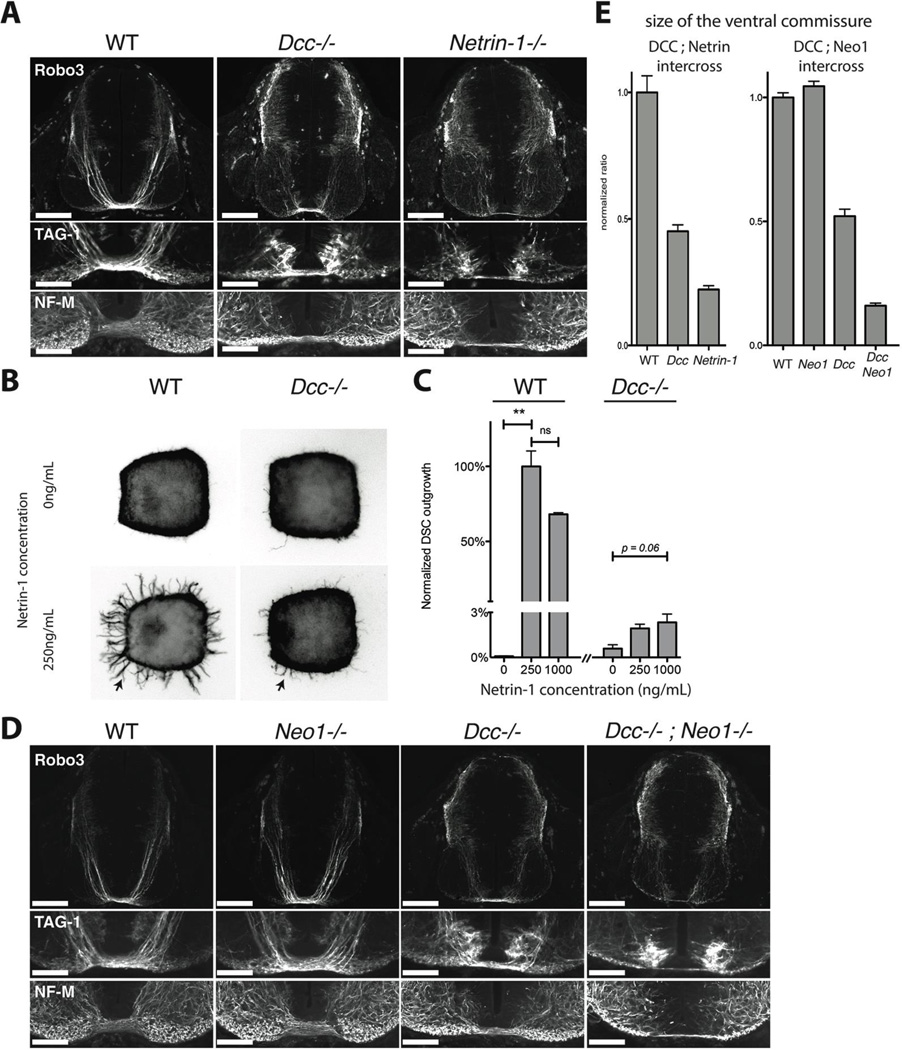
A) Cross sections of E11 wild-type, Dcc−/− and Netrin-1−/− littermate mouse embryos at the level of brachial spinal ganglia, stained for TAG-1, Robo3 and Neurofilament, Medium Chain (NF-M). Lower panels show details of the ventral commissural axon bundle. Dcc mutants have a reduced ventral commissure, but a large number of axons still cross. Netrin-1−/− embryos have a much-reduced ventral commissure. B) Axon outgrowth (arrows) in E11 wild type or Dcc−/− littermate mouse dorsal spinal cord explants cultured in 3D collagen gels with increasing concentrations of Netrin-1 and stained for Tuj1. C) Quantification of the response of axon outgrowth from wild type and Dcc−/− dorsal spinal cord explants to increasing Netrin-1 concentrations, normalized to wild type at 250ng/mL of Netrin-1. Dcc−/− mutants show a residual response to Netrin-1 application (arrow). D) Cross sections of E11 wild-type, Dcc−/−, Neo1−/− and Dcc−/−; Neo1−/− littermate mouse embryos at the level of brachial spinal ganglia, stained for TAG-1, Robo3 and Neurofilament (NF-M). Lower panels show details of the motor column and the ventral commissural axon bundle. The Dcc−/−; Neo1−/− double mutant has a much reduced ventral commissure and numerous axons in the motor column. E) Ratio of the commissural axon bundle size to the dorso-ventral spinal cord length of wild-type, Dcc−/− and Netrin1−/− embryos, normalized to wild types (left graph). Ratio of commissural axon bundle size to the dorso-ventral spinal cord length of Dcc−/−, Neo1−/− and Dcc−/−; Neo1−/− embryos normalized to wild type (right graph).The quantification shows the mean and SEM of 5 sections taken in brachial spinal cord in littermates, and is representative of 3 litters.
Scale bars are 200µm (Robo3) and 100µm (TAG-1 and NF-M).

