Abstract
The LIN28B→let-7 pathway contributes to regulation of the epithelial-mesenchymal transition (EMT) and stem cell self-renewal. The oncogenic MUC1-C transmembrane protein is aberrantly overexpressed in lung and other carcinomas; however, there is no known association between MUC1-C and the LIN28B→let-7 pathway. Here in non-small cell lung cancer (NSCLC), silencing MUC1-C downregulates the RNA binding protein LIN28B and coordinately increases the miRNA let-7. Targeting MUC1-C function with a dominant-negative mutant or a peptide inhibitor provided confirming evidence that MUC1-C induces LIN28B→let-7 signaling. Mechanistically, MUC1-C promotes NF-κB p65 chromatin occupancy of the LIN28B first intron and activates LIN28B transcription, which is associated with suppression of let-7. Consistent with let-7-mediated inhibition of HMGA2 transcripts, targeting of MUC1-C also decreases HMGA2 expression. HMGA2 has been linked to stemness, and functions as a competing endogenous RNA (ceRNA) of let-7-mediated regulation of the TGFβ co-receptor TGFBR3. Accordingly, targeting MUC1-C suppresses HMGA2 mRNA and protein, which is associated with decreases in TGFBR3, reversal of the EMT phenotype and inhibition of self-renewal capacity. These findings support a model in which MUC1-C activates the ⇑LIN28B→⇓let-7→⇑HMGA2 axis in NSCLC and thereby promotes EMT traits and stemness.
Keywords: MUC1, LIN28, let-7, HMGA2, NSCLC, EMT
Introduction
LIN28A and LIN28B are RNA binding proteins that inhibit the biogenesis of let-7 miRNAs in embryonic stem cells (1). In turn, let-7 miRNAs suppress LIN28A and LIN28B expression in a bistable switch that regulates stem cell self-renewal (1). LIN28A and LIN28B function as oncoproteins that promote cell proliferation and transformation (2–5). LIN28A/LIN28B expression has also been found in diverse malignancies and is associated with decreased survival in ovarian, colon and hepatocellular cancer (3, 6, 7). MYC induces LIN28B expression in multiple tumor models by binding to the LIN28B promoter (5). Activation of LIN28B expression may also be associated with translocation or amplification events (3). The let-7 miRNA family consists of 12 members located in regions of the genome that are often deleted in human cancers (8). With regard to LIN28-mediated suppression of let-7 miRNAs, LIN28A blocks processing of let-7 precursors by Dicer in the cytoplasm (9, 10). By a distinct mechanism, LIN28B sequesters primary let-7 transcripts in the nucleus and inhibits their processing (11). Suppression of let-7 expression induces reprogramming of fibroblasts to pluripotent stem cells (12, 13). Downregulation of let-7 miRNA family members has also been identified in numerous cancers and, like increased LIN28A/LIN28B expression, is associated with a poor prognosis (14–16). Restoration of let-7 expression inhibits cancer cell growth in mouse lung cancer models (17, 18). Moreover, let-7 inhibits self-renewal and tumorigenicity of human breast cancer cells (19). In addition to LIN28A and LIN28B, let-7 suppresses expression of other potential oncogenic proteins, including MYC, RAS and HMGA2 (1). Let-7 also directly inhibits IL-6 expression and thereby IL-6-mediated activation of the STAT3 pathway that links inflammation to cell transformation (2).
Mucin 1 (MUC1) is a heterodimeric protein that is aberrantly expressed in cancer cells (20, 21). Advances in our understanding of MUC1 function emerged from the demonstration that MUC1 undergoes autocleavage into two subunits that, in turn, form a heterodimer at the cell membrane (20). The extracellular MUC1 N-terminal subunit (MUC1-N) contains glycosylated tandem repeats that are characteristic of the mucin family (20). MUC1-N resides at the cell surface in a complex with the MUC1 transmembrane C-terminal subunit (MUC1-C) and is shed into a physical barrier that affords protection of mucosal epithelial cells (20). MUC1-C interacts with receptor tyrosine kinases (RTKs) in the cell membrane and promotes activation of downstream signaling, such as the PI3K→AKT and MEK→ERK pathways (21–23). MUC1-C is also imported into the nucleus where it interacts with transcription factors, such as NF-κB p65, and contributes to their transactivation functions (21, 24). In this way, recent work has shown that MUC1-C promotes NF-κB-mediated activation of the ZEB1 gene (25). In turn, the resulting increases in ZEB1 suppress miR-200c expression and induce EMT (25), a process that endows epithelial cells with stem cell characteristics (26). EMT also increases the capacity for cells to undergo invasion and to form spheres in non-adherent serum-free culture, which is dependent on the presence of self-renewing stem-like cells (26, 27). Consistent with the induction of ZEB1 and EMT, other work has demonstrated that MUC1-C contributes to the self-renewal of breast and NSCLC stem-like cells (28, 29). Other studies in NSCLC cells harboring EGFR or KRAS mutations have shown that targeting the MUC1-C pathway inhibits their growth as tumors in mice (29, 30). Inhibiting MUC1-C function therefore represents a potential approach for targeting EMT and self-renewing cancer cell populations. Notably in this regard, the MUC1-C cytoplasmic domain contains a CQC motif that is necessary for MUC1-C homodimerization and its oncogenic function. Targeting of endogenous MUC1-C with the cell-penetrating peptide GO-203, which blocks MUC1-C homodimerization, has thus been shown to be effective in inhibiting MUC1-C function (25, 28).
LIN28B has been functionally linked to stem-like cells and oncogenesis (31, 32). However, to our knowledge, there is no available evidence supporting an association between MUC1-C and the LIN28B→let-7 pathway. The present results demonstrate that MUC1-C induces LIN28B expression and thereby downregulates let-7 signaling in NSCLC cells. In turn, MUC1-C increases HMGA2 and TGFBR3 expression, and promotes EMT and stemness. Our findings thus demonstrate that silencing MUC1-C or targeting MUC1-C function suppresses activation of the LIN28B→let-7→HMGA2 axis, reverses EMT and decreases self-renewal capacity of NSCLC cells.
Materials and Methods
Cell culture
Human H1975/EGFR(L858R/T790M), H1299/wild-type EGFR and H1650/EGFR(delE746-A750) NSCLC cells (ATCC) were cultured in RPMI1640 medium supplemented with 10% heat-inactivated fetal bovine serum (FBS), 2 mM L-glutamine, 100 units/ml penicillin and 100 µg/ml streptomycin. Human MCF-7 breast cancer and LNCaP prostate cancer cells were grown in DMEM and RPMI-1640 medium, respectively, each containing FBS, glutamine and antibiotics. H1975, H1299 and H1650 cells were transduced with a lentiviral vector expressing either a MUC1 shRNA (Sigma) or a scrambled control shRNA (CshRNA; Sigma) vector as previously described (23). H1975 cells were also (i) transduced with lentiviral vectors expressing MUC1-C, MUC1-C(AQA) or LIN28B shRNA (Sigma), and (ii) transfected with a control siRNA or p65 siRNA. MCF-7 cells were stably transduced with a control lentiviral vector or one expressing MUC1-C as described (33). LNCaP cells were stably transfected with a control pHR-CMV vector or one expressing MUC1-C (34). Cells were treated with the MUC1-C inhibitor peptide GO-203, a control peptide CP-2 (35) or with the NF-κB pathway inhibitor BAY11-7085 (Santa Cruz Biotechnology).
Immunoprecipitation and immunoblot analysis
Cells were lysed using NP-40 lysis buffer containing protease inhibitor cocktail (Thermo Scientific). Soluble proteins were immunoprecipitated with anti-NF-κB p65 (Santa Cruz Biotechnology) or a control IgG. Precipitates and lysates not subjected to precipitation were analyzed by immunoblotting with anti-MUC1-C (LabVision), anti-NF-κB p65, anti-LIN28B, anti-E-cadherin, anti-vimentin, anti-HMGA2 (Cell Signaling) and anti-β-actin (Sigma). Immunoreactive complexes were detected using horseradish peroxidase-conjugated secondary antibodies (GE Healthcare) and an enhanced chemiluminescence (ECL) detection system (Perkin Elmer Health Sciences).
Quantitative real time PCR
Quantitative RT-PCR analysis was performed on cDNA synthesized with 1 µg total RNA using the Superscript cDNA synthesis system (Life Technologies). cDNA samples were then amplified using the SYBR green qPCR assay kit (Applied Biosystems) and the ABI Prism 7300 Sequence Detector (Applied Biosystems). qPCR primers used for detection of LIN28B, HMGA2, GAPDH and TGFBR3 transcripts are listed in Supplemental Table S1. Statistical significance was determined by the Student’s t-test. For detection of let-7a, let-7b and let-7c miRNAs from total RNA, the QuantiMir Small RNA Quantitation system (System Biosciences) was used as per the manufacturer’s protocol. The qPCR forward primers for detection of let-7a-c miRNAs are listed in Supplemental Table S2 and the universal reverse primer was supplied in the detection kit.
Analysis of Lin28B activation
Cells cultured in six-well plates were transfected with an empty vector, lin28b-P1-Luc containing an NF-κB binding site (GGGGCTTTC) in the first intron, or a mutant lin28b-P1(GGGGCTTTC→TCATACGAT)-Luc (2, 5) and, as an internal control, SV-40-Renilla-Luc (Promega) in the presence of Superfect transfection reagent (Qiagen). After 48 h, the transfected cells were left untreated or treated with the indicated agents, lysed with passive lysis buffer and then the lysates were analyzed using the dual luciferase assay system (Promega).
Tumorsphere culture
Adherent cells were harvested with gentle trypsinization, washed and resuspended using the MammoCult™ Human Medium Kit (Stem Cell Technologies). Single cells were confirmed under a microscope, counted and seeded in MammoCult™ medium in 6-well ultralow attachment culture plate (Corning CoStar) and cultured for 5 days. Tumorspheres of ≥100 µm were visualized and scored using a Nikon inverted TE2000 microscope. Sphere forming efficiency (SFE) was calculated by dividing the number of tumorspheres by the number of suspended cells.
Chromatin immunoprecipitation (ChIP) assays
Chromatin was solubilized from 2–3 × 10⁶ cells as described and precipitated with anti-NF-κB or a control nonimmune IgG. For re-ChIP analysis, NF-κB complexes from the primary ChIP were eluted and reimmunoprecipitated with anti-MUC1-C antibody as described (28). For real time ChIP qPCRs, the SYBR Green system was used with the ABI Prism 7300 Sequence Detector (Applied Biosystems). Data are reported as relative fold enrichment as described (36). Primers used for qPCR of the LIN28B first intron region (NBR: +313 to +415) and a control region (CR: −4964 to −4823) are listed in Supplemental Table S3.
Results
Silencing MUC1-C downregulates LIN28B expression
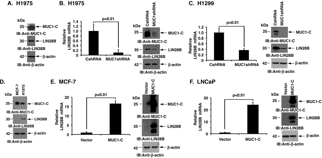
Human H1975 NSCLC cells constitutively overexpress MUC1-C and have detectable levels of LIN28B (Fig. 1A). Interestingly, stable silencing of MUC1-C in H1975 cells was associated with decreases in LIN28B expression at the mRNA (Fig. 1B, left) and protein (Fig. 1B, right) levels. Silencing MUC1-C in H1299 NSCLC cells also resulted in downregulation of LIN28B expression (Fig. 1C, left and right). Moreover, H1650 NSCLC cells responded to suppression of MUC1-C with decreases in LIN28B (Supplemental Fig. S1), indicating that MUC1-C induces LIN28B expression in diverse NSCLC cells. MUC1-C is also expressed in MCF-7 breast cancer cells (33). However, in contrast to H1975 NSCLC cells, LIN28B was low to undetectable in MCF-7 cells (Fig. 1D), indicating that MUC1-C may require the activation of downstream pathways for the induction of LIN28B expression. Accordingly, we overexpressed MUC1-C in these cells and found marked induction of LIN28B mRNA and protein (Fig. 1E, left and right). As another example, LNCaP prostate cancer cells express low levels of MUC1-C and LIN28B (Fig. 1F). Here, overexpression of MUC1-C substantially induced LIN28B mRNA and protein (Fig. 1F, left and right). These findings thus demonstrated that MUC1-C induces LIN28B expression and that a downstream MUC1-C-activated pathway, such as NF-κB p65, may be necessary for this response.
Figure 1. MUC1-C induces LIN28B expression.
A. Lysates from H1975 cells were immunoblotted with the indicated antibodies. B. H1975 cells were infected with lentiviruses to stably express a control scrambled shRNA (CshRNA) or a MUC1 shRNA. The H1975/CshRNA and H1975/MUC1shRNA cells were analyzed for LIN28B mRNA levels by qRT-PCR (left). The results are expressed as relative LIN28B mRNA levels (mean±SD of three determinations) as compared to that obtained for H1975/CshRNA cells (assigned a value of 1). Lysates from the H1975/CshRNA and H1975/MUC1shRNA cells were immunoblotted with the indicated antibodies (right). C. H1299 cells stably expressing a control scrambled shRNA (CshRNA) or a MUC1 shRNA were analyzed for LIN28B mRNA levels by qRT-PCR (left). The results are expressed as relative LIN28B mRNA levels (mean±SD of three determinations) as compared to that obtained for H1299/CshRNA cells (assigned a value of 1). Lysates from the H1299/CshRNA and H1299/MUC1shRNA cells were immunoblotted with the indicated antibodies (right). D. Lysates from MCF-7 and H1975 cells were immunoblotted with the indicated antibodies. E. MCF-7 cells stably expressing an empty vector or MUC1-C were analyzed for LIN28B mRNA levels by qRT-PCR (left). The results are expressed as relative LIN28B mRNA levels (mean±SD of three determinations) as compared to that obtained for MCF-7/vector cells (assigned a value of 1). Lysates from the MCF-7/vector and MCF-7/MUC1-C cells were immunoblotted with the indicated antibodies (right). F. LNCaP cells stably expressing an empty vector or MUC1-C were analyzed for LIN28B mRNA levels by qRT-PCR (left). The results are expressed as relative LIN28B mRNA levels (mean±SD of three determinations) as compared to that obtained for LNCaP/vector cells (assigned a value of 1). Lysates from the LNCaP/vector and LNCaP/MUC1-C cells were immunoblotted with the indicated antibodies (right).
Targeting MUC1-C homodimerization suppresses LIN28B expression
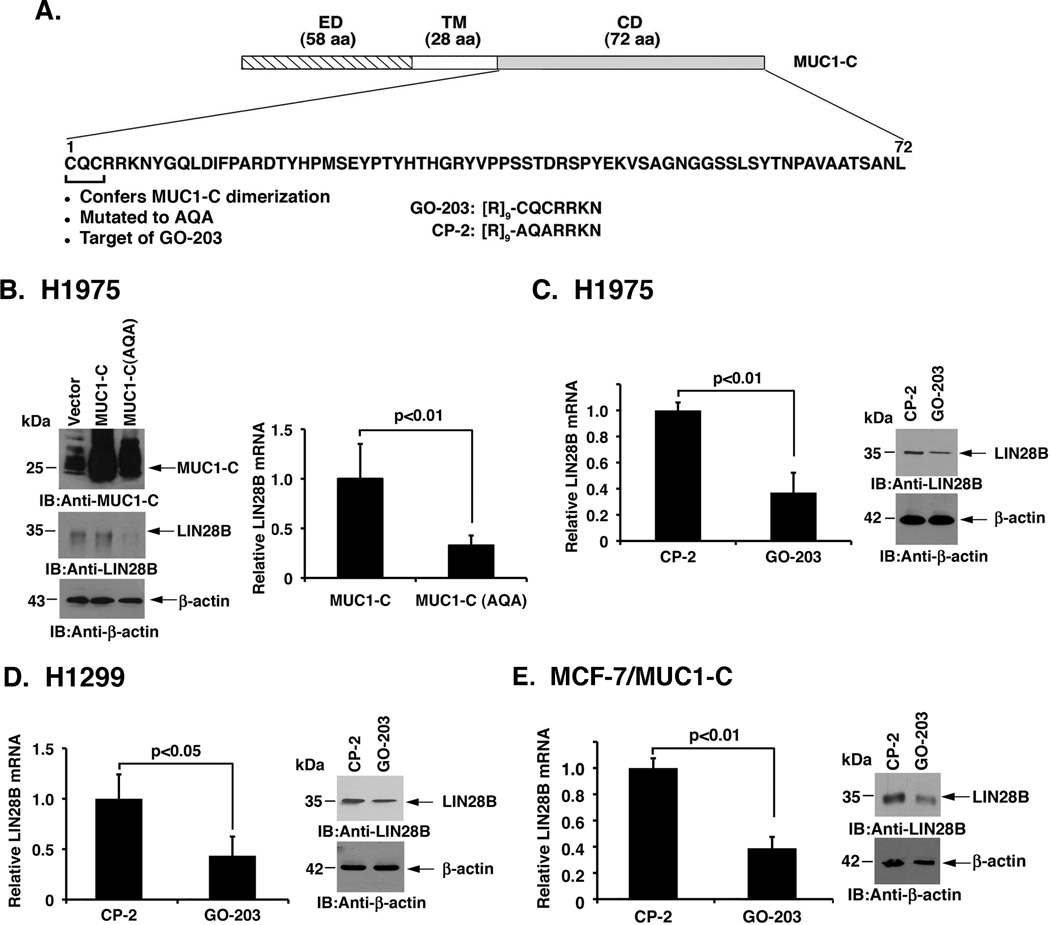
The MUC1-C cytoplasmic domain contains a CQC motif that is necessary and sufficient for the formation of MUC1-C homodimers and thereby the MUC1-C oncogenic function (Fig. 2A) (21). By extension, MUC1-C in which CQC has been mutated to AQA acts in a dominant-negative fashion (37). Accordingly, we stably overexpressed wild-type MUC1-C or the MUC1-C(CQC→AQA) mutant in H1975 cells to assess effects on LIN28B expression. In doing so and compared to a control empty vector, overexpression of MUC1-C had little if any effect on LIN28B, which as noted above is already upregulated in H1975 cells (Fig. 2B, left). By contrast, we found that MUC1-C(AQA) suppresses LIN28B expression at the protein and mRNA levels (Fig. 2B, left and right). Based on these results, we treated H1975 cells with GO-203, a cell penetrating peptide that inhibits MUC1-C homodimerization (35) (Fig. 2A). As a control, we used a peptide (CP-2) that is inactive in targeting the MUC1-C CQC motif (35) (Fig. 2A). Treatment with GO-203, but not CP-2, resulted in downregulation of LIN28B mRNA and protein (Fig. 2C, left and right). A similar decrease in LIN28B expression was obtained when treating H1299 cells with GO-203 (Fig. 2D, left and right). In further support of these results, we found that GO-203 blocks MUC1-C-induced LIN28B expression in MCF-7/MUC1-C cells (Fig. 2E). These findings thus confirmed that MUC1-C is of importance for LIN28B activation.
Figure 2. Targeting MUC1-C with a MUC1-C(AQA) mutant or the GO-203 inhibitor suppresses LIN28B expression.
A. Schematic representation of the MUC1-C subunit and the amino acid (aa) sequence of the cytoplasmic domain (CD). ED, extracellular domain. TM, transmembrane domain. The CQC motif is necessary and sufficient for MUC1-C homodimerization. Mutation of CQC to AQA thus blocks MUC1-C homodimer formation. D-aa sequences are shown for GO-203 and CP-2. B. Lysates from H1975 cells stably expressing an empty vector, MUC1-C or MUC1-C(AQA) were immunoblotted with the indicated antibodies (left). H1975/MUC1-C and H1975/MUC1-C(AQA) cells were analyzed for LIN28B mRNA levels by qRT-PCR (right). The results are expressed as relative LIN28B mRNA levels (mean±SD of three determinations) as compared to that obtained for H1975/MUC1-C cells (assigned a value of 1). C and D. H1975 (C) and H1299 (D) cells were treated with 5 µM GO-203 or CP-2 each day for 3 days and then analyzed for LIN28B mRNA levels by qRT-PCR. E. MCF-7/MUC1-C cells were treated with 5 µM GO-203 or CP-2 each day for 4 days and then analyzed for LIN28B mRNA levels by qRT-PCR. The results are expressed as relative LIN28B mRNA levels (mean±SD of three determinations) as compared to that obtained for CP-2-treated cells (assigned a value of 1) (left). Lysates from the GO-203- and CP-2-treated cells were immunoblotted with the indicated antibodies (right).
MUC1-C activates LIN28B by an NF-κB-mediated mechanism
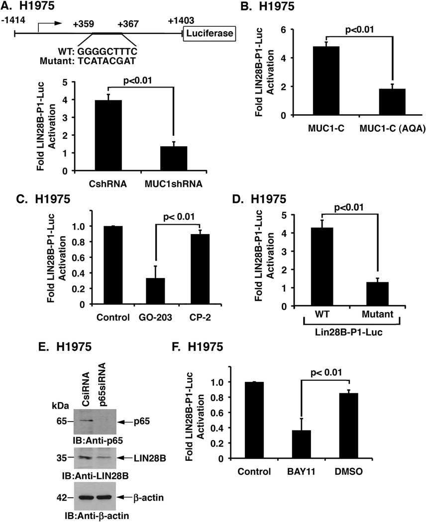
The LIN28B first intron contains an NF-κB binding site (5’-GGGGCTTTC-3’) at position +359 to +367 downstream to the transcription start site (Fig. 3A, upper panel) (2). To determine whether MUC1-C-induced LIN28B expression is dependent on NF-κB, we performed studies on H1975 cells transfected to express a LIN28B-luciferase reporter (lin28b-P1-Luc; Fig. 3A, upper panel) (2). Silencing MUC1-C was associated with a decrease in LIN28B-Luc activity as compared to that in H1975/CshRNA cells (Fig. 3A, lower panel). In addition, expression of MUC1-C(AQA) in H1975 cells (Fig. 3B) or treatment with GO-203 (Fig. 3C) resulted in a decrease in LIN28B-Luc activation. Mutation of the NF-κB-binding site to 5’-TCATACGAT-3’ (Fig. 3A, upper panel) was associated with a marked decrease in LIN28B-Luc activity in H1975 cells (Fig. 3D), indicating that MUC1-C may activate LIN28B transcription through NF-κB p65. In concert with that notion, transient silencing of NF-κB p65 in H1975 cells decreased LIN28B protein levels (Fig. 3E). Treatment of H1975 cells with the NF-κB inhibitor, BAY11-7085, also decreased LIN28B activation (Fig. 3F). Moreover, BAY11-7085 blocked MUC1-C-induced activation of LIN28B-Luc in MCF-7/MUC1-C cells (Supplemental Fig. S2), confirming that MUC1-C activates LIN28B transcription by a NF-κB p65-dependent mechanism.
Figure 3. MUC1-C activates LIN28B by an NF-κB-mediated mechanism.
A. Schema of the Lin28B-luciferase reporter (Lin28B-P1-Luc) showing location of the NF-κB binding site in the first intron relative to the transcription start site (upper panel). H1975/CshRNA and H1975/MUC1shRNA cells were transfected with the empty Luc vector or Lin28B-P1-Luc. Cells were also transfected with the SV-40-Renilla-Luc plasmid as an internal control. Luciferase activity was measured at 48 h following transfection (lower panel). The results are expressed as relative luciferase activity (mean±SD of three determinations) compared with that obtained from cells transfected with the empty Luc vector (assigned a value of 1). B. H1975/MUC1-C and H1975/MUC1-C(AQA) cells were transfected with the empty Luc vector or Lin28B-P1-Luc. Cells were also transfected with SV-40-Renilla-Luc as an internal control. Luciferase activity was measured at 48 h following transfection. Results are expressed as relative luciferase activity (mean±SD of three determinations) compared with that obtained from cells transfected with the empty Luc vector (assigned a value of 1). C. H1975 cells were co-transfected with the Lin28B-P1-Luc and SV-40-Renilla-Luc plasmids. At 48 h after transfection, cells were left untreated (Control) or treated with 5 µM GO-203 or CP-2 for an additional 36 h. Results are expressed as relative luciferase activity (mean±SD of three determinations) as compared to that obtained for the control untreated cells (assigned a value of 1). D. H1975 cells were transfected with empty Luc, wild type (WT) Lin28B-P1-Luc or mutant Lin28B-P1-Luc plasmids. Cells were also transfected with SV-40-Renilla-Luc plasmid. Luciferase activity was measured at 48 h following transfection. Results are expressed as relative luciferase activity (mean±SD of three determinations) compared with that obtained from cells transfected with the empty Luc vector (assigned a value of 1). E. H1975 cells were transfected to express a p65 siRNA or a control siRNA (CsiRNA). After 48 h, lysates were immunoblotted with the indicated antibodies. F. H1975 cells were co-transfected with Lin28B-P1-Luc and SV-40-Renilla-Luc plasmids. At 48 h following transfection, cells were left untreated (Control), treated with DMSO as vehicle control, or treated with 5 µM BAY11-7085 for an additional 12 h. Results are expressed as relative luciferase activity (mean±SD of three determinations) as compared to that obtained for the control untreated cells (assigned a value of 1).
MUC1-C occupies the LIN28B gene with NF-κB
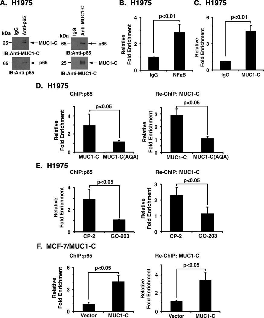
MUC1-C has been reported to interact with NF-κB p65 (24). Consistent with those findings, studies using H1975 cell lysates demonstrated that MUC1-C coprecipitates with NF-κB p65 (Fig. 4A, left and right). ChIP studies of chromatin from H1975 cells further demonstrated that NF-κB p65 occupies the LIN28B gene in the region of the first intron (Fig. 4B). Moreover, in re-ChIP analyses, we found that NF-κB p65 associates with MUC1-C on that region (Fig. 4C). Similar results were obtained using chromatin from H1299 cells, indicating that MUC1-C forms a complex with NF-κB p65 on the LIN28B first intron (Supplemental Figs. S3A and B). In concert with the LIN28B-Luc reporter studies, expression of MUC1-C(AQA) decreased occupancy of NF-κB p65 and MUC1-C (Fig. 4D). In addition, GO-203 treatment inhibited NF-κB and MUC1-C occupancy (Fig. 4E). As confirmation of these results, we found that overexpression of MUC1-C in MCF-7 cells increases occupancy of both NF-κB p65 and MUC1-C on the LIN28B first intron (Fig. 4F). These findings and those with the LIN28B-Luc reporter support a model in which MUC1-C promotes NF-κB p65 occupancy on the LIN28B first intron and thereby activates LIN28B transcription.
Figure 4. MUC1-C occupies the LIN28B gene with NF-κB.
A. Lysates from H1975 cells were precipitated with a control IgG or either anti-p65 (left) or anti-MUC1-C (right) antibody. Precipitates were immunoblotted with the indicated antibodies. B–C. Soluble chromatin from the indicated H1975 cells was precipitated with anti-NF-(B or a control IgG. The final DNA precipitates were amplified by qPCR with pairs of primers for the NF-(B binding region (NBR; +313 to +415) in intron 1 or a control region (CR; −4964 to −4823). The results (mean±SD of three independent experiments each assayed in triplicate) are expressed as the relative fold enrichment compared to that obtained with the IgG controls. D. Soluble chromatin from H1975/MUC1-C and H1975/MUC1-C(AQA) cells was precipitated with anti-NF-(B or a control IgG (left). For re-ChIP analysis, complexes were released and re-immunoprecipitated with anti-MUC1-C (right). The results (mean±SD of three independent experiments each assayed in triplicate) are expressed as the relative fold enrichment compared with the IgG controls. E. H1975 cells were left untreated or treated with 5 µM GO-203 or CP-2 each day for 3 days. Soluble chromatin was precipitated with anti-NF-(B or a control IgG (left). For re-ChIP analysis, complexes were released and re-immunoprecipitated with anti-MUC1-C (right). The results (mean±SD of three independent experiments each assayed in triplicate) are expressed as the relative fold enrichment compared with the IgG controls. F. Soluble chromatin from MCF-7/vector and MCF-7/MUC1-C cells was precipitated with anti-NF-(B or a control IgG (left). For re-ChIP analysis, complexes were released and re-immunoprecipitated with anti-MUC1-C (right). The results (mean±SD of three independent experiments each assayed in triplicate) are expressed as the relative fold enrichment compared with the IgG controls.
Targeting MUC1-C increases let-7 expression
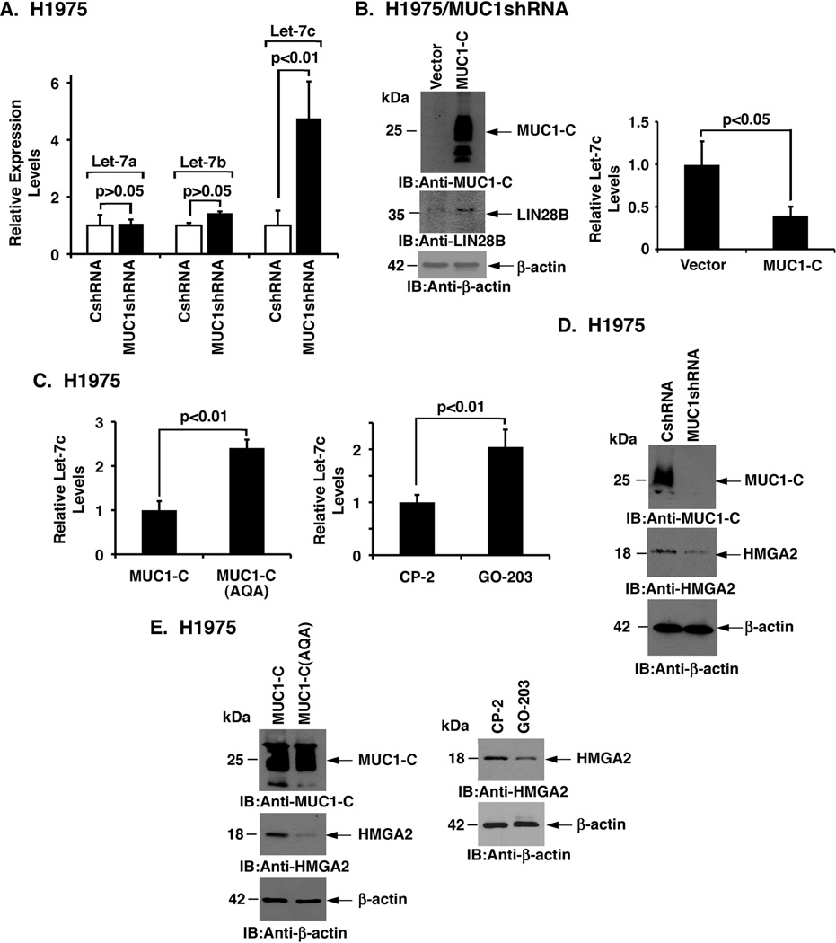
LIN28B inhibits the biogenesis of let-7 miRNAs (11). Consistent with the demonstration that MUC1-C promotes the induction of LIN28B expression, we found that silencing MUC1-C in H1975 cells results in marked upregulation of let-7c levels (Fig. 5A). By contrast, downregulation of MUC1-C had no significant effect on let-7a and let-7b expression (Fig. 5A). To confirm that this response is indeed a result of downregulating MUC1-C and not an off-target effect, we overexpressed MUC1-C in H1975/MUC1shRNA cells (Fig. 5B, left). In turn, we found upregulation of LIN28B (Fig. 5B, left) and suppression of let-7c (Fig. 5B, right). Increases in let-7c were also observed in H1975 cells (i) expressing MUC1-C(AQA) (Fig. 5C, left) or (ii) treated with GO-203 (Fig. 5C, right). In extending this analysis to H1299 cells, we found that silencing MUC1-C is associated with a predominant increase in let-7a levels and less pronounced upregulation of let-7b and let-7c expression (Supplemental Fig. S4A). Transcripts encoding the chromatin-binding HMGA2 protein are a target of let-7 inhibition (38, 39). In concert with the demonstration that silencing MUC1-C in H1975 cells increases let-7c, we found an associated decrease in HMGA2 expression (Fig. 5D). HMGA2 levels were also suppressed by MUC1-C(AQA) and treatment with GO-203 (Fig. 5E, left and right). Moreover, silencing MUC1-C in H1299 cells was similarly associated with suppression of HMGA2 (Supplemental Fig. S4B). These results provided support for MUC1-C-induced activation of the LIN28B→⇓let-7→⇑HMGA2 pathway.
Figure 5. Targeting MUC1-C increases let-7 expression.
A. H1975/CshRNA and H1975/MUC1shRNA cells were analyzed for let-7a-c miRNA levels by qRT-PCR. Results are expressed as relative let-7a-c levels (mean±SD of three determinations) as compared to that obtained for CshRNA cells (assigned a value of 1). B. H1975/MUC1shRNA cells were transfected to express a control vector or MUC1-C. Lysates were immunoblotted with the indicated antibodies (left). Total RNA was analyzed for let-7c expression (right). Results are expressed as relative let-7c levels (mean±SD of three determinations; H1975/MUC1shRNA/vector cells assigned a value of 1). C. Total RNA from H1975/MUC1-C and H1975/MUC1-C(AQA) cells (left) and from H1975 cells treated with 5 µM GO-203 or CP-2 each day for 3 days (right) was analyzed for let-7c by qRT-PCR. Results are expressed as relative let-7c levels (mean±SD of three determinations; H1975/MUC1-C cells assigned a value of 1). D. Lysates from H1975/CshRNA and H1975/MUC1shRNA cells were immunoblotted with the indicated antibodies. E. Lysates from H1975/MUC1-C and H1975/MUC1-C(AQA) cells (left) and from H1975 cells treated with 5 µM GO-203 or CP-2 each day for 3 days (right) were immunoblotted with the indicated antibodies.
MUC1-C induces EMT and tumorsphere formation
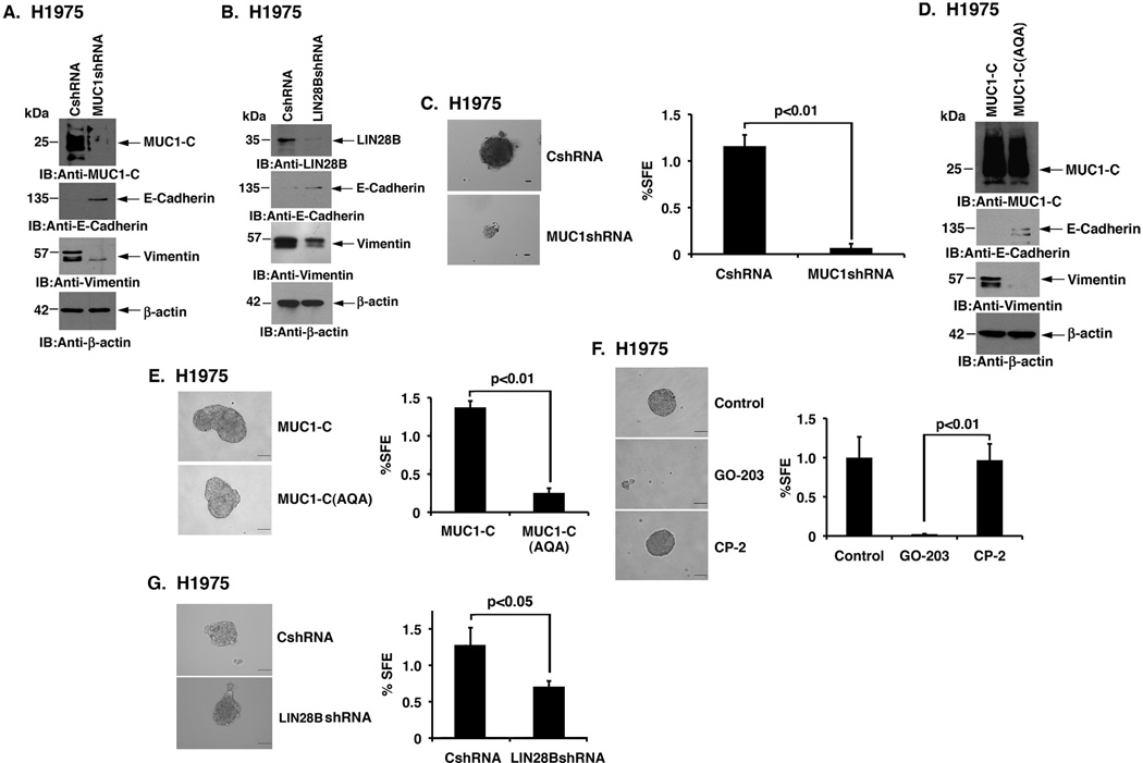
The induction of LIN28 with downregulation of let-7 promotes EMT traits and stemness (40). In addition, HMGA2 has been linked to the induction of EMT and metastasis (39, 41). These findings invoked the possibility that MUC1-C could drive EMT and stemness through LIN28B→let-7→HMGA2 signaling. Indeed, we found that silencing MUC1-C in H1975 cells reverses the EMT phenotype with increases in E-cadherin and decreases in vimentin levels (Fig. 6A). A similar response was found when silencing LIN28B (Fig. 6B), confirming that MUC1-C induces EMT through the LIN28B→let-7 pathway. EMT increases the capacity for sphere formation in non-adherent serum-free culture, a characteristic dependent on the presence of self-renewing stem-like cells (26, 27). Based on these premises, we found that silencing MUC1-C markedly inhibits H1975 sphere formation (Fig. 6C, left and right). We also found that silencing MUC1-C in H1299 cells increases E-cadherin with a concomitant decrease in vimentin expression (Supplemental Fig. S5A) and decreases sphere formation (Supplemental S5B). In extending this line of investigation, we observed that expression of the MUC1-C(AQA) mutant in H1975 cells reverses EMT (Fig. 6D) and blocks sphere formation (Fig. 6E, left and right). Moreover, GO-203 treatment was highly effective in disrupting established H1975 spheres (Fig. 6F, left and right), supporting dependence on MUC1-C function. In concert with these observations and the role of MUC1-C in driving LIN28B expression, we confirmed that downregulation of LIN28B in H1975 cells is associated with a significant decrease in tumorsphere formation (Fig. 6G). These findings demonstrate that MUC1-C induces LIN28B→⇓let-7→⇑HMGA2 signaling in association with the promotion of EMT and self-renewal.
Figure 6. MUC1-C induces EMT and tumorsphere formation.
A. Lysates from H1975/CshRNA and H1975/MUC1shRNA cells were immunoblotted with the indicated antibodies. B. H1975 cells were infected with lentiviruses expressing a control scrambled shRNA or a Lin28B shRNA. Lysates were immunoblotted with the indicated antibodies. C. Representative images of tumorspheres derived from H1975/CshRNA and H1975/MUC1shRNA cells (left). Bar represents 50 microns. The percentage sphere forming efficiency (%SFE) is expressed as the mean±SD of three determinations (right). D. Lysates from H1975/MUC1-C and H1975/MUC1-C(AQA) cells were immunoblotted with the indicated antibodies. E. Representative images of tumorspheres derived from H1975/MUC1-C and H1975/MUC1-C(AQA) cells (left). Bar represents 100 microns. The percentage SFE is expressed as the mean±SD of three determinations (right). F. Tumorspheres established from H1975 cells (left) were left untreated or treated with 5 µM GO-203 or 5 µM CP-2 for 48 h. Bar represents 100 microns. The percentage SFE is expressed as the mean±SD of three determinations (right). G. Representative images of tumorspheres derived from H1975/CshRNA and H1975/LIN28BshRNA cells (left). Bar represents 100 microns. The percentage SFE is expressed as the mean±SD of three determinations (right).
MUC1-C upregulates TGFBR3 expression in NSCLC cells
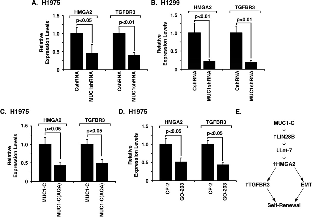
Recent work has shown that HMGA2 promotes lung cancer progression by functioning as a protein-coding gene and as a ceRNA for let-7 miRNAs (42). The present finding that MUC1-C induces HMGA2 expression in NSCLC cells thus invoked the possibility that MUC1-C could also contribute to upregulation of the TGF-β co-receptor TGFBR3, which is a target of HMGA2 ceRNA function and is coordinately expressed with HMGA2 in NSCLC tumors (42). Indeed, silencing of MUC1-C in H1975 (Fig. 7A) and H1299 (Fig. 7B) cells was associated with suppression of both HMGA2 and TGFBR3 expression. Additionally, targeting MUC1-C function with MUC1-C(AQA) (Fig. 7C) or GO-203 (Fig. 7D), which decreases HMGA2 expression, also downregulated TGFBR3 mRNA levels. These findings thus extend involvement of the MUC1-C→LIN28B→let-7 pathway to induction of the HMGA2 ceRNA function in the regulation of TGFBR3.
Figure 7. Targeting MUC1-C decreases TGFBR3 expression.
A and B. The indicated H1975 (A) and H1299 (B) cells were analyzed for HMGA2 and TGFBR3 mRNA levels by qRT-PCR. The results are expressed as relative mRNA levels (mean±SD of three determinations) as compared to that obtained for cells expressing the CshRNA (assigned a value of 1). C. H1975/MUC1-C and H1975/MUC1-C(AQA) cells were analyzed for HMGA2 and TGFBR3 mRNA levels by qRT-PCR. The results are expressed as relative mRNA levels (mean±SD of three determinations) as compared to that obtained for H1975/MUC1-C cells (assigned a value of 1). D. H1975 cells treated with 5 µM GO-203 or CP-2 each day for 3 days were analyzed for HMGA2 and TGFBR3 mRNA levels by qRT-PCR. The results are expressed as relative mRNA levels (mean±SD of three determinations) as compared to that obtained for CP-2-treated cells (assigned a value of 1). E. Schema depicting the proposed MUC1-C→⇑LIN28B→⇓let-7→⇑HMGA2→⇑TGFBR3 pathway that drives EMT and self-renewal.
Discussion
LIN28B functions as an oncogenic protein that promotes in vitro transformation and tumorigenesis in diverse models of premalignant MCF-10A breast epithelial (2), intestinal epithelial (6, 31) and neuroblastoma (32) cells. Overexpression of LIN28B has also been identified in multiple tumor types, particularly in settings of advanced disease (3). The activation of LIN28B expression in human and mouse tumor models has been linked to binding of MYC to E-boxes downstream of the LIN28B transcription start site (5). In addition to the involvement of MYC, other studies in MCF-10A cells have shown that LIN28B expression is activated by the association of NF-κB with a consensus binding site in the LIN28B first intron (2). The present work provides another perspective on the regulation of LIN28B by demonstrating that MUC1-C is necessary for LIN28B expression in NSCLC cells. Thus, silencing MUC1-C was sufficient to decrease LIN28B mRNA and protein. In support of this observation, overexpression of MUC1-C was also found to be sufficient for induction of LIN28B. To our knowledge, MUC1-C has not been previously linked to LIN28B signaling and thereby a functional interaction between these oncoproteins. With regard to the mechanistic basis for these findings, MUC1-C interacts with the NF-κB pathway by activating the IκB kinase complex and downstream signals (43). MUC1-C also binds directly to NF-κB p65 and promotes activation of NF-κB target genes, including the promoter of the MUC1 gene itself in an autoregulatory loop (24). The present studies demonstrate that MUC1-C associates with NF-κB on the LIN28B first intron, increases NF-κB occupancy in that region and promotes NF-κB-mediated activation of LIN28B transcription. Moreover, we found that suppression of NF-κB p65 blocks MUC1-C-induced LIN28B expression, thus providing evidence that MUC1-C drives the upregulation of LIN28B by an NF-κB-dependent mechanism. Our findings thereby lend support to a model in which MUC1-C-induced activation of the inflammatory NF-κB p65 pathway is linked to induction of LIN28B (Fig. 7E).
The LIN28B oncogenic function is conferred, at least in large part, by repression of let-7 processing. In this way, let-7 miRNA family members function as tumor suppressors by downregulating the expression of certain oncogenes and regulators of cell proliferation (44). As expected, silencing MUC1-C with suppression of LIN28B was associated with increases in let-7. The MUC1-C oncogenic function is dependent on the cytoplasmic domain CQC motif, which is necessary for MUC1-C homodimerization (21). Thus, expression of a MUC1-C(AQA) dominant-negative mutant in NSCLC cells or treatment with the GO-203 inhibitor reduced LIN28B expression with a concomitant derepression of let-7 expression. Inhibiting MUC1-C was also associated with decreases in HMGA2, which has been identified as a prototypic let-7 target with 6–8 let-7 binding sites in the HMGA2 3’UTR (38, 45). Let-7 destabilizes HMGA2 transcripts and thereby inhibits H1299 NSCLC cell growth, a response that is rescued by expression of the HMGA2 open reading frame without a 3’UTR (38). HMGA2 is aberrantly expressed in diverse tumors and is associated with decreased survival in patients with colorectal and lung cancer (46, 47). Notably, HMGA2 is highly expressed in metastatic NSCLC (47–49), consistent with its role in promoting tumor metastasis (41). Other work has demonstrated that HMGA2 contributes to the progression of lung cancer by functioning as a competing endogenous RNA (ceRNA) for the let-7 family members (42). Thus, in those studies, HMGA2 promoted transformation of lung cancer cells that was unrelated to the HMGA2 protein-coding function, but dependent on the presence of HMGA2 mRNA let-7 binding sites (42). The HMGA2 ceRNA was further shown to regulate expression of the TGF-β co-receptor TGFBR3, which is of importance for HMGA2 to drive lung cancer progression (42). In this context, we found that MUC1-C-induced HMGA2 expression is associated with coordinate upregulation of TGFBR3, consistent with what has been observed in NSCLC tissue (42). These findings indicate that MUC1-C activates the LIN28B→⇓let-7→⇑HMGA2→⇑TGFBR3 pathway and that MUC1-C may also contribute to induction of TGFBR3 and tumor progression by increasing HMGA2 ceRNA activity.
Overexpression of MUC1-C has been linked to the induction of EMT, stemness and self-renewal in breast and certain other cancer cells (25, 28). The LIN28B→let-7 pathway has also been shown to confer self-renewal of breast cancer stem-like cells, at least in part through the suppression of HMGA2 (19). The LIN28B-let-7-HMGA2 axis also determines the self-renewal potential of fetal stem cells (50). These findings invoked the possibility that MUC1-C contributes to EMT and self-renewal by activating LIN28B→⇓let-7→⇑HMGA2 signaling. Indeed, the present studies demonstrate that targeting MUC1-C in NSCLC cells is associated with reversal of the EMT phenotype. Similar responses were observed when silencing LIN28B, indicating that the MUC1-C→LIN28B→let-7 pathway contributes to the induction of EMT in these NSCLC cells. LIN28B functions in large part by the suppression of let-7 miRNAs (11); however, we do not exclude the possibility that LIN28B silencing could also promote EMT by let-7-independent pathways. In this way, other studies will be needed to more precisely define the role of let-7 miRNAs in LIN28B-mediated regulation of EMT, as well as TGFBR3 and self-renewal, in NSCLC cells. MUC1-C has been shown to confer EMT in renal cancer cells through WNT/β-catenin and induction of SNAIL (51) and in breast cancer cells by activation of ZEB1→miR-200c in breast cancer cells (25). These findings and those reported here thus indicate that MUC1-C can promote the induction of EMT by diverse pathways that appear to be dependent on cell context. The association between EMT and self-renewal in non-adherent serum-free culture (26, 27) provided the basis for further determining whether MUC1-C also promotes the capacity of NSCLC cells to form spheres. Accordingly, we found that silencing MUC1-C or expression of the MUC1-C(AQA) mutant markedly inhibits NSCLC sphere formation. In addition, targeting MUC1-C with the GO-203 inhibitor, which blocks activation of LIN28B→let-7→HMGA2 signaling, was highly effective in disrupting established NSCLC spheres. These findings thus provide the first evidence that MUC1-C activates the LIN28B→⇓let-7→⇑HMGA2 pathway and is necessary for EMT and self-renewal of NSCLC cells. Of note, let-7 repression can upregulate diverse cell-cycle regulators, such as MYC, RAS and cyclin D1, as well as the PI3K→AKT pathway (1). In this regard, other studies have shown that MUC1-C upregulates cyclin D1 expression (52) and activates PI3K→AKT signaling in NSCLC cells (35). The present studies have focused on MUC1-C induced expression of HMGA2; therefore, our results do not exclude the possibility that MUC1-C affects the expression of other let-7 targets in NSCLC and other carcinoma cells.
Supplementary Material
Acknowledgments
Financial Support: Research reported in this publication was supported by the National Cancer Institute of the National Institutes of Health under award numbers CA166480 and CA97098.
Abbreviations
- MUC1
Mucin 1
- MUC1-C
MUC1 transmembrane C-terminal subunit
- HMGA2
high-mobility group A2 protein
- NSCLC
non-small cell lung cancer
- EMT
epithelial-mesenchymal transition
- EGFR
epidermal growth factor receptor
- ChIP
chromatin immunoprecipitation
- ceRNA
competing endogenous RNA
- TGFBR3
TGFβ co-receptor 3
Footnotes
Conflict of Interest Disclosure: D.K. holds equity in Genus Oncology and is a consultant to the company. The other authors disclosed no potential conflicts of interest.
Implications: A novel pathway is defined in which MUC1-C drives LIN28B→let-7→HMGA2 signaling, EMT and self-renewal in NSCLC.
References
- 1.Shyh-Chang N, Daley GQ. Lin28: primal regulator of growth and metabolism in stem cells. Cell Stem Cell. 2013;12(4):395–406. doi: 10.1016/j.stem.2013.03.005. [DOI] [PMC free article] [PubMed] [Google Scholar]
- 2.Iliopoulos D, Hirsch HA, Struhl K. An epigenetic switch involving NF-kappaB, Lin28, Let-7 MicroRNA, and IL6 links inflammation to cell transformation. Cell. 2009;139(4):693–706. doi: 10.1016/j.cell.2009.10.014. [DOI] [PMC free article] [PubMed] [Google Scholar]
- 3.Viswanathan SR, Powers JT, Einhorn W, et al. Lin28 promotes transformation and is associated with advanced human malignancies. Nat Genet. 2009;41(7):843–848. doi: 10.1038/ng.392. [DOI] [PMC free article] [PubMed] [Google Scholar]
- 4.West JA, Viswanathan SR, Yabuuchi A, et al. A role for Lin28 in primordial germ-cell development and germ-cell malignancy. Nature. 2009;460(7257):909–913. doi: 10.1038/nature08210. [DOI] [PMC free article] [PubMed] [Google Scholar]
- 5.Chang TC, Zeitels LR, Hwang HW, et al. Lin-28B transactivation is necessary for Myc-mediated let-7 repression and proliferation. Proc Natl Acad Sci USA. 2009;106(9):3384–3389. doi: 10.1073/pnas.0808300106. [DOI] [PMC free article] [PubMed] [Google Scholar]
- 6.King CE, Cuatrecasas M, Castells A, Sepulveda AR, Lee JS, Rustgi AK. LIN28B promotes colon cancer progression and metastasis. Cancer Res. 2011;71(12):4260–4268. doi: 10.1158/0008-5472.CAN-10-4637. [DOI] [PMC free article] [PubMed] [Google Scholar]
- 7.Cheng SW, Tsai HW, Lin YJ, et al. Lin28B is an oncofetal circulating cancer stem cell-like marker associated with recurrence of hepatocellular carcinoma. PloS One. 2013;8(11):e80053. doi: 10.1371/journal.pone.0080053. [DOI] [PMC free article] [PubMed] [Google Scholar]
- 8.Calin GA, Sevignani C, Dumitru CD, et al. Human microRNA genes are frequently located at fragile sites and genomic regions involved in cancers. Proc Natl Acad Sci USA. 2004;101(9):2999–3004. doi: 10.1073/pnas.0307323101. [DOI] [PMC free article] [PubMed] [Google Scholar]
- 9.Hagan JP, Piskounova E, Gregory RI. Lin28 recruits the TUTase Zcchc11 to inhibit let-7 maturation in mouse embryonic stem cells. Nat Struct Mol Biol. 2009;16(10):1021–1025. doi: 10.1038/nsmb.1676. [DOI] [PMC free article] [PubMed] [Google Scholar]
- 10.Heo I, Joo C, Kim YK, et al. TUT4 in concert with Lin28 suppresses microRNA biogenesis through pre-microRNA uridylation. Cell. 2009;138(4):696–708. doi: 10.1016/j.cell.2009.08.002. [DOI] [PubMed] [Google Scholar]
- 11.Piskounova E, Polytarchou C, Thornton JE, et al. Lin28A and Lin28B inhibit let-7 microRNA biogenesis by distinct mechanisms. Cell. 2011;147(5):1066–1079. doi: 10.1016/j.cell.2011.10.039. [DOI] [PMC free article] [PubMed] [Google Scholar]
- 12.Martinez NJ, Gregory RI. MicroRNA gene regulatory pathways in the establishment and maintenance of ESC identity. Cell Stem Cell. 2010;7(1):31–35. doi: 10.1016/j.stem.2010.06.011. [DOI] [PMC free article] [PubMed] [Google Scholar]
- 13.Melton C, Judson RL, Blelloch R. Opposing microRNA families regulate self-renewal in mouse embryonic stem cells. Nature. 2010;463(7281):621–626. doi: 10.1038/nature08725. [DOI] [PMC free article] [PubMed] [Google Scholar]
- 14.Boyerinas B, Park SM, Hau A, Murmann AE, Peter ME. The role of let-7 in cell differentiation and cancer. Endocr Relat Cancer. 2010;17(1):F19–F36. doi: 10.1677/ERC-09-0184. [DOI] [PubMed] [Google Scholar]
- 15.Shell S, Park SM, Radjabi AR, et al. Let-7 expression defines two differentiation stages of cancer. Proc Natl Acad Sci USA. 2007;104(27):11400–11405. doi: 10.1073/pnas.0704372104. [DOI] [PMC free article] [PubMed] [Google Scholar]
- 16.Takamizawa J, Konishi H, Yanagisawa K, et al. Reduced expression of the let-7 microRNAs in human lung cancers in association with shortened postoperative survival. Cancer Res. 2004;64(11):3753–3756. doi: 10.1158/0008-5472.CAN-04-0637. [DOI] [PubMed] [Google Scholar]
- 17.Esquela-Kerscher A, Trang P, Wiggins JF, et al. The let-7 microRNA reduces tumor growth in mouse models of lung cancer. Cell Cycle. 2008;7(6):759–764. doi: 10.4161/cc.7.6.5834. [DOI] [PubMed] [Google Scholar]
- 18.Trang P, Medina PP, Wiggins JF, et al. Regression of murine lung tumors by the let-7 microRNA. Oncogene. 2010;29(11):1580–1587. doi: 10.1038/onc.2009.445. [DOI] [PMC free article] [PubMed] [Google Scholar]
- 19.Yu F, Yao H, Zhu P, et al. let-7 regulates self renewal and tumorigenicity of breast cancer cells. Cell. 2007;131(6):1109–1123. doi: 10.1016/j.cell.2007.10.054. [DOI] [PubMed] [Google Scholar]
- 20.Kufe D. Mucins in cancer: function, prognosis and therapy. Nature Reviews Cancer. 2009;9(12):874–885. doi: 10.1038/nrc2761. [DOI] [PMC free article] [PubMed] [Google Scholar]
- 21.Kufe D. MUC1-C oncoprotein as a target in breast cancer: activation of signaling pathways and therapeutic approaches. Oncogene. 2013;32(9):1073–1081. doi: 10.1038/onc.2012.158. [DOI] [PMC free article] [PubMed] [Google Scholar]
- 22.Raina D, Uchida Y, Kharbanda A, et al. Targeting the MUC1-C oncoprotein downregulates HER2 activation and abrogates trastuzumab resistance in breast cancer cells. Oncogene. 2013 Aug 5; doi: 10.1038/onc.2013.308. advance online publication. [DOI] [PMC free article] [PubMed] [Google Scholar]
- 23.Alam M, Ahmad R, Rajabi H, Kharbanda A, Kufe D. MUC1-C oncoprotein activates ERK→C/EBPβ-mediated induction of aldehyde dehydrogenase activity in breast cancer cells. J Biol Chem. 2013;288(43):30829–30903. doi: 10.1074/jbc.M113.477158. [DOI] [PMC free article] [PubMed] [Google Scholar]
- 24.Ahmad R, Raina D, Joshi MD, Kawano T, Kharbanda S, Kufe D. MUC1-C oncoprotein functions as a direct activator of the NF-κB p65 transcription factor. Cancer Res. 2009;69:7013–7021. doi: 10.1158/0008-5472.CAN-09-0523. [DOI] [PMC free article] [PubMed] [Google Scholar]
- 25.Rajabi H, Alam M, Takahashi H, et al. MUC1-C oncoprotein activates the ZEB1/miR-200c regulatory loop and epithelial-mesenchymal transition. Oncogene. 2013;33(13):1680–1689. doi: 10.1038/onc.2013.114. [DOI] [PMC free article] [PubMed] [Google Scholar]
- 26.Mani SA, Guo W, Liao MJ, et al. The epithelial-mesenchymal transition generates cells with properties of stem cells. Cell. 2008;133(4):704–715. doi: 10.1016/j.cell.2008.03.027. [DOI] [PMC free article] [PubMed] [Google Scholar]
- 27.Dontu G, Abdallah WM, Foley JM, et al. In vitro propagation and transcriptional profiling of human mammary stem/progenitor cells. Genes Dev. 2003;17(10):1253–1270. doi: 10.1101/gad.1061803. [DOI] [PMC free article] [PubMed] [Google Scholar]
- 28.Alam M, Rajabi H, Ahmad R, Jin C, Kufe D. Targeting the MUC1-C oncoprotein inhibits self-renewal capacity of breast cancer cells. Oncotarget. 2014;5(9):2622–2634. doi: 10.18632/oncotarget.1848. [DOI] [PMC free article] [PubMed] [Google Scholar]
- 29.Kharbanda A, Rajabi H, Jin C, Alam M, Wong K, Kufe D. MUC1-C confers EMT and KRAS independence in mutant KRAS lung cancer cells. Oncotarget. 2014 doi: 10.18632/oncotarget.2360. (in press). [DOI] [PMC free article] [PubMed] [Google Scholar]
- 30.Kharbanda A, Rajabi H, Jin C, et al. Targeting the oncogenic MUC1-C protein inhibits mutant EGFR-mediated signaling and survival in non-small cell lung cancer cells. Clin Canc Res. 2014 doi: 10.1158/1078-0432.CCR-13-3168. (in press). [DOI] [PMC free article] [PubMed] [Google Scholar]
- 31.Madison BB, Liu Q, Zhong X, et al. LIN28B promotes growth and tumorigenesis of the intestinal epithelium via Let-7. Genes Dev. 2013;27(20):2233–2245. doi: 10.1101/gad.224659.113. [DOI] [PMC free article] [PubMed] [Google Scholar]
- 32.Molenaar JJ, Domingo-Fernandez R, Ebus ME, et al. LIN28B induces neuroblastoma and enhances MYCN levels via let-7 suppression. Nat Genet. 2012;44(11):1199–1206. doi: 10.1038/ng.2436. [DOI] [PubMed] [Google Scholar]
- 33.Kharbanda A, Rajabi H, Jin C, Raina D, Kufe D. MUC1-C oncoprotein induces tamoxifen resistance in human breast cancer. Mol Cancer Res. 2013;11(7):714–723. doi: 10.1158/1541-7786.MCR-12-0668. [DOI] [PMC free article] [PubMed] [Google Scholar]
- 34.Rajabi H, Ahmad R, Jin C, et al. MUC1-C oncoprotein confers androgen-independent growth of human prostate cancer cells. Prostate. 2012;72(15):1659–1668. doi: 10.1002/pros.22519. [DOI] [PMC free article] [PubMed] [Google Scholar]
- 35.Raina D, Kosugi M, Ahmad R, et al. Dependence on the MUC1-C oncoprotein in non-small cell lung cancer cells. Mol Cancer Therapeutics. 2011;10(5):806–816. doi: 10.1158/1535-7163.MCT-10-1050. [DOI] [PMC free article] [PubMed] [Google Scholar]
- 36.Wang Q, Carroll JS, Brown M. Spatial and temporal recruitment of androgen receptor and its coactivators involves chromosomal looping and polymerase tracking. Mol Cell. 2005;19(5):631–642. doi: 10.1016/j.molcel.2005.07.018. [DOI] [PubMed] [Google Scholar]
- 37.Kufe D. Functional targeting of the MUC1 oncogene in human cancers. Cancer Biol Ther. 2009;8(13):1201–1207. doi: 10.4161/cbt.8.13.8844. [DOI] [PMC free article] [PubMed] [Google Scholar]
- 38.Lee YS, Dutta A. The tumor suppressor microRNA let-7 represses the HMGA2 oncogene. Genes Dev. 2007;21(9):1025–1030. doi: 10.1101/gad.1540407. [DOI] [PMC free article] [PubMed] [Google Scholar]
- 39.Fusco A, Fedele M. Roles of HMGA proteins in cancer. Nat Rev Cancer. 2007;7(12):899–910. doi: 10.1038/nrc2271. [DOI] [PubMed] [Google Scholar]
- 40.Tam WL, Weinberg RA. The epigenetics of epithelial-mesenchymal plasticity in cancer. Nat Med. 2013;19(11):1438–1449. doi: 10.1038/nm.3336. [DOI] [PMC free article] [PubMed] [Google Scholar]
- 41.Morishita A, Zaidi MR, Mitoro A, et al. HMGA2 is a driver of tumor metastasis. Cancer Res. 2013;73(14):4289–4299. doi: 10.1158/0008-5472.CAN-12-3848. [DOI] [PMC free article] [PubMed] [Google Scholar]
- 42.Kumar MS, Armenteros-Monterroso E, East P, et al. HMGA2 functions as a competing endogenous RNA to promote lung cancer progression. Nature. 2014;505(7482):212–217. doi: 10.1038/nature12785. [DOI] [PMC free article] [PubMed] [Google Scholar] [Retracted]
- 43.Ahmad R, Raina D, Trivedi V, et al. MUC1 oncoprotein activates the IκB kinase β complex and constitutive NF-κB signaling. Nat Cell Biol. 2007;9:1419–1427. doi: 10.1038/ncb1661. [DOI] [PMC free article] [PubMed] [Google Scholar]
- 44.Bussing I, Slack FJ, Grosshans H. let-7 microRNAs in development, stem cells and cancer. Trends Mol Med. 2008;14(9):400–409. doi: 10.1016/j.molmed.2008.07.001. [DOI] [PubMed] [Google Scholar]
- 45.Mayr C, Hemann MT, Bartel DP. Disrupting the pairing between let-7 and Hmga2 enhances oncogenic transformation. Science. 2007;315(5818):1576–1579. doi: 10.1126/science.1137999. [DOI] [PMC free article] [PubMed] [Google Scholar]
- 46.Wang X, Liu X, Li AY, et al. Overexpression of HMGA2 promotes metastasis and impacts survival of colorectal cancers. Clin Cancer Res. 2011;17(8):2570–2580. doi: 10.1158/1078-0432.CCR-10-2542. [DOI] [PMC free article] [PubMed] [Google Scholar]
- 47.Sarhadi VK, Wikman H, Salmenkivi K, et al. Increased expression of high mobility group A proteins in lung cancer. J Pathol. 2006;209(2):206–212. doi: 10.1002/path.1960. [DOI] [PubMed] [Google Scholar]
- 48.Di Cello F, Hillion J, Hristov A, et al. HMGA2 participates in transformation in human lung cancer. Mol Cancer Res. 2008;6(5):743–750. doi: 10.1158/1541-7786.MCR-07-0095. [DOI] [PMC free article] [PubMed] [Google Scholar]
- 49.Meyer B, Loeschke S, Schultze A, et al. HMGA2 overexpression in non-small cell lung cancer. Mol Carcinog. 2007;46(7):503–511. doi: 10.1002/mc.20235. [DOI] [PubMed] [Google Scholar]
- 50.Copley MR, Babovic S, Benz C, et al. The Lin28b-let-7-Hmga2 axis determines the higher self-renewal potential of fetal haematopoietic stem cells. Nat Cell Biol. 2013;15(8):916–925. doi: 10.1038/ncb2783. [DOI] [PubMed] [Google Scholar]
- 51.Gnemmi V, Bouillez A, Gaudelot K, et al. MUC1 drives epithelial-mesenchymal transition in renal carcinoma through Wnt/beta-catenin pathway and interaction with SNAIL promoter. Cancer Lett. 2013 doi: 10.1016/j.canlet.2013.12.029. [DOI] [PubMed] [Google Scholar]
- 52.Rajabi H, Ahmad R, Jin C, et al. MUC1-C oncoprotein induces TCF7L2 activation and promotes cyclin D1 expression in human breast cancer cells. J Biol Chem. 2012;287(13):10703–10713. doi: 10.1074/jbc.M111.323311. [DOI] [PMC free article] [PubMed] [Google Scholar]
Associated Data
This section collects any data citations, data availability statements, or supplementary materials included in this article.


