Abstract
We report a rare case of immunoglobulin G4 (IgG4)–related sclerosing cholangitis without other organ involvement. A 69-year-old-man was referred for the evaluation of jaundice. Computed tomography revealed thickening of the bile duct wall, compressing the right portal vein. Endoscopic retrograde cholangiopancreatography showed a lesion extending from the proximal confluence of the common bile duct to the left and right hepatic ducts. Intraductal ultrasonography showed a bile duct mass invading the portal vein. Hilar bile duct cancer was initially diagnosed and percutaneous transhepatic portal vein embolization was performed, preceding a planned right hepatectomy. Strictures persisted despite steroid therapy. Therefore, partial resection of the common bile duct following choledochojejunostomy was performed. Histologic examination showed diffuse and severe lymphoplasmacytic infiltration, and abundant plasma cells, which stained positive for anti-IgG4 antibody. The final diagnosis was IgG4 sclerosing cholangitis. Types 3 and 4 IgG4 sclerosing cholangitis remains a challenge to differentiate from cholangiocarcinoma. A histopathologic diagnosis obtained with a less invasive approach avoided unnecessary hepatectomy.
Key words: Immunoglobulin G4, Cholangitis, Cholangiocarcinoma, Autoimmune pancreatitis, Hepatectomy, Diagnosis
Serum immunoglobulin G4–related sclerosing cholangitis (IgG4-SC) is a type of autoimmune pancreatitis associated with elevated serum IgG4 levels.1,2 Types 3 and 4 IgG4-SC are difficult to differentiate from bile duct cancer, and hepatectomy has been reported sporadically in such situations.3,4 We describe a rare case of a patient with IgG4-SC but without pancreatic lesions. Accurate diagnosis was made, without the need for performing a partial hepatectomy.
Case Report
The patient was a 69-year-old man referred for the evaluation of jaundice and steatorrhea persisting for 1 week. He had a prior history of hypertension, diabetes mellitus, and benign prostatic hypertrophy, with no previous pancreatic disease or autoimmune diseases. Physical examination revealed normal vital signs, scleral icterus, and jaundice, with no abnormalities on abdominal examination. Laboratory studies showed elevated total bilirubin, liver, and biliary enzyme levels. Endoscopic retrograde cholangiopancreatography was performed for the evaluation and treatment of obstructive jaundice. Despite further evaluation, the diagnosis of cholangiocarcinoma could not be ruled out. Therefore, the patient was transferred to the Department of Surgery of Jichi Medical University Hospital for further evaluation and management.
Further laboratory studies showed that the white blood cell count was 7300/μL, and the hemoglobin level was 10.3 g/dL, which is consistent with anemia. The platelet count was 102 × 103/μL, indicating thrombocytopenia. Serum chemistries showed total bilirubin level of 8.01 mg/dL, serum aspartate aminotransferase level of 839 U/L, and serum alanine aminotransferase level of 500 U/L. The serum level of glutamyl transpeptidase was elevated to 230 U/L. Serum amylase was not elevated (85 U/L). Serum IgG and IgE levels were increased to 1744 mg/dL and 536 mg/dL, respectively, and serum IgG4 level was increased to 381 mg/dL. Tests conducted for the presence of antinuclear antibody were positive. Those conducted for the presence of perinuclear antineutrophil cytoplasmic antibodies were negative. Serum carcinoembryonic antigen level was 2.2 ng/mL and serum CA 19-9 level was elevated to 42 U/mL. Serologic examinations for infectious diseases were negative for both hepatitis B surface antibody and hepatitis C virus antibody.
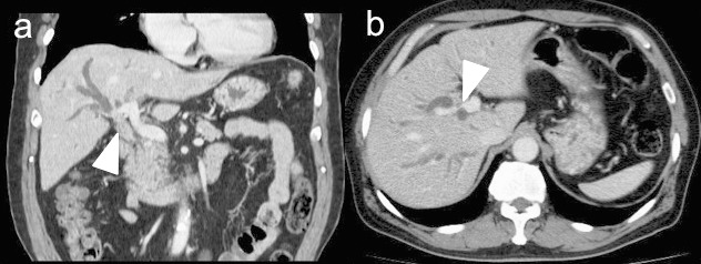
A multidetector computed tomography scan of the abdomen showed thickening of the bile duct wall associated with the enhancement of the right hepatic duct, extending from the proximal confluence of the common hepatic duct to the left and right hepatic ducts. In addition, dilatation of the peripheral intrahepatic bile ducts was present. The right portal vein was compressed by the lesion, which was considered a possible indication of a carcinoma with invasion (Fig. 1). Abdominal ultrasonography showed thickening of the common bile duct wall, without swelling of the pancreas. Magnetic resonance cholangiopancreatography revealed dilation extending from the distal common bile duct to the left and right intrahepatic bile ducts. The main pancreatic duct and the confluence of the bile duct and pancreatic duct showed no abnormalities.
Fig. 1.

(a) Computed tomographic imaging shows a thickened wall of the common bile duct enhanced with contrast media (arrow) and narrowing of right portal vain. (b) Peripheral hepatic ducts are dilated because of obstruction. The right portal vain was compressed by a bile duct mass (arrow).
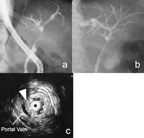
Endoscopic retrograde cholangiopancreatography showed a sharply angulated stricture extending from the proximal portion of the confluence of the 3 ducts to the bifurcation of the left and right hepatic ducts (Fig. 2a). Intraductal ultrasonography showed portal vein stenosis invaded by a notched lesion with a circular-asymmetric appearance in the main bile duct (Fig. 2c). Histopathologic examination of biopsy samples showed atypical epithelial fragments with dysplastic cells. Cytologic examination of the bile revealed class III cells. At the time of transhepatic percutaneous drainage, the bile duct wall was extremely indurated and difficult to enter with the needle. Therefore, invasion by a bile duct cancer was suspected. An indwelling percutaneous transcutaneous hepatic drainage tube was inserted from the proximal side of the anterior superior segmental duct (B8) to relieve jaundice. Based on these results, the preoperative diagnosis of an upper bile duct cancer extending to the hepatic hilum was established. Right hepatectomy was planned.
Fig. 2.

(a) Endoscopic retrograde cholangiopancreatography shows a common bile duct stricture from the level of the proximal common bile duct to the intrahepatic ducts. (b) Percutaneous transhepatic cholangiography shows persistent common bile duct stenosis after steroid treatment. (c) Intraductal ultrasonography shows an intraductal mass lesion (arrow) extending with stenosis of the portal vain.
Percutaneous transhepatic portal vein embolization was performed approximately 1 month before the planned surgery, and prednisolone (1 g) pulse therapy was performed once after percutaneous transhepatic portal vein embolization as a routine treatment. Cholangiography showed that the stricture had slightly improved 2 weeks after percutaneous transhepatic portal vein embolization (Fig. 2b).
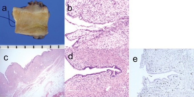
Finally, because we could not be rule out IgG4-SC or cholangiocarcinoma, we performed exploratory laparotomy and planed bile duct resection to determine whether the upper bile duct tissue was malignant. At operation, the hepatogastric ligament was fibrous and indurated, making it difficult to dissect. Induration was present; however, the bile duct wall appeared softer than would be expected in the presence of an invasive tumor. There was no palpable mass in the common bile duct, suggesting the absence of a malignancy. Thereafter, partial resection of the upper bile duct was performed, and operative rapid pathologic diagnosis method revealed bile duct wall thickening and diffuse granulomatous changes with no evidence of malignancy (Fig. 3b). Therefore, we did not perform right hepatectomy.
Fig. 3.

(a) Histopathologic examination of the resected common bile duct shows a thickened wall and smooth mucosa without mass formation. (b) Frozen section shows infiltrating inflammatory cells and granulomatous changes without malignant cells (×100). (c, d) Microscopic findings show fibrous tissue with an inflammatory cell infiltrate in the bile duct wall (hematoxylin and eosin, ×10 and ×100). (e) Immunostaining (×100) of IgG4-positive cells.
Histopathologic examination showed that the bile duct wall was grossly thickened with smooth mucosa (Fig. 3a). Hematoxylin and eosin staining mainly revealed fibrous tissue in all layers of the bile duct wall, with inflammatory cell infiltration, and with lymphocytes and plasma cells around the bile duct and nerves (Fig. 3c and 3d). Immunostaining showed more than 50 IgG4-positive cells per high-power field, and IgG4-SC was diagnosed (Fig. 3e). After surgery, we started steroid treatment from 20 mg of prednisolone oral prescription and gradually tapered every 6 months. Finally, we stopped prednisolone 2 years later. The serum IgG4 level remained normal without symptoms or abnormalities in a laboratory data and imaging diagnosis during 4 years of follow-up.
Discussion
IgG4-SC is a subtype of autoimmune pancreatitis. Recent reports have shown that this disease occurs mainly in the elderly in association with high serum IgG4 levels and with IgG4-positive plasma cell infiltration of tissue, leading to fibrosis of various organs, such as the pancreas, salivary glands, bile ducts, and retroperitoneum; the disease usually responds well to steroid therapy.5–8
Clinically, IgG4-SC is often accompanied by obstructive jaundice due to common bile duct strictures.9–12 Although these strictures are characteristic on imaging studies, all or part of the bile duct may be involved in IgG4-SC. Sclerotic changes in the intrahepatic and extrahepatic bile ducts were found in one half of patients with IgG4-SC.11
In general, diffuse pancreatic enlargement or a pancreatic mass with autoimmune pancreatitis is common in patients with IgG4-SC. However, cases of isolated biliary tract involvement without pancreatic lesions have also been reported.11,12 Moreover, types 3 and 4 IgG4-SC, in the absence of pancreatitis, may mimic cholangiocarcinoma and can be difficult to distinguish from malignant tumors. Therefore, major surgical resections, such as a hepatectomy or pancreaticoduodenectomy, have been reported in several patients with this disease.3,4,10,11
Hilar cholangiocarcinoma is usually referred to as Klatskin tumor. Curative treatment necessitates bile duct resection with en bloc hepatectomy. Delays in surgery are not acceptable for planned curative treatment of hilar cholangiocarcinoma. However, 5% to 24% of patients undergoing surgery with a preoperative diagnosis of hilar bile duct cancers have been ultimately found to have benign disease, with strictures mainly caused by postinflammatory changes.13–17 Therefore it is necessary to differentiate types 3 and 4 IgG4-SC from cholangiocarcinoma, and thereby avoid unnecessary hepatectomy.
The diagnostic criteria for autoimmune pancreatitis according to the HISORt criteria are: (1) histologic evidence of IgG4-positive cells, (2) evidence of pancreatitis on imaging studies, (3) high serum IgG4 concentrations, (4) other organ findings, and (5) a response to steroids.5 In Japan, similar diagnostic criteria have been proposed. Both sets of criteria emphasize the importance of histologic findings, but they include the importance of a response to steroids as a criterion.18,19
Intraductal ultrasonography plays an important role in the diagnosis of types 3 and 4 IgG4-SC, which often shows a smooth, circular-symmetric, and homogeneous bile duct wall. Noda et al20 reported that intraductal ultrasonography and biopsy provide useful information for the diagnosis of cholangiocarcinoma and IgG4-SC after endoscopic retrograde cholangiopancreatography in a single session. Endoscopic biopsy to establish a histopathologic diagnosis is useful and minimally invasive; however, it yields only a small number of cells from a limited area. The sensitivity of biopsy for the diagnosis of cholangiocarcinoma ranges from 54% to 86%.21,22 Transpapillary biopsy demonstrated abundant IgG4-positive plasma cells in only 18% of patients with IgG4-SC.23 Therefore, transpapillary biopsy may not be useful for the diagnosis of IgG4-SC, even with IgG4 immunostaining.23–25
To avoid delaying treatment in cases of cholangiocarcinoma, we generally have not conducted a steroid trial for IgG4-SC patients. Moreover, the present case was not involved in other organs and right portal vein was compressed, which was strongly suspicious of a carcinoma. However, after only one round of steroid pulse treatment, the bile duct stricture in this patient improved; therefore, we may make a better diagnosis if we do undertake the steroid trial. Thus, steroid treatment after percutaneous transhepatic portal vein embolization may be useful in the diagnosis of bile duct stenosis that has value for hilar bile duct type of IgG4-SC, to estimate steroid effects.26 If steroid pulse treatment is effective, we should start conducting routine steroid trials in these cases, which would greatly improve our diagnostic success.
In summary, types 3 and 4 IgG4-SCs without involvement of other organs are challenging to diagnose because of the difficulty of differentiating them from hilar cholangiocarcinoma. Diagnostic criteria emphasize the importance of histologic findings and transpapillary biopsy. Taken together, a histopathologic diagnosis obtained with a less invasive approach may avoid unnecessary hepatectomy. We reported a rare case of IgG4-SC.
Acknowledgments
The authors would like to thank Enago (http://www.enago.jp) for the English language review.
References
- 1.Finkelberg DL, Sahani D, Deshpande V, Brugge WR. Autoimmune pancreatitis. N Engl J Med. 2006;355(25):2670–2676. doi: 10.1056/NEJMra061200. [DOI] [PubMed] [Google Scholar]
- 2.Ghazale A, Chari ST, Zhang L, Smyrk TC, Takahashi N, Levy MJ, et al. Immunoglobulin G4-associated cholangitis: clinical profile and response to therapy. Gastroenterology. 2008;134(3):706–715. doi: 10.1053/j.gastro.2007.12.009. [DOI] [PubMed] [Google Scholar]
- 3.Maeda M, Shimada K. A case of IgG4-related sclerosing cholangitis mimicking an intrahepatic cholangiocellular carcinoma. Jpn J Clin Oncol. 2012;42(2):153. doi: 10.1093/jjco/hys003. [DOI] [PubMed] [Google Scholar]
- 4.Erdogan D, Kloek JJ, ten Kate FJ, Rauws EA, Busch OR, Gouma DJ, et al. Immunoglobulin G4-related sclerosing cholangitis in patients resected for presumed malignant bile duct strictures. Br J Surg. 2008;95(6):727–734. doi: 10.1002/bjs.6057. [DOI] [PubMed] [Google Scholar]
- 5.Chari ST, Smyrk TC, Levy MJ, Topazian MD, Takahashi N, Zhang L, et al. Diagnosis of autoimmune pancreatitis: the Mayo Clinic experience. Clin Gastroenterol Hepatol. 2006;4(8):1010–1016. doi: 10.1016/j.cgh.2006.05.017. quiz 934. [DOI] [PubMed] [Google Scholar]
- 6.Hamano H, Kawa S, Horiuchi A, Unno H, Furuya N, Akamatsu T, et al. High serum IgG4 concentrations in patients with sclerosing pancreatitis. N Engl J Med. 2001;344(10):732–738. doi: 10.1056/NEJM200103083441005. [DOI] [PubMed] [Google Scholar]
- 7.Erkelens GW, Vleggaar FP, Lesterhuis W, van Buuren HR, van der Werf SD. Sclerosing pancreato-cholangitis responsive to steroid therapy. Lancet. 1999;354(9172):43–44. doi: 10.1016/s0140-6736(99)00603-0. [DOI] [PubMed] [Google Scholar]
- 8.Nowatari T, Kobayashi A, Fukunaga K, Oda T, Sasaki R, Ohkohchi N. Recognition of other organ involvement might assist in the differential diagnosis of IgG4-associated sclerosing cholangitis without apparent pancreatic involvement: report of two cases. Surg Today. 2012;42(11):1111–1115. doi: 10.1007/s00595-012-0278-6. [DOI] [PubMed] [Google Scholar]
- 9.Takikawa H, Takamori Y, Tanaka A, Kurihara H, Nakanuma Y. Analysis of 388 cases of primary sclerosing cholangitis in Japan; presence of a subgroup without pancreatic involvement in older patients. Hepatol Res. 2004;29(3):153–159. doi: 10.1016/j.hepres.2004.03.006. [DOI] [PubMed] [Google Scholar]
- 10.Nishino T, Toki F, Oyama H, Oi I, Kobayashi M, Takasaki K, et al. Biliary tract involvement in autoimmune pancreatitis. Pancreas. 2005;30(1):76–82. [PubMed] [Google Scholar]
- 11.Nakazawa T, Ohara H, Sano H, Ando T, Aoki S, Kobayashi S, et al. Clinical differences between primary sclerosing cholangitis and sclerosing cholangitis with autoimmune pancreatitis. Pancreas. 2005;30(1):20–25. [PubMed] [Google Scholar]
- 12.Hamano H, Kawa S, Uehara T, Ochi Y, Takayama M, Komatsu K, et al. Immunoglobulin G4-related lymphoplasmacytic sclerosing cholangitis that mimics infiltrating hilar cholangiocarcinoma: part of a spectrum of autoimmune pancreatitis? Gastrointest Endosc. 2005;62(1):152–157. doi: 10.1016/s0016-5107(05)00561-4. [DOI] [PubMed] [Google Scholar]
- 13.Are C, Gonen M, D'Angelica M, DeMatteo RP, Fong Y, Blumgart LH, et al. Differential diagnosis of proximal biliary obstruction. Surgery. 2006;140(5):756–763. doi: 10.1016/j.surg.2006.03.028. [DOI] [PubMed] [Google Scholar]
- 14.Hadjis NS, Collier NA, Blumgart LH. Malignant masquerade at the hilum of the liver. Br J Surg. 1985;72(8):659–661. doi: 10.1002/bjs.1800720826. [DOI] [PubMed] [Google Scholar]
- 15.Gerhards MF, Vos P, van Gulik TM, Rauws EA, Bosma A, Gouma DJ. Incidence of benign lesions in patients resected for suspicious hilar obstruction. Br J Surg. 2001;88(1):48–51. doi: 10.1046/j.1365-2168.2001.01607.x. [DOI] [PubMed] [Google Scholar]
- 16.Wetter LA, Ring EJ, Pellegrini CA, Way LW. Differential diagnosis of sclerosing cholangiocarcinomas of the common hepatic duct (Klatskin tumors) Am J Surg. 1991;161(1):57–62. doi: 10.1016/0002-9610(91)90361-g. discussion 62–63. [DOI] [PubMed] [Google Scholar]
- 17.Fujita T, Kojima M, Kato Y, Gotohda N, Takahashi S, Konishi M, et al. Clinical and histopathological study of “follicular cholangitis”: sclerosing cholangitis with prominent lymphocytic infiltration masquerading as hilar cholangiocarcinoma. Hepatol Res. 2010;40(12):1239–1247. doi: 10.1111/j.1872-034X.2010.00716.x. [DOI] [PubMed] [Google Scholar]
- 18.Ohara H, Okazaki K, Tsubouchi H, Inui K, Kawa S, Kamisawa T, et al. Clinical diagnostic criteria of IgG4-related sclerosing cholangitis 2012. J Hepatobiliary Pancreat Sci. 2012;19(5):536–542. doi: 10.1007/s00534-012-0521-y. [DOI] [PubMed] [Google Scholar]
- 19.Okazaki K, Uchida K, Ikeura T, Takaoka M. Current concept and diagnosis of IgG4-related disease in the hepato-bilio-pancreatic system. J Gastroenterol. 2013;48(3):303–314. doi: 10.1007/s00535-012-0744-3. [DOI] [PMC free article] [PubMed] [Google Scholar]
- 20.Noda Y, Fujita N, Kobayashi G, Ito K, Horaguchi J, Takazawa O, et al. Intraductal ultrasonography before biliary drainage and transpapillary biopsy in assessment of the longitudinal extent of bile duct cancer. Dig Endosc. 2008;20(2):73–78. [Google Scholar]
- 21.Weber A, von Weyhern C, Fend F, Schneider J, Neu B, Meining A, et al. Endoscopic transpapillary brush cytology and forceps biopsy in patients with hilar cholangiocarcinoma. World J Gastroenterol. 2008;14(7):1097–1101. doi: 10.3748/wjg.14.1097. [DOI] [PMC free article] [PubMed] [Google Scholar]
- 22.Higashizawa T, Tamada K, Tomiyama T, Wada S, Ohashi A, Satoh Y, et al. Biliary guidewire facilitates bile duct biopsy and endoscopic drainage. J Gastroenterol Hepatol. 2002;17(3):332–336. doi: 10.1046/j.1440-1746.2002.02691.x. [DOI] [PubMed] [Google Scholar]
- 23.Naitoh I, Nakazawa T, Ohara H, Ando T, Hayashi K, Tanaka H, et al. Endoscopic transpapillary intraductal ultrasonography and biopsy in the diagnosis of IgG4-related sclerosing cholangitis. J Gastroenterol. 2009;44(11):1147–1155. doi: 10.1007/s00535-009-0108-9. [DOI] [PubMed] [Google Scholar]
- 24.Nakazawa T, Naitoh I, Hayashi K, Okumura F, Miyabe K, Yoshida M, et al. Diagnostic criteria for IgG4-related sclerosing cholangitis based on cholangiographic classification. J Gastroenterol. 2012;47(1):79–87. doi: 10.1007/s00535-011-0465-z. [DOI] [PubMed] [Google Scholar]
- 25.Nakazawa T, Naitoh I, Hayashi K. Usefulness of intraductal ultrasonography in the diagnosis of cholangiocarcinoma and IgG4-related sclerosing cholangitis. Clin Endosc. 2012;45(3):331–336. doi: 10.5946/ce.2012.45.3.331. [DOI] [PMC free article] [PubMed] [Google Scholar]
- 26.Kubo N, Suzuki H, Kobayashi T, Araki K, Sasaki S, Wada W, et al. Usefulness of steroid administration for diagnosis of IgG4-related sclerosing cholangitis. Int Surg. 2012;97(2):145–149. doi: 10.9738/CC78.1. [DOI] [PMC free article] [PubMed] [Google Scholar]


