Abstract
This study aims to present the management of priapism in adult men in Port Harcourt, Nigeria. All patients who presented with priapism in 2 hospitals in Port Harcourt from July 2007 to April 2014 were prospectively studied. Treatment was assigned based on clinical presentation. Data analyzed included: age on clinical presentation, risk factor, mode, and outcome of management. There were 18 patients aged 17 to 60 years (median age: 30 years). Three patients (16.7%) presented with stuttering priapism. Most of the patients presented after 24 hours of onset. Sixteen patients (89.9%) had hematological disorders. Five patients (27.8%) took suspected aphrodisiac medications. Seven patients (38.9%) were managed conservatively. The rest achieved detumescence following glandulo-cavernous shunting. Erectile function after treatment was satisfactory in 5 patients (27.8%). The commonest cause of priapism in Port Harcourt was hematological disorder. Most of the patients presented late. Prevalence of erectile dysfunction after treatment was high.
Key words: Priapism, Sickle cell anemia, Tumescence
Priapism is a potentially painful medical condition in which the erect penis or clitoris does not return to its flaccid state, despite the absence of both physical and psychological stimulation, within 4 hours.1 Priapism has been described as a genuine erectile dysfunction in which erection persists without sexual stimulation.2 It is a rare condition with overall incidence of 1.5 cases per 100,000 person-years.3 However, it is an important urologic emergency because erectile tissue damage may occur leading to loss of functional erections.4 The time interval between the onset of symptoms and presentation for medical intervention impacts on the outcome of management.
Priapism is associated with various risk factors such as: antipsychotics drugs, local aphrodisiacs; hematologic malignancies, and congenital vascular disorders. Hemoglibinopathies constitute the major cause in countries where sickle cell disease (SCD) is endemic.5
Depending on the predisposing factor, priapism may be ischemic, nonischemic, or stuttering. Ischemic priapism is also referred to as veno-occlusive or low flow priapism. Ischemic priapism is characterized by painful persistent erection, reduced or absent cavernosal blood flow, and abnormal cavernous blood gases. The corpora cavernosa are rigid and painful.2,3
Nonischemic priapism is of arterial origin and is said to be a high-flow type. It is due to unregulated cavernous arterial inflow. The phallus in this case may not be rigid or painful and may not require emergency surgical intervention.2
In stuttering priapism, the patient has an unwanted painful erection which occurs repeatedly with periods of detumescence. These patients are thought to have recurrent ischemic priapism.4,5
Studies on priapism in the African populations focused on risk factors. This article is to present the results of a 7-year prospective study of priapism in 2 institutions in Port Harcourt, Nigeria, with the aim of documenting the relationship between the outcome of treatment and interval between onset of priapism and intervention in an African population.
Materials and Methods
All patients who presented at the University of Port Harcourt Teaching Hospital and Potter's Touch Medical Consultants (PTMC) between June 2007 and April 2014 were included in the study. University of Port Harcourt Teaching Hospital is a tertiary hospital while PTMC is a private urology practice. Both hospitals are in the metropolis of Port Harcourt, Nigeria. The information documented for analysis for each patient included: the primary complaint, age, duration of priapism before presentation, reason for delayed presentation, presence or absence of pain, the degree of angulations of the phallus, and suspected precipitating or predisposing factors. Also documented were: type of treatment and supportive care given, duration of treatment before achievement of detumescence, and subsequent erection after treatment. A diagnosis of priapism was based on the clinical presentation, with tender rigid phallus involving the whole penile shaft. Investigations included: hemoglobin genotype, white cell count, and platelet count. The angle of tumescence was measured as the angle between the dorsum of the erect penis and the anterior abdominal wall. Each patient had individualized care, depending on the precipitating factor and severity of the presentation. Detumescence was achieved either conservatively or by surgical intervention. The latter was by glandulo-cavernous shunt using the technique described by Ebbehoj.6,30 Under local infiltration of 1% plain xylocaine through the glans penis into the corpus cavernosum on 1 side, a size 15 surgical blade is used to create a shunt between the glans penis and the corpus carvenosum. Altered blood in the cavernosum is evacuated. Thereafter, normal saline is used for irrigation. Only 1 patient had shunts on both corpora cavernosa, after 1 side failed.
The results were analyzed, descriptively assisted with bar charts, and pie chart. The patients were followed up on monthly outpatient clinic visits.
Results
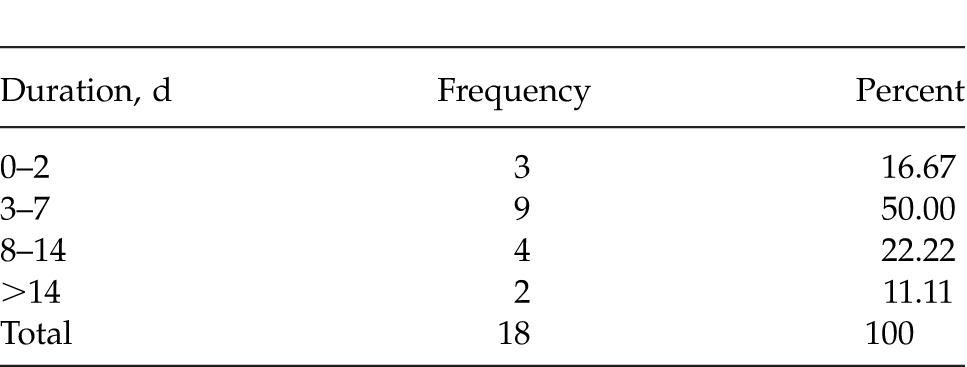
A total of 18 men presented with priapism during the period under review. All the patients were men. The ages ranged from 17 to 60 years with a median of 30 years. The median duration of symptoms before presentation was 6 days (range: 1–35 days). Major contributors to this delay included ignorance and the fact that some of the patients went for unorthodox treatment before they came to the hospital. All the patients presented with pain. All the patients had rigid and tender phalluses suggestive of ischemic priapism. Three of the patients (16.7%) presented with stuttering priapism. The duration of symptoms before presentation is summarized in Table 1.
Table 1.
Duration of symptoms before presentation

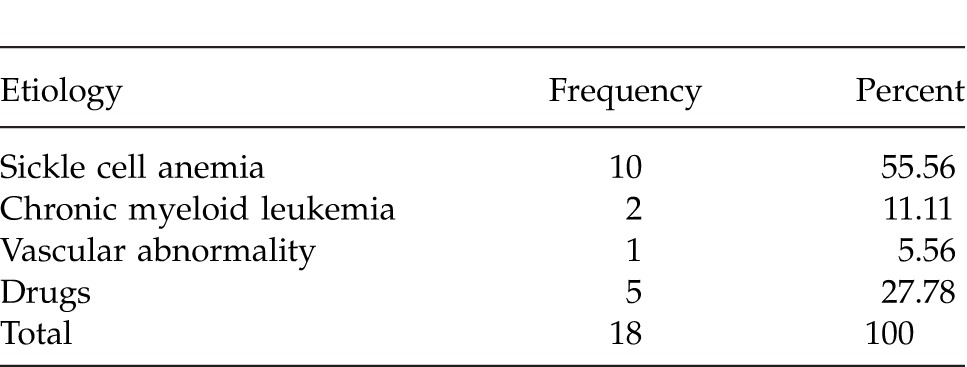
One of the cases in this study had a vascular disorder with reduced blood flow in the iliac vein on Doppler ultrasound. None of the patients had priapism precipitated by sexual intercourse. Five patients (27.8 %) had a history of ingestion of drugs: 2 patients took cannabis; 2 ingested local herbs; and 1 patient took bromcriptine for treatment of hyperprolactinemia. Three of the patients who took some drugs also had underlying hematological problems such as chronic myeloid leukemia (CML) and sickle cell anemia. Table 2 shows the main etiological factors.
Table 2.
Major etiology of priapism

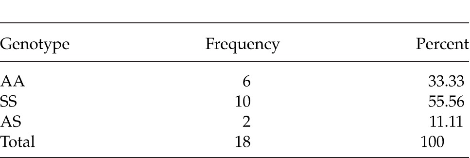
Ten (55.6%) of the cases had hemoglobin genotype SS. Two patients (11.1%) had genotype AS while 6 (33.3%) were of genotype AA as shown in Table 3. One of the 18 patients (5.6%) had thrombocytosis, while 2 (11.1 %) of them had a high white cell count. These were later diagnosed as CML. Eight patients (44.4%) were also found to be anemic. Seven patients (38.9%) had normal hemoglobin levels. Only 2 of these patients did not have a background hematological disorder. The remaining 16 (89.9%) had a hematological disorder such as sickle cell anemia and chronic myeloid leukemia. The mean angle of tumescence in these patients was 51.4° (range: 15–70°).
Table 3.
Hemoglobin genotype distribution

Seven patients (38.9%) were managed conservatively with intravenous fluids, nonsteroidal anti-inflammatory drugs, epinephrine, and stilbesterol. They achieved detumescence within 1 to 7 days of treatment. Eleven of the patients (61.1%) had glandulo-cavernous shunt before detumescence was achieved. Three of the 7 managed conservatively were of hemoglobin SS (abbreviated as HBSS is a homozygous inheritance of the mutant hemoglobin S and is associated with distortion of the red cell with increased deoxynation) genotype who presented with stuttering priapism.
The outcome of treatment assessed by degree of patient's satisfaction with subsequent penile erection showed that 4 (27.8%) of the patients had satisfactory erection after detumescence. Seven of the patients (38.9%) had weak erection while 6 patients (33.3%) could not achieve erections at all after 3 months' observation.
The patients with no erections had a median duration of 10.5 days before presentation (range: 5–35 days). Those with weak erections had a median duration of 7 days (range: 3–24 days). The patients with satisfactory outcome had a median duration of 1 day (range: 1–7 days). Outcome of erectile function after treatment was ranked against the duration in days before intervention using the nonparametric Spearman's test. The correlation is weak though it is statistically significant.
One patient with a vascular disorder was treated by glandulo-cavernous shunt. He developed ED subsequently. For this, he had a rigid implant procedure in India, with a satisfactory result.
Discussion
The median age range of our patients was 30 years. Previous reports indicate that the peak periods are in the first and fourth decades of life.6–8 This bimodal distribution can be explained by the difference in the etiological factors. Younger age groups are more often associated with sickle cell disease, while older groups tend to be secondary to pharmacologic agents.9 In this study, only adult patients were treated. Since this study did not include pediatric patients, a bimodal peak could not be reflected. The observed age distribution is close to the fourth decade previously documented in adults.10
Although priapism has been observed in females, all patients seen in this study were men.1,6 This picture is also common in many studies in Nigeria because priapism of the clitoris is very rare.7,11,12 It may be rarer in Nigeria because of clitoridectomy in female genital mutilation. Clitoral priapism is associated with specific classes of medications, diseases that alter clitoral blood flow, or others associated with small to large vessel disease.1,6
Late presentation as reported in this study was also the finding in other reports from Nigeria.12,14 This is in contrast with a report from New York where the mean duration of priapism before presentation was 22 hours, with over 60% presenting within 24 hours of onset.14 Poverty and ignorance are factors that may also contribute to the delay in seeking for help.11,12
Priapism results from a disturbance of the regulatory mechanisms that initiate and maintain penile flaccidity.15 All the patients in this study presented with low-flow (ischemic) priapism. This picture is similar to reports elsewhere.7,11
Blood dyscrasias have been considered as a major risk factor for priapism.5 Dyscrasias that have been implicated include: sickle cell disease, thrombotic conditions, and hematologic malignancies (e.g., leukemia and multiple myeloma).16 From this study, the highest incidence was among HbSS: 71.4%. This is in accordance with findings in other parts of Nigeria.11,12 However, it differs from reports from Canada where pharmacological agents and idiopathic cases were predominant.16 Principally, the Port Harcourt community is a sickle cell–endemic community. It has been documented that the lifetime prevalence of ischemic priapism in SCD ranges from 2% to 35% and high flow (nonischemic) priapism is not common in SCD.5 Priapism secondary to SCD has been reported in up to 23% of adult sickle cell patients and 63% of the pediatric age group.18
Anatomical factors and a low-flow state contribute to making the erectile tissues in the SCD patient to be more prone to ischemia and pain. However, dysregulation of the nitric oxide pathway and its downstream signaling may be the stronger causative agent for development of priapism in sickle cell patients.20 Some markers of hemolysis have also been associated with the risk of priapism in sickle cell patients (e.g., lactate dehydrogenase, serum bilirubin, and reticulocyte counts).20
Hematologic malignancy, CML, was contributory in 14.3% of the patients. Priapism is another mode of presentation of CML.21,22
Venous outflow obstructions of the corporal bodies and increased intracavernous blood viscosity have been suggested as likely mechanisms for priapism associated with hematologic dyscrasias such as multiple myeloma, CML, and thrombocytosis.5 Priapism may occur in up to 50% of cases of CML.23 Hyperleukocytosis is linked to the causation of priapism in patients with leukemia. Some studies have shown that a leukocyte count greater than 100 × 109/L or more is a major contributor of an elevation of the whole-blood viscosity.13,14 The elevated blood viscosity resulting from high white blood cell count and platelet count may have contributed to the priapism observed. When priapism occurs in hematologic disorders, it may resolve with treatment of the primary disorder or progress to become recurrent with or without some degree of erectile impairment.5 Among the 3 patients (16.7%) in this series who had recurrent or stuttering presentation, 2 of them were SCD and the other had CML.
An important cause of priapism is ingestion of predisposing drugs. Many drugs have been linked to priapism. These include: oral medications used for erectile dysfunction (ED), intracavernosal drug for ED, antidepressants, antipsychotics, blood thinners, and recreational drugs.6,13,14,24 Antipsychotic drugs linked to priapism have adrenergic blocking effects.14 The etiology of priapism in 5 (28.6%) of the cases in this study were associated with drugs: 2 were due to cannabis abuse; 2 ingested local herbs thought to have suspected aphrodisiac potentials; and 1 patient ingested bromcriptine for hyperprolactinemia. Three of these 5 patients also had background hematological disorders, such as SCD, and CML. Perhaps, the drugs were mere triggering agents. In this report, none of the patients with priapism presented secondary to antipsychotic agents.
In rats, cannabinoids can potentially modulate blood flow in central and peripheral nervous systems by inhibiting norepinepherine and increasing vagal tone. Thus, increased parasympathetic activity may cause penile tumescence.24 This may explain the cause of priapism in the patients precipitated by cannabis smoking. Reports from Enugu, Nigeria, showed that a local aphrodisiac agent was the commonest cause of priapism in that region.14 Although the chemical content of the herbal concoction drank by 2 of our patients is unknown, it is possible that the local herbs may have aphrodisiac properties, thereby leading to the observed priapism. Our observation is similar to reports by Omisanjo12 in Lagos where aphrodisiacs did not play as much role in the etiology as in Enugu.14 The drugs were not taken for sexual performance enhancement, unlike in the cases reported in Enugu.
Alpha adrenergic blockers used for treatment of hypertension and lower urinary tract symptoms (LUTS) have also been reported to induce priapism.26 Alpha sympathomimetic blockade results in extended relaxation of the carvenosal smooth muscles and venous stasis in the corpus cavernosum.21 Although these drugs are prescribed to a large population of patients with LUTS, none of the patients presented with alpha blocker–induced priapism.
One patient who had hyperprolactinemia was treated with oral bromcriptine. He developed priapism after 4 hours of therapy. This resolved when the drug was discontinued. Bromcriptine lowers prolactin level by stimulation of dopamine receptors in the pituitary gland. Stimulation of dopamine receptors enhances sexual interest.27 It has been postulated that pro-erection properties of dopaminergic neurons occur by activating oxytocinergic neurons in the paraventricular nuclei and that the release of oxytocin produces erection.28
The patient who presents with priapism should be properly examined and investigated to determine the cause. Treatment should be prompt and the urologist must be involved early in the management of these patients to improve outcome.4 The aim of management is to achieve detumescence as soon as possible in order to preserve erectile function. Priapism is a urological emergency. Once a diagnosis is made, treatment should be commenced immediately. This is to avoid irreversible ultrastructural changes as a result of stasis and cavernosal tissue ischemia.29 Delay in presentation and treatment may lead to impaired function of the phallus. Therefore, it is important to decompress the corpora cavernosa to relieve the ischemia and pain.4 After diagnosis, the 2-pronged approach to treatment aims to secure detumescence of the turgid penis, and simultaneously treat any underlying disease. Detumescence may be achieved conservatively or by surgical intervention, depending on the predisposing cause and clinical presentation.
Seven patients (38.9%) were managed conservatively with analgesics, intravenous fluids, epinephrine, sedatives and withdrawal of the precipitating drugs (bromcriptine and cannabis). There was a statistical difference between the outcome of erectile function after treatment and the duration of priapism before intervention. This relationship between erectile function has been documented elsewhere. According to Van Der Horst,31 the overall rate of erectile dysfunction can be as high as 59%.31 El-Bahnasawy and colleagues32 reported only 43% of their patients had erection after prolonged duration of priapism. In their study, the median range of duration was 48 hours. In this study, with a median duration of 5 days before intervention, only 27.8% had normal erection after treatment as opposed to the report above. One explanation is that the patients in this study presented later than those in the study of El-Bahnasawy and colleagues. The longer the priapism, the greater the incidence of ischemic necrosis with the resultant fibrosis and erectile dysfunction.31 One patient in this study with erectile dysfunction following priapism received a semirigid penile implant with satisfaction. Penile prosthesis is a known modality of treatment for post-priapism erectile dysfunction.33
Study Limitations
The small sample size in this study limits its wide applicability. Patients with priapism may not present to the hospital for financial reasons and social stigmatization. This explains the small size of our data. It was because of the size of our sample that we used “median” and not “mean” to calculate the averages of duration of priapism before treatment as well as the ages of the patients. Median is less sensitive to extreme scores and is probably a better indicator of where the middle of the class is, especially for smaller sample sizes. Mean is the arithmetic average often used with larger sample sizes.34,35
Conclusion
Hematological disorders and some drugs of addiction were associated with priapism in adult men in Port Harcourt. Most of the patients presented late primarily due to ignorance. The outcome of treatment was related to the time interval between onset of priapism and treatment. Education of men and the population at risk on the need to seek immediate treatment will enhance the preservation of erectile function.
References
- 1.Gharahbaghaian L. Clitoral priapism with no known risk factor. West J Emerg Med. 2008;9(4):235–237. [PMC free article] [PubMed] [Google Scholar]
- 2.Montague DK, Jarrow J, Broderick GA, Omochowski RR, Heaton JP, Lue TF, et al. American Urologic Association guideline on the management of priapism. J Urol. 2003;170((4 pt 1)):1318–1324. doi: 10.1097/01.ju.0000087608.07371.ca. [DOI] [PubMed] [Google Scholar]
- 3.Eland IA, van der Lei J, Stricker BH, Sturkenboom MJ. Incidence of priapism in the general population. Urology. 2001;57(5):970–972. doi: 10.1016/s0090-4295(01)00941-4. [DOI] [PubMed] [Google Scholar]
- 4.Burnett AL. Therapy insight: priapism associated with haematologic dyscrasias. Nat Clin Pract Urol. 2005;2(9):449–456. doi: 10.1038/ncpuro0277. [DOI] [PubMed] [Google Scholar]
- 5.Cherian J, Rao AR, Thwaini A, Kapasi F, Shergill IS, Samman R. Medical and surgical management of priapism. Postgrad Med J. 2006;82(964):89–94. doi: 10.1136/pgmj.2005.037291. [DOI] [PMC free article] [PubMed] [Google Scholar]
- 6.Medina CA. Clitoral priapism: A rare condition presenting as a case of vulvar pain. Obstet Gynecol. 2002;100((5 pt 2)):1089–1091. doi: 10.1016/s0029-7844(02)02084-7. [DOI] [PubMed] [Google Scholar]
- 7.Adetayo FO. Outcome of management of acute prolonged priapism in patients with homozygous sickle cell disease. West Afr J Med. 2009;28(4):234–239. [PubMed] [Google Scholar]
- 8.Ochoa UO, Hermida P. Priapism, our priapism our experience. Arch Esp Urol. 1998;51(3):269–276. [PubMed] [Google Scholar]
- 9.Al-Qudah HS Al-Omar O, Parraga-Marquez M. Priapism. Available at: http://emedicine.medscape.com/article/437237-overview. Accessed September 09, 2014. [Google Scholar]
- 10.Mohamed MA, Hassan MA, Badaway HA. Priapism in a series of 459 patients presented for office evaluation of erectile dysfunction. El-Minia Med Bull. 2007;18(1):352–353. [Google Scholar]
- 11.Akanbi AA, Lateef BA, Olasunkanmi SA. Clinical presentation and management of priapism in a Nigeria tertiary hospital. J Surg Sci. 2009;1:103–106. [Google Scholar]
- 12.Omisanjo O, Ikuerowo A, Abdusalam A, Esho J. Clinical characteristics and outcome of management of priapism at the Lagos state university teaching hospital. Internet J Urol. 2012;9(3) doi: 10.4103/1596-3519.142287. [DOI] [PubMed] [Google Scholar]
- 13.Badmus TA, Adediran IA, Adesunkanmi ARK, Katung IA. Priapism in Southwestern Nigeria. East Afr Med J. 2003;80(10):518–524. doi: 10.4314/eamj.v80i10.8754. [DOI] [PubMed] [Google Scholar]
- 14.Aghaji AE. Priapism in adult Nigerians. BJU Int. 2000;85(4):493–495. doi: 10.1046/j.1464-410x.2000.00561.x. [DOI] [PubMed] [Google Scholar]
- 15.Abber JC, Lue TF, Luo JA, Juenemann KP, Tanagho EA. Priapism induced by chlorpromazine and trazodone: mechanism of action. J Urol. 1987;137(5):1039–1042. doi: 10.1016/s0022-5347(17)44355-2. [DOI] [PubMed] [Google Scholar]
- 16.Gregory AB, Ates K, Trinity JB, Hussein G, Ajay N, Rany S. Priapism: pathogenesis, epidemiology and management. J Sex Med. 2010;7:476–500. doi: 10.1111/j.1743-6109.2009.01625.x. (1 pt 2) [DOI] [PubMed] [Google Scholar]
- 17.Paulter SE, Brock GB. Priapism. From Priapus to the present time. Urol Clin North Am. 2001;28(2):391–403. doi: 10.1016/s0094-0143(05)70147-6. [DOI] [PubMed] [Google Scholar]
- 18.Nelson JH, 3rd, Winter CC. Priapism: evolution of management in 48 patients in a 22-year series. J Urol. 1977;117(4):455–458. doi: 10.1016/s0022-5347(17)58497-9. [DOI] [PubMed] [Google Scholar]
- 19.Crane GM, Bennett NE. Priapism in sickle cell anaemia: emerging mechanistic understanding and better preventive strategies. Anemia. 2011;2011:297364. doi: 10.1155/2011/297364. [DOI] [PMC free article] [PubMed] [Google Scholar]
- 20.Nolan VG, Wyszynski DF, Steinberg MH. Haemolysis-associated priapism in sickle cell disease. Blood. 2005;106(9):3264–3267. doi: 10.1182/blood-2005-04-1594. [DOI] [PMC free article] [PubMed] [Google Scholar]
- 21.Ekeke ON, Omunakwe HE, Nwauche CA. Chronic myeloid leukaemia presenting as priapism. Port Harcourt Med J. 2012;6(4):484–487. [Google Scholar]
- 22.Tazi I. Priapism as the first manifestation of chronic myeloid leukaemia. Ann Saudi Med. 2009;29(5):412. doi: 10.4103/0256-4947.55176. [DOI] [PMC free article] [PubMed] [Google Scholar]
- 23.Morano SG. The treatment of long-standing priapism in chronic myeloid leukemia at onset. Ann Haematol. 2000;79(11):27–32. doi: 10.1007/s002770000199. [DOI] [PubMed] [Google Scholar]
- 24.Reeves RR, Mack JE. Priapism associated with two atypical antipsychotic agents. Pharmacotherapy. 2002;22(8):1070–1073. doi: 10.1592/phco.22.12.1070.33613. [DOI] [PubMed] [Google Scholar]
- 25.Hillad CJ. Endocannabinoids and vascular function. J Pharmacol Exp Ther. 2000;294(1):27–32. [PubMed] [Google Scholar]
- 26.Spagnul SJ, Cabral PH, Verndl DO, Glina S. Adrenegic a-blockers: An infrequent and overlooked cause of priapism. Int J Impot Res. 2011;23(3):95–98. doi: 10.1038/ijir.2011.12. [DOI] [PubMed] [Google Scholar]
- 27.Sullivan G, Lukoff D. Sexual side effects of antipsychotic medication: evaluation and interventions. Hosp Community Psychiatry. 1990;41(11):1238–1241. doi: 10.1176/ps.41.11.1238. [DOI] [PubMed] [Google Scholar]
- 28.Melis MR, Succu S. PIP3EA and PD168077, two selective dopamine D4 receptor agonists, induce penile erection in male rats: site and mechanism of action in the brain. Eur J Neurosci. 2006;24(7):2021–2030. doi: 10.1111/j.1460-9568.2006.05043.x. [DOI] [PubMed] [Google Scholar]
- 29.Spycher MA, Hauri D. The ultrastructure of the erectile tissue in priapism. J Urol. 1986;135(1):142–147. doi: 10.1016/s0022-5347(17)45549-2. [DOI] [PubMed] [Google Scholar]
- 30.Ebbehoj J. A new operation for priapism. Scand J Plastic Reconstr Surg. 1974;8(3):241–242. doi: 10.3109/02844317409084400. [DOI] [PubMed] [Google Scholar]
- 31.Van der Horst C, Stuebinger H, Seif C, Melchior D, Martenez-Portillo FJ, Juenemann KP. Priapism - etiology, pathophysiology and management. Int Braz J Urol. 2003;29(5):301–400. doi: 10.1590/s1677-55382003000500002. [DOI] [PubMed] [Google Scholar]
- 32.El-Bahnasawy MS, Dawood A, Farouk A. Low-flow priapism: risk factors for erectile dysfunction. BJU Int. 2002;89(3):285–290. doi: 10.1046/j.1464-4096.2001.01510.x. [DOI] [PubMed] [Google Scholar]
- 33.Durazi MH, Jalal AA. Penile prosthesis implantation for treatment of postpriapism erectile dysfunction. Urol J. 2008;5(2):115–119. [PubMed] [Google Scholar]
- 34.Northwest Evaluation Association. MAP assessments: our scale and norms. Available at: http://www.nwea.org/node/4723. Accessed June 25, 2013. [Google Scholar]
- 35.Te Kete Ipurangi. Statistical concepts. Available at: http://assessment.tki.org.nz/Using-evidence-for-learning/Concepts. Accessed May 21, 2013. [Google Scholar]


