Abstract
Background
Group 2 Innate lymphoid cells (ILC2s) are innate cells that produce the TH2 cytokines IL-5 and IL-13. The importance of these cells has recently been demonstrated in experimental models of parasitic diseases but there is a paucity of data on ILC2s in the context of human parasitic infections and in particular of the blood dwelling parasite Schistosoma haematobium.
Methodology/Principal Findings
In this case-control study human peripheral blood ILC2s were analysed in relation to infection with the helminth parasite Schistosoma haematobium. Peripheral blood mononuclear cells of 36 S. haematobium infected and 36 age and sex matched uninfected children were analysed for frequencies of ILC2s identified as Lin-CD45+CD127+CD294+CD161+. ILC2s were significantly lower particularly in infected children aged 6–9 years compared to healthy participants. Curative anti-helminthic treatment resulted in an increase in levels of the activating factor TSLP and restoration of ILC2 levels.
Conclusion
This study demonstrates that ILC2s are diminished in young helminth infected children and restored by removal of the parasites by treatment, indicating a previously undescribed association between a human parasitic infection and ILC2s and suggesting a role of ILC2s before the establishment of protective acquired immunity in human schistosomiasis.
Author Summary
Understanding how immune responses are generated is critical for vaccine development. There are comparatively few studies on the interface between the innate and adaptive immune system in generating protective immune responses. Infections with helminth parasites, a cause of neglected tropical diseases, have a huge collective impact on public health in affected developing countries. Helminths are associated with a complex type 2 immune response mediated by cytokines characteristically produced by adaptive T helper 2 cells (TH2). However in recent years a newly described type of innate immune cells has been shown to produce TH2 cytokines. This cell type was subsequently called ‘Group 2 innate lymphoid cells’ (ILC2s). The importance of these cells has been demonstrated in experimental models of helminth infection as well as in allergic diseases. The present study describes changes in human ILC2s during the human helminth infection schistosomiasisas (bilharzia). Our study shows that the proportions of ILC2s, were lower in young, but not older infected children when compared to uninfected participants, suggesting a role for ILC2s before the establishment of helminth protective acquired immunity. Furthermore ILC2s were restored after curative treatment of the helminth infection. Human mechanistic studies will determine if the association between ILC2s and schistosome infection is causal or a marker of resistance to infection.
Introduction
Innate lymphoid cells are a recently described cell type that has transformed our understanding of the role of innate immune responses in the generation of adaptive immune responses. Group 2 Innate lymphoid cells (ILC2s) produce the classical TH2 cytokines IL-5 and IL-13 [1–3] and have been shown to be crucial for protective immune responses in experimental helminth infection [4–6]. ILC2s play an important role as an early source of IL-13 and are crucial for timely worm expulsion in mice infected with the murine helminth parasite Nippostrongylus brasiliensis. However, to date there is only one study in humans which characterised ILCs in a small population of patients with diverse filarial infections [7].
The interaction of helminth parasites and the host immune system has been extensively studied in experimental models and naturally infected humans. In experimental models, the immune responses to helminths, including schistosome infections, is polarized towards a type 2 immune response (reviewed in [8–10]). However, research over the last decade has shown that the establishment of helminth infections requires modulation of host type 2 responses, which is mediated by several different mechanisms including regulatory T cells, regulatory B cells and the immuno-modulatory cytokines IL-10 and TGF-β [11–15]. To a large extent these observations have been supported by evidence from human studies [16–18]. Type 2 immune responses have been shown to involve innate immune cells including dendritic cells, basophils and alternatively activated macrophages [19–21]. However in human, TH2 responses to helminth infections are less pronounced [22, 23]. In addition, humans infected with helminths show alterations of cellular responses such as regulatory T cells, dendritic cells and CD4+ T Cell responses [16, 24, 25] and these responses vary with age (and thereby with history of infection with helminths). Thus, the specific characteristics of the human type 2 response to helminths are more complex than in experimental models meaning that observations from the experimental setting must be validated in natural human infections.
Human ILC2s are negative for lineage markers (Lin-), but positive for the hematopoietic cell marker CD45 [26]. IL-7 is essential for the development of ILC2s [5, 27] and therefore CD127 (IL-7Rα) is a main marker for characterising ILC2s. Furthermore human ILC2s express the ‘chemokine receptor homologous molecule expressed on TH2 cells’ (CRTH2 = CD294) a marker of TH2 cells [28] and expressed the NK cell receptor CD161 (NKR-P1A) [26], whereas CD117 (c-kit) is mainly expressed on ILC3 and only on a subset of ILC2s [2].
ILC2s were initially described in fetal and adult gut and lung, but have also been identified in peripheral blood [26], but the function of blood ILC2s has not yet been investigated in detail. Changes in proportions of blood ILC2s could reflect either a global modulation of ILC2s or changes in the potential to migrate to tissues.
ILC2s are dependent on the TH2-associated transcription factors GATA-3 [29, 30] and RORα [27, 31] and the ‘thymic stromal lymphopoietin’ TSLP enhances GATA-3 expression and subsequent production of IL-4, IL-5 and IL-13, especially if supplied in combination with IL-33, but less efficiently with IL-25 [32]. Changes of systemic IL-33 during helminth infection have been recently reported [33].
The presence of ILC2s in human peripheral blood has been demonstrated in healthy individuals and in patients with mild or severe asthma [34, 35]. Furthermore, in mouse models ILC2s as well as IL-25 and IL-33, have been shown to be involved in allergic immune responses [36–41].
The aim of this study was to determine if levels of blood ILC2s change in the context of a parasitic infection in a human population, thereby providing evidence that ILC2s are important in human parasitic diseases. The study focused on the helminth parasite of the genus Schistosoma (blood-flukes) which causes schistosomiasis, a neglected tropical disease affecting about 240 million people mainly in Sub-Saharan Africa [42]. The most prevalent form is urogenital schistosomiasis caused by Schistosoma haematobium. The heaviest burden of this helminthic disease occurs in children, who typically acquire infection in the first year of life [43, 44]. Infection accumulates with age, and in most populations peaks around the age of 9–14 years, followed by a decrease in infection [45, 46] which has been attributed to the development of protective acquired immunity [47, 48]. Therefore immune epidemiological analyses covering these different stages of infection have been shown to be important in the analysis of the immune response towards helminth infections [16, 24, 25].
In this study we describe for the first time, alterations of ILC2 proportions in human blood during natural infection with schistosomes in a heterogeneous population (different ages to capture the different infection dynamics of rising, peaking and declining infection levels) and the effect of removing the helminth parasites through curative anti-helminthic drug treatment on ILC2 proportions. To elucidate pathways involved in changes of ILC2s, levels of the ILC2 activating factors TSLP and IL-33 were analysed.
Methods
Ethical statement
The study received institutional and ethical approval from the Ethical Review Board of the University of Zimbabwe and the Medical Research Council of Zimbabwe respectively. Permission to conduct the study in the selected area was obtained from the Provincial Medical Director, the District Educational Officer and School Heads. The aims and procedures of the study were explained to participants and their parents/guardians (in the case of children) in the local language, Shona, with written informed consent and assent obtained from the participants or parents/guardian before enrolment into the study and before administration of the anti-helminthic drug. Participants were free to drop out at any time of the study and parents/guardian could withdraw their children from the study. After sample collection, anti-helminthic treatment with the standard dose of praziquantel was offered to all participants and administered by a local physician.
Study area and design
This study was conducted in Magaya village in the Murehwa district of the Mashonaland East Province of Zimbabwe (31°91’E; 17°63’S) as part of a larger study investigating the immuno-epidemiology of human urogenital schistosomiasis of which several aspects have been published [24, 25, 49, 50]. Samples used within this study were collected between September and November 2008. Previous studies in this area and national surveys indicated low prevalence’s of S. mansoni and soil-transmitted helminths (STH), whereas the S. haematobium prevalence was high (>50%) [51, 52]. The area is mesoendemic for Plasmodium infection [53].
Magaya is a rural village where subsistence farming is the predominant occupation. Due to lack of safe water and sanitation facilities, participants are in frequent contact with fresh water, which harbours the parasite life stage (cercariae) infective to the human host. Frequency of water contacts of participants, their history of anti-helminthic treatment and residential history in the schistosome endemic area were documented by questionnaires.
Study group
To be included in the cross-sectional part of the study, participants had to meet the following criteria: a) been lifelong residents of the study area, b) not have previously received anti-helminthic treatment, c) be negative for S. mansoni, STH, Plasmodium falciparum (ensuring that the confounding effects of these parasites were excluded from the study) and HIV, d) have provided at least two urine and two stool samples on consecutive days for parasitological analysis and a blood sample sufficient for PBMC and plasma isolation. From the participants who fulfilled these criteria a group of 72 people were selected covering an age range of 6–18 years with 24 people in each of following three age groups: 6–9, 10–13, 14–18 years. Within each age group equal numbers of uninfected and infected people were selected providing a S. haematobium prevalence of 50%. Furthermore participants were age and sex matched between the uninfected and infected groups. Egg positive samples were chosen to have comparable infection intensities between the three age groups. The resulting study group is shown in Table 1.
Table 1. Description of the selected study group.
| Age group (years) | S. haematobium Status | Sample size (N) | Mean age (years) | S. haematobium Infection intensity: Mean / Median (range) | M/F |
|---|---|---|---|---|---|
| 6–9 | egg ve- | 12 | 7.83 | 0 | 6/6 |
| egg ve+ | 12 | 7.92 | 28.3 / 16.0 (2.7–158.3) | 6/6 | |
| Total | 24 | 7.88 | 14.1 / 1.3 (0–158.3) | 12/12 | |
| 10–13 | egg ve- | 12 | 11.58 | 0 | 4/8 |
| egg ve+ | 12 | 12.75 | 34.4 / 23.3 (3.0–132.0) | 7/5 | |
| Total | 24 | 12.17 | 17.2 / 1.5 (0–132.0) | 11/13 | |
| 14–18 | egg ve- | 12 | 15.33 | 0 | 5/7 |
| egg ve+ | 12 | 14.92 | 31.6 / 21.5 (1.7–100.3) | 5/7 | |
| Total | 24 | 15.12 | 15.8 / 0.8 (0–100.3) | 10/14 | |
| Total (6–18) | Prevalence 50% | 72 | 11.64 | 15.2 / 0.8 (0–158.0) | 33/39 |
All participants were negative for HIV, soil transmitted helminths and S. mansoni; M—male, F—female, infection intensity: eggs / 10 mL urine; ve- negative, ve+ positive
The effect of removing the parasites on ILC2s and levels of the activating factors TSLP and IL-33 were determined in a cohort study where participants were treated with the anti-helminthic drug praziquantel at the recommended dose of 40 mg/mL and followed up 6 weeks later (before re-infections reach patency) as recommended [54]. To be included in this study participants had to meet the following criteria a) been included in the cross-sectional study described above, b) been positive for infection with S. haematobium parasites at baseline, c) been treated with the anti-helminthic drug praziquantel, d) have provided at least two urine and two stool samples for a parasitology check and have been confirmed negative for infection with S. haematobium parasites 6 weeks after treatment, e) provided a blood sample for isolation of PBMC and plasma at the 6 week post-treatment survey. Twelve participants, 9 males and 3 females, with a mean age of 9.58 years (range 6–13 years) and a mean infection intensity of 43.0 eggs/10 mL (range 3.0–158.3) before treatment fulfilled these criteria.
Parasitology, blood collection and isolation of PBMC
At least two urine and two stool samples were collected over three consecutive days (between 9am and 1pm). Infection with S. haematobium was determined by filtration of 10 mL urine and microscopic analysis following the standard urine filtration procedure [55]. Infection with S. mansoni and STH was determined in stool samples using the Kato-Katz method [56], with the results confirmed in a random subset of stool samples by the formol-ether concentration technique [57].
Up to 20 mL of venous blood was collected into heparinised blood collection tubes and a further five mL into EDTA coated tubes. Blood was analysed for HIV using the rapid test ‘DoubleCheckGoldTM HIV 1&2’ (Orgenics) and positive samples were re-tested using ‘Determine HIV ½ Ag/Ab Combo’ (InvernessMedical). Blood smears were stained with Giemsa, microscopically examined for Plasmodium falciparum and checked using a serological test (Paracheck-PF®, Orchid Biomedical Systems). Heparinised blood was used for the isolation of PBMCs through density gradient centrifugation LymphoprepTM (Axis-Shield). PBMC were cryopreserved in 10% DMSO (Sigma) and 90% fetal calf serum (Lonza).
Phenotyping ILC2 and plasma analysis
1x10⁶ PBMCs were surface stained using the following anti-human antibodies: FITC-conjugated lineage cocktail (BD Bioscience) containing CD3, CD14, CD16, CD19, CD20, CD56, FITC-conjugated anti-CD11c (clone 3.9), FITC-conjugated anti-CD123 (clone 6H6), e450-conjugated anti-CD161 (HP-3G10), APC-e780-conjugated anti-CD127 (eBioRDR5; all eBioscience), VioGreen-conjugated anti-CD45 (clone 5B1), APC-conjugated anti-CD117 (A3C6E2), PE-conjugated anti-CD294 (BM16; all Miltenyi Biotec). At least 4x10⁵ stained PBMCs were acquired on a FACSCantoIITM (BD Bioscience) and analysed using FlowJo7 software (TreeStar). Previous experiments confirmed that cryopreserved PBMCs did not include any basophils and mast cells using this phenotyping approach. Plasma levels of TSLP and IL-33 by ELISA were analysed using commercially available kits (eBioscience and RnD systems respectively). IL-4, IL-5 and IL-13 were measured using established protocols [50, 58].
Statistical analysis
Since the data did not meet the assumptions of parametric tests, all statistical analyses were carried out using non-parametric tests. For the comparison of two groups (egg negative versus egg positive, male versus female), Mann-Whitney U test was applied, for multiple groups the Kruskal-Wallis test followed by a post-hoc comparison (Mann-Whitney U test with Bonferroni correction) was used. For comparison of paired data (pre- versus post-treatment) the Wilcoxon signed-rank test was applied. Correlations were tested using a Spearman's rho analysis. All statistical analysis was carried out using IBM SPSS v19 and p-values were taken as significant if ≤ 0.05.
Results
Characterisation of human ILC2s
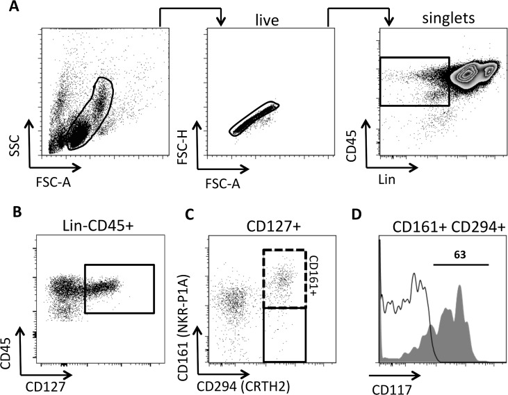
To identify human peripheral blood ILC2s, Mjösberg and co-workers [26] depleted PBMCs from T cells, B cells and monocytes, followed by a flow cytometric analysis of the remaining cells. This approach was not practical here, due to a much larger sample size used in the present study, and as cell numbers were limited. We therefore directly stained whole PBMCs with a lineage cocktail, which also included the NK cell marker CD56. PBMCs were gated on live cells and duplicates excluded (Fig. 1A). Gated single cells were analysed by a Lin cocktail and CD45, which showed a Lin- negative population expressing CD45. Within this population a CD45hi and a CD45int population, could be clearly distinguished.
Fig 1. Characterisation of human ILC2 by flow cytometry.
(A) Human PBMC were gated on live cells using forward (FSC) and side (SSC) scatter. Gated live cells were analysed using the area against height of the FSC to discriminate singlet’s from duplicates. Singlet’s were subsequently analysed by a lineage cocktail (Lin) and CD45 to gate on Lin-CD45+ cells (as indicated in the box) (B). Lin-CD45+ cells were gated on CD127+ cells, which were analysed for CD294 and CD161 (C). CD127+CD294+CD161+ were analysed for CD117 (grey histogram) versus isotype control (open histogram) (D). Flow charts as presented are from the PBMC analysis of a 9 year old female who was negative for S. haematobium eggs.
To identify human ILC2s, Lin-CD45+ PBMCs were gated on CD127+ (Fig. 1B). Within Lin-CD45+CD127+ cells a defined population of CD294+CD161+ cells could be identified (Fig. 1C). CD294+CD161+ cells contained both a CD117+ and a CD117- subset (Fig. 1D). In reverse CD127+CD117+ ILCs did not exclusively define CD294+ and CD161+ cells (Median of CD294+CD161+ in CD127+CD117+: 45.6, range 13.1–74.7) and it is known that CD117+ compromise both ILC2s and ILC3s. Of note, CD127+CD294+CD161+ subsets are all CD45hi. In summary, although not completely homogenous Lin-CD45+CD127+CD294+CD161+ cells define a population of human blood ILC2s, hence CD127+CD294+CD161+ ILC2s were used in subsequent analyses.
Proportions of ILC2s are diminished during parasitic helminth infection
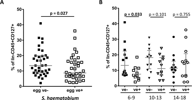
Next, the effect of current schistosome status on the proportions of blood ILC2s was analysed. The analyses showed that CD127+CD294+CD161+ ILC2s were all significantly lower in schistosome-infected individuals (p = 0.027) when levels of these cells were expressed as percentages of lin-CD45+CD127+ (Fig. 2A). This was also true if ILC2s were expressed as percentages of lin-CD45+ (Median: 4.8 egg negative (ve-), 3.1 egg positive (ve+) p = 0.044). In addition proportions of ILC2s were negatively correlated to the intensity of infection determined by egg count (r = −0.300, p = 0.005 using non-parametric Spearman correlation). Of note, CD127+CD117+ ILCs show a comparable pattern (Median: 13.7 egg ve-, 7.5 egg ve+, p = 0.001).
Fig 2. Proportions of ILC2s identified by CD127+CD294+CD161+ are lower in S. haematobium egg positive young children.
(A) The participants were compared between S. haematobium egg negative (ve-, N = 36) and egg positive (ve+, N = 36) individuals by a non-parametric Mann-Whitney U test. (B) The cohort was divided into three age groups (age in years) and egg positive (ve+, open symbols) were compared to egg negative (ve-, closed symbols) people. Grey lines indicate median and the interquartile range. ILC2s proportions were analysed using a Kruskal-Wallis test followed by a multiple comparison of egg positive versus egg negative children.
To determine if these differences were consistent across age groups undergoing different schistosome infection dynamics, the population was divided into three age groups, 6–9, 10–13 and 14–18 year olds reflecting rising, peaking and declining infection levels. This analysis showed that ILC2s in infected participants were significantly lower than those in uninfected people of the youngest age group (Fig. 2B). This difference was not significant in children aged 10–13 years and proportions of ILC2s were comparable in the oldest age group aged 14–18 years. Comparable results were obtained if ILC2s levels were correlated to infection intensity. The youngest age group showed a strong negative correlation between infection intensity and ILC2 proportions (r = −0.483, p = 0.008), whereas a weaker negative correlation was observed in 10–13 old participants (r = −0.403, p = 0.025) and with no significant correlation in the 14–18 years old (r = −0.045, p = 0.418).
CD127 (IL-7Rα) was used for identification of ILC2s, allowing the actual expression levels (MFI) of CD127 on ILC2s to be measured. Expression levels of CD127 did not show significant variation between infected and uninfected people for all age groups (6–9 years: p = 0.514; 10–13 years: p = 0.114; 14–18 years: p = 0.551).
Of note, levels of the systemic effector TH2 cytokines IL-4, IL-5 and IL-13 did not show any association with peripheral ILC2s (i.e. ILC2s to IL-4: r = −0.113 (p = 0.346); to IL-5 r = 0.138 (p = 0.248 and to IL-13 r = 0.016 (p = 0.894) using non-parametric Spearman correlation). Systemic TH2 cytokines were positively associated to infection intensity only in the oldest age group (IL-4: r = 0.488, p = 0.008; IL-5: r = 0.400, p = 0.027; IL-13: r = 0.497, p = 0.007). Detailed data for the TH2 cytokines are presented in S1 Fig with IL-5 and IL-13 only significant in children 14–18 years of age.
Curative anti-helminthic treatment restores ILC2 levels
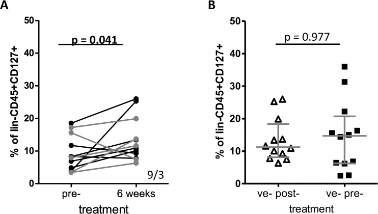
To test if the differences in the ILC2 proportions were related to the presence of schistosome parasites, schistosome adult worms were removed by curative treatment and analysed in 12 children aged 6–13 years who were S. haematobium positive prior to treatment and had cleared infection 6 weeks later were followed up and proportions of ILC2s were determined. The overall proportions of their ILC2 cells increased significantly post-treatment as shown in Fig. 3A.
Fig 3. Proportions of ILC2s increase 6 weeks after curative anti-helminthic treatment with praziquantel in children aged 6–13 years.
(A) The proportions of ILC2s of 12 individuals who were positive pre-treatment and had cleared S. haematobium infections 6 weeks post-treatment were analysed using a non-parametric Wilcoxon signed-rank test. Numbers in the lower-right corner indicate positive/negative ranks. Grey lines are individuals 10–13 years whereas black lines present children 6–9 years of age. (B) The proportions of ILC2s compared between the 12 children aged 6–13 years who had completely cleared their S. haematobium infections 6 weeks post-treatment (open triangles) and the 12 participants aged 14–18 years who were already S. haematobium negative before treatment (black squares). Grey lines indicate median and the interquartile range.
Older egg negative participants have putatively developed a protective immune response and have been referred to in other studies as endemic normal and a high worm specific IgE/IgG4 ratio is a marker for schistosome-specific resistance [25]. Curative treatment by chemotherapy has been shown to accelerate the development of a protective immune response [59], hence post-treatment levels of ILC2 were compared to egg ve- individuals aged 14–18 years. Post-treatment, levels of ILC2 rose up to levels observed in untreated 14–18 year old participants who were egg negative prior to treatment (Fig. 3B).
After dividing these 12 children into two groups of 6–9 and 10–13 years of age, ILC2s increased in 5 out of 7 and in 4 out of 5 children, respectively.
The increase of ILC2 levels were inversely related to the pre-treatment levels of these cells, with the lowest pre-treatment proportions exhibiting the highest magnitude of change post-treatment. We have previously reported this phenomenon in changes in antibody levels in children exposed to S. mansoni infection [59].
Curative anti-helminthic treatment increases TSLP levels
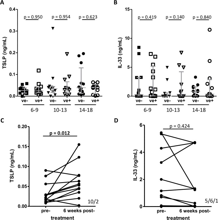
Maintenance and activation of ILC2s has been reported to depend on the cytokines IL-25, IL-33 and TSLP. Pre-treatment plasma levels of IL-33 and TSLP were measured, but no significant difference was observed when egg negative and egg positive children were compared regardless of the age group investigated (Fig. 4A, B). However TSLP levels increased significantly 6 weeks after curative treatment (Fig. 4C). In contrast, the level of IL-33 remained unchanged following treatment (Fig. 4D).
Fig 4. Comparison of plasma TSLP and IL-33 levels in relation to S. haematobium infection.
Plasma levels of (A) TSLP and (B) IL-33 were measured by ELISA and data were divided into three age group and S. haematobium egg negative (ve-, closed symbols) compared to egg positive (ve+, open symbols) individuals. Grey lines indicate median and the interquartile range. Levels were compared using a Kruskal-Wallis test followed by a multiple comparison of egg positive versus egg negative children. Plasma levels of (C) TSLP and (D) IL-33 from 12 individuals (as described for Fig. 3) were compared pre- and 6 week’s post-treatment. P-values are obtained from a Wilcoxon signed-rank test and positive/negative/unchanged ranks are shown in the lower-right corner of panel (C) and (D).
Discussion
In parasitic infections, ILC2s have been described in experimental models of helminth infections demonstrating their role as an early source of the cytokine IL-13 and in worm expulsion [4, 6, 60–62]. Given these results, this study aimed to determine if ILC2s populations are altered during human parasite infections using an immuno-epidemiological approach reflecting the complex dynamics during human parasite infections and specifically focusing on young children with accumulating infection. Hence, included in the study are young children who have yet to acquire schistosome infection (negative for schistosome eggs and parasite-specific antibodies), infected young and older children (schistosome egg positive) and older children putatively resistant to schistosome infection (schistosome egg negative but positive for schistosome-specific IgG and IgE [63]).
First, this study confirms that ILC2s can be identified in human peripheral blood using Lin-CD45+CD127+ cells in combination with CD161+ and CD294+ in a population of young African children.
This study, shows for the first time that proportions of human blood ILC2s are diminished during an infection with trematodes. The strongest effect was observed in the youngest age group. Reductions in proportions of ILC2s could theoretically arise in two ways; due to a reduction in generation/maintenance of the cells in people infected with S. haematobium parasites. Alternatively, ILC2s could migrate and accumulate at the site of infection or to the tissues where eggs get trapped, initiating a localised immune response thereby leading to a reduction of the cells in peripheral blood. Furthermore reduced levels of CD127 could indicate a reduced responsiveness to IL-7 and thereby ILC2 survival. However, expression levels did not show any variation which supports reduced responsiveness to IL-7. In this present study ILC2s are quantified as proportions. Changes in ILC2 proportions due to an increase of other cell types within Lin-CD45+ cells cannot be excluded. However, our results are consistent with those from a previous study on blood ILC2s, indicating that in a TH2 mediated disease, ILC2s are lower in patients compared to controls [35]. In contrast a recent study showed in 21 people with diverse filarial infections (Loa loa, Wuchereria bancrofti and with Onchocerca volvulus) an increase of ILCs characterised by CD127+CD117+ [7]. However infected people were aged 25–66 years and therefore significantly older than even in the oldest age group investigated in this study, in which ILC2s were also slightly higher in egg positive compared to egg negative people, which may become significant if older people analysed.
Interestingly, reduced ILC2s were mainly observed in the youngest children, whereas in older children (14–18 years) there were no differences between egg negative and positive individuals. These differences are likely to be related to the dynamics of infection and the immune response due to ILC2s may have different dynamics compared to the development of protective acquired immunity. Age-related changes of cellular, antibody and cytokine pattern in the context of schistosome infection have been reported previously [24, 64, 65] and such pattern are supported by theoretical models of helminth infections [66]. These changes have been taken to represent population changes from susceptibility to infection the development of protective acquired immunity [66]. Parasitological and immuno-epidemiological studies show that the egg negative children in the youngest age group in this population have yet to acquire infection (no parasite specific—IgM responses which are indicative of exposure to parasite life stages). The remaining study population are either currently infected (indicated by the presence of parasite eggs) or have had previous infection and are now putatively immune (indicated by the absence of parasite eggs in combination with the presence of parasite-specific immune responses associated with resistance to infection such as anti-worm antibodies (IgE, IgG1, IgE/IgG4 ratio) and parasite specific cellular responses (IL-5) as previously reported [25, 58, 67]). Thus, in this population, the youngest egg negative children are the only ones who have not yet experienced a schistosome infection, and the observed changes in ILC2s in this age group upon infection, suggests that these cells are important in the early phase of the immune response, when the TH2 pathway is initially triggered, rather than in the later phases of infection, when TH2 responses become established. This is consistent with our results from studies of the myeloid dendritic cells in this population [24], which show a similar pattern. Indeed it has been shown that ILC2s, in particular, provide an early source of IL-13 during experimental helminth infection [4]. Dynamic changes in the immune response to schistosomes are well documented and age-related changes of cellular immune responses have been previously reported in this and other study populations [16, 24, 25].
Clearance of S. haematobium infection following curative treatment with the anti-helminthic drug praziquantel caused an increase in ILC2 proportions, recovering to levels comparable to egg negative 14–18 year old participants, who have already developed natural protective immunity against parasites [25]. This is in keeping with earlier studies showing that chemotherapy accelerates the development of protective acquired immunity against schistosomes [59] as well as studies from this population showing that chemotherapy induced cellular responses associated with resistance against re-infection [50]. However due to the small sample size post-treatment, this result requires further verification.
Both IL-33 and TSLP are important in ILC2 activation and propagation. There was no significant association with systemic levels of IL-33 or TSLP with schistosome infection prior to treatment. Hence systemic levels of these cytokines do not explain the alterations in blood ILC2s and do not indicate a different activation status. However, if changes in ILC2 proportion are due to migration, IL-33 and TSLP might still play a role if regulated locally (in which case local levels of the cytokines would not necessarily be reflected systemically). We have obtained supportive evidence for the importance IL-33 by analysing the effect of a specific IL-33 single nucleotide polymorphisms (SNP) showing that allele variation influences schistosome infection intensity without influencing systemic IL-33 levels (manuscript in preparation).
In addition, systemic TH2 cytokines IL-4, IL-5 and IL-13 did not show significant associations with peripheral ILC2s. However, if changes in ILC2s are due to migration, ILC2 activation could be still initiated locally and play an important role in the immune response. This is supported by the finding that systemic TH2 cytokines are only associated to infection in the oldest age group, indicating that a classical TH2 response is more important and reflected on systemic levels in older chronically infected individuals. Further studies are required to analyse the activation status of ILC2s in infected people relative to their expression levels of GATA-3, IL-5 and IL-13 as well as receptors for IL-33 and TSLP. Recently is has been reported that in mouse models IL-9 and amphiregulin are important in the function of ILC2s [68, 69]. However, data in humans are sparce and both IL-9 and amphiregulin were not incorporated when this study was carried out.
Increased levels of TSLP were observed 6 weeks after treatment, which suggests that TSLP may be involved in the increase of ILC2s following treatment, but no change in IL-33 was observed after treatment. This latter result contradicts previous reports of an increase in IL-33 several weeks after treatment [33]. Further studies are required to clarify the role of IL-33 in human infections with S. haematobium and should also include IL-25, which was not analysed during this study due to limitations in blood levels that could be collected safely from the younger children.
In summary, this is the first study to report peripheral blood ILC2s being diminished in young children suggesting that ILC2s may play a role in the immune responses to human helminth infection before the establishment of schistosome protective immunity. Clearly further mechanistic studies are required to determine how ILC2 levels are diminished in helminth infection and how they are restored following removal of the helminth infection by curative treatment. Nonetheless, this study validates the paradigms demonstrated in experimental studies of an association between parasitic infection and ILC2 cells.
Supporting Information
(PDF)
IL-4 (A), IL-5 (B) and IL-13 (C) were analysed by ELISA and data devided by age group and S. haematobium egg negative (ve-, closed symbols) are compared to egg positive (ve+, open symbols). Grey lines indicate Median and the interquartile range and levels were compared using a Kruskal-Wallis test followed by a multiple comparsion.
(TIF)
Acknowledgments
We thank the participants, their parents and teachers from Magaya village for taking part in this study. We also thank the Provincial Medical Director of Mashonaland East, the Environmental Health Workers, nursing staff at Chitate and Chitowa Clinics and Murehwa Hospital. We are very grateful for the co-operation of the Ministry of Health and Child Welfare in Zimbabwe. For their technical support, we would like to thank the National Institutes of Health Research Zimbabwe and members of the Department of Biochemistry at the University of Zimbabwe. We also thank Professor Rick M. Maizels at the University of Edinburgh for his comments on the manuscript and Seth Amanfo and Welcome M. Wami (University of Edinburgh) for proofreading the manuscript.
Data Availability
We do not have ethical clearance from the Medical Research Council of Zimbabwe (MRCZ) to make the raw data available. Other users would have to apply for permission to use the data from the MRCZ who granted us permission to conduct the study. The Medical Research Council of Zimbabwe (MRCZ) can be contacted via their online form: http://www.mrcz.org.zw/index.php?option=com_contact&view=contact&id=4&Itemid=10 or the email address mrcz@mrcz.org.zw.
Funding Statement
This work was supported by the Wellcome Trust (Grant no WT082028MA; http://www.wellcome.ac.uk) and FM is funded by the Thrasher Research Fund (www.thrasherresearch.org). The funders had no role in study design, data collection and analysis, decision to publish, or preparation of the manuscript.
References
- 1. Sonnenberg GF, Mjosberg J, Spits H, Artis D. SnapShot: innate lymphoid cells. Immunity. 2013;39(3):622–e1. Epub 2013/09/10. 10.1016/j.immuni.2013.08.021 [DOI] [PubMed] [Google Scholar]
- 2. Spits H, Artis D, Colonna M, Diefenbach A, Di Santo JP, Eberl G, et al. Innate lymphoid cells—a proposal for uniform nomenclature. Nature reviews Immunology. 2013;13(2):145–9. Epub 2013/01/26. 10.1038/nri3365 [DOI] [PubMed] [Google Scholar]
- 3.Hazenberg MD, Spits H. Human innate lymphoid cells. Blood. 2014. Epub 2014/04/30. [DOI] [PubMed]
- 4. Neill DR, Wong SH, Bellosi A, Flynn RJ, Daly M, Langford TK, et al. Nuocytes represent a new innate effector leukocyte that mediates type-2 immunity. Nature. 2010;464(7293):1367–70. Epub 2010/03/05. 10.1038/nature08900 [DOI] [PMC free article] [PubMed] [Google Scholar]
- 5. Moro K, Yamada T, Tanabe M, Takeuchi T, Ikawa T, Kawamoto H, et al. Innate production of T(H)2 cytokines by adipose tissue-associated c-Kit(+)Sca-1(+) lymphoid cells. Nature. 2010;463(7280):540–4. Epub 2009/12/22. 10.1038/nature08636 [DOI] [PubMed] [Google Scholar]
- 6. Price AE, Liang HE, Sullivan BM, Reinhardt RL, Eisley CJ, Erle DJ, et al. Systemically dispersed innate IL-13-expressing cells in type 2 immunity. Proceedings of the National Academy of Sciences of the United States of America. 2010;107(25):11489–94. 10.1073/pnas.1003988107 [DOI] [PMC free article] [PubMed] [Google Scholar]
- 7. Boyd A, Ribeiro JM, Nutman TB. Human CD117 (cKit)+ Innate Lymphoid Cells Have a Discrete Transcriptional Profile at Homeostasis and Are Expanded during Filarial Infection. PLoS One. 2014;9(9):e108649 10.1371/journal.pone.0108649 [DOI] [PMC free article] [PubMed] [Google Scholar]
- 8. Finkelman FD, Shea-Donohue T, Morris SC, Gildea L, Strait R, Madden KB, et al. Interleukin-4- and interleukin-13-mediated host protection against intestinal nematode parasites. Immunological reviews. 2004;201:139–55. [DOI] [PubMed] [Google Scholar]
- 9. Pearce EJ, MK C, Sun J, JT J, McKee AS, Cervi L. Th2 response polarization during infection with the helminth parasite Schistosoma mansoni . Immunological reviews. 2004;201:117–26. Epub 2004/09/14. [DOI] [PubMed] [Google Scholar]
- 10. Anthony RM, Rutitzky LI, Urban JF Jr., Stadecker MJ, Gause WC. Protective immune mechanisms in helminth infection. Nature reviews Immunology. 2007;7(12):975–87. Epub 2007/11/17. [DOI] [PMC free article] [PubMed] [Google Scholar]
- 11. Taylor MD, van der Werf N, Harris A, Graham AL, Bain O, Allen JE, et al. Early recruitment of natural CD4+ Foxp3+ Treg cells by infective larvae determines the outcome of filarial infection. European journal of immunology. 2009;39(1):192–206. Epub 2008/12/18. 10.1002/eji.200838727 [DOI] [PubMed] [Google Scholar]
- 12. Grainger JR, Smith KA, Hewitson JP, McSorley HJ, Harcus Y, Filbey KJ, et al. Helminth secretions induce de novo T cell Foxp3 expression and regulatory function through the TGF-{beta} pathway. J Exp Med. 2010;207(11):2331–41. Epub 2010/09/30. 10.1084/jem.20101074 [DOI] [PMC free article] [PubMed] [Google Scholar]
- 13. Amu S, Saunders SP, Kronenberg M, Mangan NE, Atzberger A, Fallon PG. Regulatory B cells prevent and reverse allergic airway inflammation via FoxP3-positive T regulatory cells in a murine model. The Journal of allergy and clinical immunology. 2010;125(5):1114–24 e8. 10.1016/j.jaci.2010.01.018 [DOI] [PubMed] [Google Scholar]
- 14. Wilson MS, Cheever AW, White SD, Thompson RW, Wynn TA. IL-10 blocks the development of resistance to re-infection with Schistosoma mansoni . PLoS pathogens. 2011;7(8):e1002171 Epub 2011/08/11. 10.1371/journal.ppat.1002171 [DOI] [PMC free article] [PubMed] [Google Scholar]
- 15. Walsh KP, Brady MT, Finlay CM, Boon L, Mills KHG. Infection with a Helminth Parasite Attenuates Autoimmunity through TGF-β-Mediated Suppression of Th17 and Th1 Responses. The Journal of Immunology. 2009;183(3):1577–86. 10.4049/jimmunol.0803803 [DOI] [PubMed] [Google Scholar]
- 16. Nausch N, Midzi N, Mduluza T, Maizels RM, Mutapi F. Regulatory and activated T cells in human Schistosoma haematobium infections. PLoS One. 2011;6(2):e16860 10.1371/journal.pone.0016860 [DOI] [PMC free article] [PubMed] [Google Scholar]
- 17. van der Vlugt LE, Labuda LA, Ozir-Fazalalikhan A, Lievers E, Gloudemans AK, Liu KY, et al. Schistosomes induce regulatory features in human and mouse CD1d(hi) B cells: inhibition of allergic inflammation by IL-10 and regulatory T cells. PLoS One. 2012;7(2):e30883 10.1371/journal.pone.0030883 [DOI] [PMC free article] [PubMed] [Google Scholar]
- 18. van den Biggelaar AH, Borrmann S, Kremsner P, Yazdanbakhsh M. Immune responses induced by repeated treatment do not result in protective immunity to Schistosoma haematobium: interleukin (IL)-5 and IL-10 responses. J Infect Dis. 2002;186(10):1474–82. [DOI] [PubMed] [Google Scholar]
- 19. Phythian-Adams AT, Cook PC, Lundie RJ, Jones LH, Smith KA, Barr TA, et al. CD11c depletion severely disrupts Th2 induction and development in vivo. J Exp Med. 2010;207(10):2089–96. 10.1084/jem.20100734 [DOI] [PMC free article] [PubMed] [Google Scholar]
- 20. Kim S, Prout M, Ramshaw H, Lopez AF, LeGros G, Min B. Cutting edge: basophils are transiently recruited into the draining lymph nodes during helminth infection via IL-3, but infection-induced Th2 immunity can develop without basophil lymph node recruitment or IL-3. Journal of immunology. 2010;184(3):1143–7. 10.4049/jimmunol.0902447 [DOI] [PMC free article] [PubMed] [Google Scholar]
- 21. Herbert DR, Holscher C, Mohrs M, Arendse B, Schwegmann A, Radwanska M, et al. Alternative macrophage activation is essential for survival during schistosomiasis and downmodulates T helper 1 responses and immunopathology. Immunity. 2004;20(5):623–35. [DOI] [PubMed] [Google Scholar]
- 22. Wilson S, Jones FM, Mwatha JK, Kimani G, Booth M, Kariuki HC, et al. Hepatosplenomegaly is associated with low regulatory and Th2 responses to schistosome antigens in childhood schistosomiasis and malaria coinfection. Infect Immun. 2008;76(5):2212–8. 10.1128/IAI.01433-07 [DOI] [PMC free article] [PubMed] [Google Scholar]
- 23. Mduluza T, Ndhlovu PD, Midzi N, Scott JT, Mutapi F, Mary C, et al. Contrasting cellular responses in Schistosoma haematobium infected and exposed individuals from areas of high and low transmission in Zimbabwe. Immunol Lett. 2003;88(3):249–56. [DOI] [PubMed] [Google Scholar]
- 24. Nausch N, Louis D, Lantz O, Peguillet I, Trottein F, Chen IY, et al. Age-related patterns in human myeloid dendritic cell populations in people exposed to Schistosoma haematobium infection. PLoS neglected tropical diseases. 2012;6(9):e1824 10.1371/journal.pntd.0001824 [DOI] [PMC free article] [PubMed] [Google Scholar]
- 25. Nausch N, Bourke CD, Appleby LJ, Rujeni N, Lantz O, Trottein F, et al. Proportions of CD4+ memory T cells are altered in individuals chronically infected with Schistosoma haematobium . Sci Rep. 2012;2:472 10.1038/srep00472 [DOI] [PMC free article] [PubMed] [Google Scholar]
- 26. Mjosberg JM, Trifari S, Crellin NK, Peters CP, van Drunen CM, Piet B, et al. Human IL-25- and IL-33-responsive type 2 innate lymphoid cells are defined by expression of CRTH2 and CD161. Nature immunology. 2011;12(11):1055–62. 10.1038/ni.2104 [DOI] [PubMed] [Google Scholar]
- 27. Wong SH, Walker JA, Jolin HE, Drynan LF, Hams E, Camelo A, et al. Transcription factor RORalpha is critical for nuocyte development. Nature immunology. 2012;13(3):229–36. 10.1038/ni.2208 [DOI] [PMC free article] [PubMed] [Google Scholar]
- 28. Cosmi L, Annunziato F, Galli MIG, Maggi RME, Nagata K, Romagnani S. CRTH2 is the most reliable marker for the detection of circulating human type 2 Th and type 2 T cytotoxic cells in health and disease. European journal of immunology. 2000;30(10):2972–9. [DOI] [PubMed] [Google Scholar]
- 29. Hoyler T, Klose CS, Souabni A, Turqueti-Neves A, Pfeifer D, Rawlins EL, et al. The transcription factor GATA-3 controls cell fate and maintenance of type 2 innate lymphoid cells. Immunity. 2012;37(4):634–48. 10.1016/j.immuni.2012.06.020 [DOI] [PMC free article] [PubMed] [Google Scholar]
- 30. Liang HE, Reinhardt RL, Bando JK, Sullivan BM, Ho IC, Locksley RM. Divergent expression patterns of IL-4 and IL-13 define unique functions in allergic immunity. Nature immunology. 2012;13(1):58–66. 10.1038/ni.2182 [DOI] [PMC free article] [PubMed] [Google Scholar]
- 31. Halim TY, MacLaren A, Romanish MT, Gold MJ, McNagny KM, Takei F. Retinoic-acid-receptor-related orphan nuclear receptor alpha is required for natural helper cell development and allergic inflammation. Immunity. 2012;37(3):463–74. 10.1016/j.immuni.2012.06.012 [DOI] [PubMed] [Google Scholar]
- 32. Mjosberg J, Bernink J, Golebski K, Karrich JJ, Peters CP, Blom B, et al. The transcription factor GATA3 is essential for the function of human type 2 innate lymphoid cells. Immunity. 2012;37(4):649–59. 10.1016/j.immuni.2012.08.015 [DOI] [PubMed] [Google Scholar]
- 33. Wilson S, Jones FM, Fofana HK, Landoure A, Kimani G, Mwatha JK, et al. A late IL-33 response after exposure to Schistosoma haematobium antigen is associated with an up-regulation of IL-13 in human eosinophils. Parasite Immunology. 2013;35(7–8):224–8. 10.1111/pim.12029 [DOI] [PMC free article] [PubMed] [Google Scholar]
- 34. Kim BS, Siracusa MC, Saenz SA, Noti M, Monticelli LA, Sonnenberg GF, et al. TSLP elicits IL-33-independent innate lymphoid cell responses to promote skin inflammation. Sci Transl Med. 2013;5(170):170ra16. 10.1126/scitranslmed.3005374 [DOI] [PMC free article] [PubMed] [Google Scholar]
- 35. Barnig C, Cernadas M, Dutile S, Liu X, Perrella MA, Kazani S, et al. Lipoxin A4 regulates natural killer cell and type 2 innate lymphoid cell activation in asthma. Sci Transl Med. 2013;5(174):174ra26. 10.1126/scitranslmed.3005127 [DOI] [PMC free article] [PubMed] [Google Scholar]
- 36. Hurst SD, Muchamuel T, Gorman DM, Gilbert JM, Clifford T, Kwan S, et al. New IL-17 family members promote Th1 or Th2 responses in the lung: in vivo function of the novel cytokine IL-25. Journal of immunology. 2002;169(1):443–53. [DOI] [PubMed] [Google Scholar]
- 37. Kondo Y, Yoshimoto T, Yasuda K, Futatsugi-Yumikura S, Morimoto M, Hayashi N, et al. Administration of IL-33 induces airway hyperresponsiveness and goblet cell hyperplasia in the lungs in the absence of adaptive immune system. Int Immunol. 2008;20(6):791–800. 10.1093/intimm/dxn037 [DOI] [PubMed] [Google Scholar]
- 38. Halim TY, Krauss RH, Sun AC, Takei F. Lung natural helper cells are a critical source of Th2 cell-type cytokines in protease allergen-induced airway inflammation. Immunity. 2012;36(3):451–63. 10.1016/j.immuni.2011.12.020 [DOI] [PubMed] [Google Scholar]
- 39. Kim HY, Chang YJ, Subramanian S, Lee HH, Albacker LA, Matangkasombut P, et al. Innate lymphoid cells responding to IL-33 mediate airway hyperreactivity independently of adaptive immunity. The Journal of allergy and clinical immunology. 2012;129(1):216–27 e1–6. 10.1016/j.jaci.2011.10.036 [DOI] [PMC free article] [PubMed] [Google Scholar]
- 40. Barlow JL, Bellosi A, Hardman CS, Drynan LF, Wong SH, Cruickshank JP, et al. Innate IL-13-producing nuocytes arise during allergic lung inflammation and contribute to airways hyperreactivity. The Journal of allergy and clinical immunology. 2012;129(1):191–8 e1–4. 10.1016/j.jaci.2011.09.041 [DOI] [PubMed] [Google Scholar]
- 41. Klein Wolterink RG, Kleinjan A, van Nimwegen M, Bergen I, de Bruijn M, Levani Y, et al. Pulmonary innate lymphoid cells are major producers of IL-5 and IL-13 in murine models of allergic asthma. European journal of immunology. 2012;42(5):1106–16. 10.1002/eji.201142018 [DOI] [PubMed] [Google Scholar]
- 42. World Health Organisation. Schistosomiasis. Geneva: World Health Organisation, 2013. Contract No.: 8. [Google Scholar]
- 43. Mafiana CF, Ekpo UF, Ojo DA. Urinary schistosomiasis in preschool children in settlements around Oyan Reservoir in Ogun State, Nigeria: implications for control. Trop Med Int Health. 2003;8(1):78–82. [DOI] [PubMed] [Google Scholar]
- 44. Garba A, Barkire N, Djibo A, Lamine MS, Sofo B, Gouvras AN, et al. Schistosomiasis in infants and preschool-aged children: Infection in a single Schistosoma haematobium and a mixed S. haematobium-S. mansoni foci of Niger. Acta Trop. 2010;115(3):212–9. 10.1016/j.actatropica.2010.03.005 [DOI] [PubMed] [Google Scholar]
- 45. Stothard JR, Sousa-Figueiredo JC, Betson M, Green HK, Seto EY, Garba A, et al. Closing the praziquantel treatment gap: new steps in epidemiological monitoring and control of schistosomiasis in African infants and preschool-aged children. Parasitology. 2011;138(12):1593–606. 10.1017/S0031182011001235 [DOI] [PMC free article] [PubMed] [Google Scholar]
- 46. Mutapi F, Ndhlovu PD, Hagan P, Spicer JT, Mduluza T, Turner CM, et al. Chemotherapy accelerates the development of acquired immune responses to Schistosoma haematobium infection. J Infect Dis. 1998;178:289–93. [DOI] [PubMed] [Google Scholar]
- 47. Clarke VD. The influence of acquired resistance in the epidemiology of bilharziasis. Cent Afr J Med. 1966;12(6):1–30. Epub 1966/06/01. PubMed PMID: . [PubMed] [Google Scholar]
- 48. Woolhouse MEJ, Taylor P, Matanhire D, Chandiwana SK. Acquired immunity and epidemiology of Schistosoma haematobium . Nature. 1991;351:757–9. [DOI] [PubMed] [Google Scholar]
- 49. Appleby LJ, Nausch N, Midzi N, Mduluza T, Allen JE, Mutapi F. Sources of heterogeneity in human monocyte subsets. Immunol Lett. 2013;152(1):32–41. 10.1016/j.imlet.2013.03.004 [DOI] [PMC free article] [PubMed] [Google Scholar]
- 50. Bourke CD, Nausch N, Rujeni N, Appleby LJ, Mitchell KM, Midzi N, et al. Integrated analysis of innate, Th1, Th2, Th17, and regulatory cytokines identifies changes in immune polarisation following treatment of human schistosomiasis. The Journal of infectious diseases. 2013;208(1):159–69. 10.1093/infdis/jis524 [DOI] [PMC free article] [PubMed] [Google Scholar]
- 51.Ndhlovu P, Chimbari M, Ndmba J, Chandiwana SK. 1992 national schistosomiasis survey, Blair report for Zimbabwe. Blair Research Institute, 1996 May 1996. Report No.
- 52. Midzi N, Mhlanga G, Manangazira P, Mduluza T, Tshuma C, Chimbari M, et al. Report on the National Soil Transmitted Helminthiasis and Schistosomiasis Survey. Harare: National Institute of Health Research, 2010. [Google Scholar]
- 53. Reilly LJ, Magkrioti C, Cavanagh DR, Mduluza T, Mutapi F. Effect of treating Schistosoma haematobium infection on Plasmodium falciparum-specific antibody responses. BMC Infect Dis. 2008;8(1):158. [DOI] [PMC free article] [PubMed] [Google Scholar]
- 54.World Health Organization. Preventive chemotherapy in human helminthiasis. Geneva: 2009.
- 55. Mott KE. A reusable polyamide filter for diagnosis of S. haematobium infection by urine filtration. Bull Soc Pathol Exot. 1983;76:101–4. [PubMed] [Google Scholar]
- 56. Katz N, Chaves A, Pellegrino J. A simple device for quantitative stool thick smear technique in schistosomiasis mansoni. Rev Inst MedTrop de Sao Paulo 1972;14:397–400. [PubMed] [Google Scholar]
- 57. Ritchie LS. An ether sedimentation technique for routine stool examinations. Bull U S Army Med Dep. 1948;8(4):326 [PubMed] [Google Scholar]
- 58. Milner T, Reilly L, Nausch N, Midzi N, Mduluza T, Maizels R, et al. Circulating cytokine levels and antibody responses to human Schistosoma haematobium: IL-5 and IL-10 levels depend upon age and infection status. Parasite Immunology. 2010;32(11–12):710–21. 10.1111/j.1365-3024.2010.01220.x [DOI] [PMC free article] [PubMed] [Google Scholar]
- 59. Mutapi F, Hagan P, Woolhouse ME, Mduluza T, Ndhlovu PD. Chemotherapy-induced, age-related changes in antischistosome antibody responses. Parasite Immunol. 2003;25(2):87–97. [DOI] [PubMed] [Google Scholar]
- 60. Fallon PG, Ballantyne SJ, Mangan NE, Barlow JL, Dasvarma A, Hewett DR, et al. Identification of an interleukin (IL)-25-dependent cell population that provides IL-4, IL-5, and IL-13 at the onset of helminth expulsion. The Journal of experimental medicine. 2006;203(4):1105–16. [DOI] [PMC free article] [PubMed] [Google Scholar]
- 61. Yasuda K, Muto T, Kawagoe T, Matsumoto M, Sasaki Y, Matsushita K, et al. Contribution of IL-33-activated type II innate lymphoid cells to pulmonary eosinophilia in intestinal nematode-infected mice. Proceedings of the National Academy of Sciences of the United States of America. 2012;109(9):3451–6. 10.1073/pnas.1201042109 [DOI] [PMC free article] [PubMed] [Google Scholar]
- 62. Smith KA, Harcus Y, Garbi N, Hammerling GJ, MacDonald AS, Maizels RM. Type 2 innate immunity in helminth infection is induced redundantly and acts autonomously following CD11c(+) cell depletion. Infection and immunity. 2012;80(10):3481–9. 10.1128/IAI.00436-12 [DOI] [PMC free article] [PubMed] [Google Scholar]
- 63. Viana IC, CorreaOliveira R, Dos Santos Carvalho O, Lara Massara C, Colosimo E, Colley D, et al. Comparison of antibody isotype responses to Schistosoma mansoni antigens by infected and putative resistant individuals living in an endemic area. Parasite Immunology. 1995;17(6):297–304. [DOI] [PubMed] [Google Scholar]
- 64. Ndhlovu P, Cadman H, Vennervald BJ, Christensen NO, Chidimu N, Chandiwana SK. Age-related antibody profiles in Schistosoma haematobium in a rural community in Zimbabwe. Parasite Immunology. 1996;18:181–91. [DOI] [PubMed] [Google Scholar]
- 65. Webster M, LibrandaRamirez BDL, Aligui GD, Olveda RM, Ouma JH, Kariuki HC, et al. The influence of sex and age on antibody isotype responses to Schistosoma mansoni and Schistosoma japonicum in human populations in Kenya and the Philippines. Parasitology. 1997;114(Pt4):383–93. [DOI] [PubMed] [Google Scholar]
- 66. Woolhouse MEJ. A theoretical framework for the immunoepidemiology of helminth infection. Parasite Immunol. 1992;14:563–78. [DOI] [PubMed] [Google Scholar]
- 67. Rujeni N, Nausch N, Bourke CD, Midzi N, Mduluza T, Taylor DW, et al. Atopy Is Inversely Related to Schistosome Infection Intensity: A Comparative Study in Zimbabwean Villages with Distinct Levels of Schistosoma haematobium Infection. Int Arch Allergy Immunol. 2012;158(3):288–98. 10.1159/000332949 [DOI] [PMC free article] [PubMed] [Google Scholar]
- 68. Turner JE, Morrison PJ, Wilhelm C, Wilson M, Ahlfors H, Renauld JC, et al. IL-9-mediated survival of type 2 innate lymphoid cells promotes damage control in helminth-induced lung inflammation. The Journal of experimental medicine. 2013;210(13):2951–65. 10.1084/jem.20130071 [DOI] [PMC free article] [PubMed] [Google Scholar]
- 69. Salimi M, Barlow JL, Saunders SP, Xue L, Gutowska-Owsiak D, Wang X, et al. A role for IL-25 and IL-33-driven type-2 innate lymphoid cells in atopic dermatitis. The Journal of experimental medicine. 2013;210(13):2939–50. 10.1084/jem.20130351 [DOI] [PMC free article] [PubMed] [Google Scholar]
Associated Data
This section collects any data citations, data availability statements, or supplementary materials included in this article.
Supplementary Materials
(PDF)
IL-4 (A), IL-5 (B) and IL-13 (C) were analysed by ELISA and data devided by age group and S. haematobium egg negative (ve-, closed symbols) are compared to egg positive (ve+, open symbols). Grey lines indicate Median and the interquartile range and levels were compared using a Kruskal-Wallis test followed by a multiple comparsion.
(TIF)
Data Availability Statement
We do not have ethical clearance from the Medical Research Council of Zimbabwe (MRCZ) to make the raw data available. Other users would have to apply for permission to use the data from the MRCZ who granted us permission to conduct the study. The Medical Research Council of Zimbabwe (MRCZ) can be contacted via their online form: http://www.mrcz.org.zw/index.php?option=com_contact&view=contact&id=4&Itemid=10 or the email address mrcz@mrcz.org.zw.


