Abstract
Metal oxide nanoparticles have been suggested as good candidates for the development of antibacterial agents. Cerium oxide (CeO2) and iron oxide (Fe2O3) nanoparticles have been utilized in a number of biomedical applications. Here, the antibacterial activity of CeO2 and Fe2O3 nanoparticles were evaluated on a panel of gram positive and gram negative bacteria in both the planktonic and biofilm cultures. Additionally, the effect of combining CeO2 and Fe2O3 nanoparticles with the broad spectrum antibiotic ciprofloxacin on tested bacteria was investigated. Thus, minimum inhibitory concentrations (MICs) of CeO2 and Fe2O3 nanoparticles that are required to inhibit bacterial planktonic growth and bacterial biofilm, were evaluated, and were compared to the MICs of the broad spectrum antibiotic ciprofloxacin alone or in the presence of CeO2 and Fe2O3 nanoparticles. Results of this study show that both CeO2 and Fe2O3 nanoparticles fail to inhibit bacterial growth and biofilm biomass for all the bacterial strains tested. Moreover, adding CeO2 or Fe2O3 nanoparticles to the broad spectrum antibiotic ciprofloxacin almost abolished its antibacterial activity. Results of this study suggest that CeO2 and Fe2O3 nanoparticles are not good candidates as antibacterial agents, and they could interfere with the activity of important antibiotics.
Keywords: Cerium oxide, Iron oxide, Nanoparticles, Antibacterial activity, Ciprofloxacin, Biofilm bacteria
Introduction
The introduction of antimicrobial agents had a vital role in decreasing the total deaths from infectious diseases during the mid-twentieth century (Cohen 2000). However, the emergence of bacterial resistance to antibacterial drugs has become a serious problem for public health (Kurek et al. 2011). Many traditionally used antibiotics are not effective anymore in managing drug-resistant bacteria. This might lead to the re-emergence of once controlled microbial diseases (Cohen 2000). Additionally, many bacteria escape most antibiotic treatments and host defense systems by forming a protective matrix of exopolymeric substances called biofilm (Subbiahdoss et al. 2012; Weir et al. 2008). Moreover, the horizontal gene transfer between bacteria within biofilms can increase the spread of antibiotic resistance (Fux et al. 2005; Weir et al. 2008). Adding to the antibiotic resistance problem is the decline in the development of new antibacterial agents, with only few newly approved agents introduced to the pharmaceutical market (Donadio et al. 2010). Therefore, there is a great need to develop new antibacterial agents.
Over the last decade, many researchers have been evaluating potential antibacterial effect of metals in their nanoparticle form. Metals including zinc, silver, and copper have been used as antibacterial agents for long time (Subbiahdoss et al. 2012). The advantage of using metals in their nanoparticle form is that these particles can be prepared to have very small diameter, and therefore to have a high surface area to volume ratio. It is thought that the high surface area to volume ratios and the resultant unique chemico-physical properties of the nanoparticles could contribute to their antimicrobial activities (Huh and Kwon 2011; Pal et al. 2007). Moreover, antibacterial nanoparticles influence several structures and biological pathways found in a wide range of pathogenic bacteria. This makes it harder for bacteria to develop resistance against nanoparticles (Nel et al. 2009; Huang et al. 2008; Pal et al. 2007).
Cerium oxide (CeO2) nanoparticles are metal oxide nanoparticles that have been exploited in a number of biomedical applications. For example, they have been used as a UV light absorber in sunscreens (Wu et al. 2010). CeO2 nanoparticles exhibit an antioxidant activity at physiological pH, and were shown to protect cells against oxidative stress, inflammation, or damage caused by radiation (Tarnuzzer et al. 2005; Niu et al. 2007; Perez et al. 2008). Studies on the antibacterial activity of CeO2 nanoparticles have shown mixed results as well. While some studies have suggested antibacterial activity for CeO2 nanoparticles (Shah et al. 2012), others have indicated no toxic effect of CeO2 nanoparticles on bacteria (Negahdary et al. 2012; Pelletier et al. 2010; Thill et al. 2006).
Iron Oxide nanoparticles represent another nanoparticle that has been utilized in biomedical research due to its biocompatibility, ease to functionalize for many applications, and magnetic characteristics (Gupta and Gupta 2005). Clinical and experimental applications of iron oxide nanoparticles include its usage in magnetic resonance imaging (MRI) as a contrast agent (Babes et al. 1999), magnetic fluid hyperthermia (Khandhar et al. 2012; Gonzales-Weimuller et al. 2009), targeted drug therapy as a drug carrier (Chertok et al. 2008), immunoassays, detoxification of biological fluids, tissue repair, and cell separation (Gupta and Gupta 2005; Pareta et al. 2008). Previous studies have suggested the antibacterial activity of iron oxide nanoparticles in the form of Fe3O4 against some bacteria including Staphylococcus aureus and Staphylococcus epidermidis (Taylor and Webster 2009; Tran et al. 2010). A study by Ravikumar et al., reported antibacterial effect for Fe3O4 nanoparticles against Pseudomonas aeruginosa, Klebsiella pneumoniae and Streptococcus pyogenes, but had no effect on Escherichia coli, Streptococcus viridans, Acinetobacter sp. and other Klebsiella sp. (Ravikumar et al. 2011). A recent study by Gokulakrishnan et al. investigated the antibacterial activity of Fe2O3 nanoparticles on a number of bacteria in their planktonic forms (Gokulakrishnan et al. 2012). In the present study, antibacterial activity of CeO2 and Fe2O3 nanoparticles on a larger panel of gram-positive and gram-negative bacteria in both the planktonic and biofilm cultures were evaluated. Moreover, the effect of combining CeO2 and Fe2O3 nanoparticles with the broad spectrum antibiotic ciprofloxacin on tested bacteria was investigated.
Materials and methods
Bacterial strains and maintenance
Bacterial strains were obtained from the American Type Culture Collection (ATCC, Manassas, VA, USA). The following microorganisms were included in this study: E. coli (ATCC 25922), P. aeruginosa (ATCC 27853), methicillin-sensitive S. aureus (MSSA) (ATCC 29213), methicillin-resistant S. aureus (MRSA) (ATCC 43300), Streptococcus pneumoniae (ATCC 25923), vancomycin-sensitive Enterococcus faecalis (VSE) (ATCC 19433), vancomycin-resistant E. faecalis (VRE) (ATCC 51299), Acinetobacter baumannii (ATCC 17978), Proteus mirabilis (ATCC 12459), K. pneumoniae (ATCC 13833), S. pyogenes (ATCC 19615), Haemophilus influenzae (ATCC 29247), S. epidermidis (ATCC 12228), Enterobacter aerogenes (ATCC 29751), Citrobacter freundii (ATCC 8090), and Enterobacter cloacae (ATCC 13047). The organisms were stored at −70 °C in trypticase-soy broth with 20 % glycerol (BBL Microbiology Systems, Cockeysville, Md., USA) until ready for batch susceptibility testing. They were thawed and passed 3 times to assure purity and viability.
Preparation of the CeO2 and γ-Fe2O3 nanoparticles
CeO2 and Fe2O3 nanoparticles prepared as previously described by Aljarrah et al. (2012). Briefly, equimolar amounts (0.1 M) of Ce(NO3)3.6H2O and FeCl3·6H2O (Sigma Aldrich, St. Louis, MO, USA, >99 %) were added into a two separate 100 ml glass flasks containing 50 ml of 0.2 M glycine (Sigma Aldrich, 99 %). Each solution was rigorously mixed to generate a 0.1 M Fe3+ and 0.1 M Ce4+ solutions and were, then, transferred into two separate 100 ml Teflon-lined stainless steel vessels. The vessels were tightly sealed and heated to 150 °C for 10 h. They were, then, slowly cooled to room temperature. Precipitated powders were washed several times using deionized water and absolute ethanol. The precipitates were sonicated for 5 min prior to filtering, annealed at 250 °C in oxygen for 2 h, cooled to room temperature, and dried in air for 10 h.
The morphology and the microstructure of samples was observed using field emission scanning electron microscope (FE-SEM, JEOL, Peabody, MA, USA). The crystal structure of the samples was measured using an X-ray diffractometer (XRD, Shimadzu 6000, Kyoto, Japan) with CuKα (λ = 1.5418 Å) radiation in the 2θ range of 20–70°. The scan rate was 5°/min.
Biofilm formation and screening
Bacterial biofilms were prepared as described previously (Masadeh et al. 2013; Cernohorska and Votava 2008). Briefly, 100 μl of bacterial suspension from each of the bacterial strains tested were cultivated in polypropylene tubes containing 2 ml of Trypticase Soy Broth (TSB) supplemented with 1 % glucose for 48 h at 37 °C. Culture media was refreshed after 24 h of incubation. In order to screen for biofilm formation, some of the cultivated tubes were stained as described previously (Christensen et al. 1985). Briefly, after being emptied from their content, culture tubes were stained with trypan blue or safranin. Biofilms were judged by the appearance of a visible film that lined the walls of the tube. Observations were carried out by three independent observers. Biofilms were scored as absent (score 0), weak (score 1), moderate (score 2), or strong (score 3).The average scores were used.
Minimum inhibitory concentration (MIC)
The MICs of the broad spectrum antibiotic ciprofloxacin, CeO2 nanoparticles, Fe2O3 nanoparticles, ciprofloxacin mixed with CeO2 nanoparticles, and ciprofloxacin mixed with Fe2O3 nanoparticles were evaluated. MICs were determined using broth macrodilution method according to the Clinical and Laboratory Standards Institute guidelines (Clinical and Laboratory Standards Institute (CLSI) 2012). Aliquots of 10 μl from each bacterial strain tested were inoculated in 10 ml of Muller-Hinton Broth (MHB), and incubated for 24 h at 37 °C. After 24 h, bacterial suspensions from each bacterial strain tested were adjusted to 0.5 McFarland (1.5 × 10⁸ colony forming units (CFU)/ml).
Five different samples were performed in aqueous solution: bacterial suspension, ciprofloxacin suspension, CeO2 nanoparticles suspension, Fe2O3 nanoparticles suspension, ciprofloxacin mixed with CeO2 nanoparticles, and ciprofloxacin mixed with Fe2O3 nanoparticles. Nanoparticles samples were sonicated for 1 h to get a well-dispersed and clear suspension. All samples were incubated for 1 h at 37 °C with gentle shaking every 15 min prior to biofilm cultures. Adjusted bacterial suspensions (100 μl) were added to ciprofloxacin, CeO2 nanoparticles, Fe2O3 nanoparticles, ciprofloxacin mixed with CeO2 nanoparticles, and ciprofloxacin mixed with Fe2O3 nanoparticles. Bacterial suspensions were incubated at 37 °C for 24 h for planktonic cultures, and for 48 h for biofilm cultures as described above. To determine the MIC of ciprofloxacin treatment, the same amount of adjusted bacterial suspension (0.5 McFarland) from each examined bacterial strain were added to serial dilutions of ciprofloxacin. Ciprofloxacin concentrations from 0.015 to 0.96 μg/ml were tested. To determine the MIC of CeO2 and Fe2O3 nanoparticles treatment, adjusted bacterial suspension (0.5 McFarland) from each examined bacterial strain, were added to twofold serial dilutions of CeO2 and Fe2O3 nanoparticles. CeO2 and Fe2O3 nanoparticles concentrations from 8.25 to 528 μg/ml were tested. Similarly, to determine the MIC of ciprofloxacin in the presence of CeO2 or Fe2O3 nanoparticles adjusted bacterial suspension (0.5 McFarland) from each examined bacterial strain were added to serial dilutions of ciprofloxacin that contains either CeO2 or Fe2O3 nanoparticles. MICs were determined as the lowest concentration of ciprofloxacin or nanoparticles at which there was no growth, a faint haze or fewer than 3 discrete colonies. Tubes of bacterial suspensions without nanoparticles served as control. As a particle control, tubes of CeO2 or Fe2O3 nanoparticles solutions were added to tubes containing only MHB at the same concentrations that were used above. Plates were read in triplicate and the higher MIC value was recorded (Clinical and Laboratory Standards Institute (CLSI) 2012).
Statistical analysis
Analysis was performed using GraphPad Prism software (version 4.0, GraphPad software, La Jolla, CA, USA). One-way ANOVA followed by Dunnett’s post-test were used to determine significant difference. P values <0.05 was considered significant.
Results
Nanoparticles synthesis and characterization
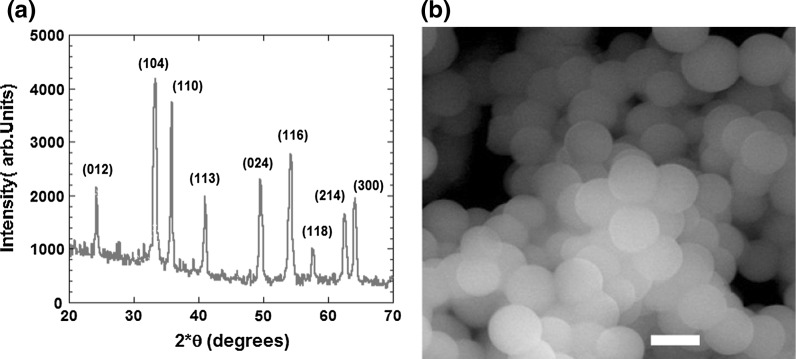
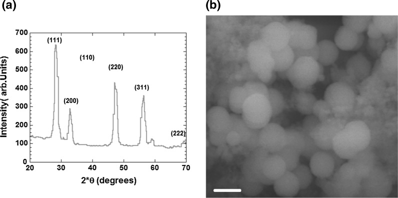
Figure 1a shows X-ray powder diffraction patterns of the prepared γ-Fe2O3 nanoparticles. The X-ray powder diffraction patterns of the material proved its crystalline nature and all the peaks matched well with standard γ-Fe2O3 reflections. The sharpness of XRD peaks reveals high crystallinity of the nanoparticles. No traces of other phases have been detected in the pattern. SEM image of prepared γ-Fe2O3 (Fig. 1b) indicates the presence of spherical-shaped nanoparticles. The grain boundaries are clean and round with no presence of other phases or salts in microstructure. The mean size of the particles varies from 40 to 50 nm. Figure 2a, b shows the XRD and SEM data for the CeO2 nanoparticles, respectively. Similarly, XRD peaks show quite high degree of crystallinity of the nanoparticles. No traces of other phases have been detected in the pattern. The SEM shows the presence of spherical-shaped CeO2 nanoparticles of homogeneous morphology with a grain size from 25 to 50 nm. However, traces of salt washing residues with smaller nanoparticle size are present between the grains and on the grain boundaries of CeO2 nanoparticles.
Fig. 1.
a XRD of γ-Fe2O3 nanoparticles, all major diffraction peaks are indexed, b SEM of γ-Fe2O3 nanoparticles with a scale bar of 50 nm
Fig. 2.
a XRD of CeO2 nanoparticles, all major diffraction peaks are indexed, b SEM of CeO2 nanoparticles with a scale bar of 50 nm
It has been noticed that the pH values of the colloidal solutions plays an important role in the precipitation process and was controlled before and after the hydrothermal process. However, in many cases (especially for the Fe colloidal solution), it is not easy to precipitate specific iron oxide particles directly in the desired size and shape. The size and shape of the nanoparticles can be tailored with relative success by adjusting pH, ionic strength, temperature, nature of the salts (chlorides, sulfates, and nitrates), or the Fe2+/Fe3+ concentration ratio (Issa et al. 2013). Moreover, based on the careful handling of the hydrothermal process carried out in the current study, we believe that final annealing in oxygen for resultant iron oxide powder facilitates oxidation of Fe3O4 to γ-Fe2O3 and produce a monodisperse, porous and magnetic γ-Fe2O3 nanoparticles. This was supported by the X-ray and the SEM data shown in Figs. 1 and 2.
CeO2 and Fe2O3 nanoparticles have no inhibitory effect on a panel of planktonic gram positive and gram negative bacteria
In order to assess the antibacterial effect of CeO2 and Fe2O3 nanoparticles against planktonic bacteria, the MICs of CeO2 and Fe2O3 nanoparticles were evaluated on a panel of planktonic gram positive and gram negative bacteria. Both CeO2 and Fe2O3 nanoparticles showed no antibacterial effect against all planktonic bacterial strains that were tested (Table 1). MICs of ciprofloxacin against all the bacterial strains tested were used as control. Ciprofloxacin treatment inhibited the planktonic bacterial growth of all selected strains (Table 1). Further controls included bacterial suspensions without nanoparticles, as well as, CeO2 and Fe2O3 nanoparticles suspensions without bacteria.
Table 1.
A comparison between the minimum inhibitory concentrations (MIC; μg/mL) of ciprofloxacin, CeO2 nanoparticles, Fe2O3 nanoparticles, and ciprofloxacin in the presence of CeO2 or Fe2O3 nanoparticles against planktonic gram positive and gram negative bacterial cultures
| Ciprofloxacin MIC (μg/ml) | CeO2 NP MIC (μg/ml) | Fe2O3 NP MIC (μg/ml) | Ciprofloxacin + CeO2 NP MIC (μg/ml) | Ciprofloxacin + Fe2O3 NP MIC (μg/ml) | |
|---|---|---|---|---|---|
| Gram +ve planktonic bacterial cultures | |||||
| MSSA | 0.10 ± 0.04 | 50 ± 20 | 20 ± 0.0 | 50 ± 20 | 30 ± 10 |
| MRSA | 0.40 ± 0.20 | 70 ± 0.0 | 60 ± 20 | 30 ± 10 | 50 ± 20 |
| S. pneumonia | 0.40 ± 0.20 | 110 ± 40 | 60 ± 20 | 70 ± 0.0 | 50 ± 20 |
| VSE | 0.40 ± 0.20 | 50 ± 20 | 30 ± 10 | 20 ± 0.0 | 30 ± 10 |
| VRE | 0.40 ± 0.20 | 110 ± 40 | 70 ± 0.0 | 60 ± 20 | 60 ± 20 |
| S. pyogenes | 0.20 ± 0.07 | 30 ± 10 | 50 ± 20 | 30 ± 0.0 | 30 ± 10 |
| S. epidermidis | 0.20 ± 0.07 | 20 ± 5 | 40 ± 0.0 | 10 ± 0.0 | 20 ± 5 |
| Gram −ve planktonic bacterial cultures | |||||
| E. coli | 0.03 ± 0.01 | 50 ± 10 | 20 ± 5 | 30 ± 10 | 20 ± 5 |
| P. aeruginosa | 0.08 ± 0.04 | 20 ± 5 | 50 ± 20 | 30 ± 10 | 20 ± 5 |
| A. baumannii | 0.20 ± 0.07 | 70 ± 0.0 | 110 ± 40 | 40 ± 0.0 | 60 ± 20 |
| P. mirabilis | 0.20 ± 0.07 | 30 ± 10 | 220 ± 80 | 8 ± 0.0 | 140 ± 0.0 |
| K. pneumonia | 0.70 ± 0.30 | 140 ± 0.0 | 110 ± 40 | 70 ± 0.0 | 50 ± 20 |
| H. influenzae | 0.40 ± 0.20 | 360 ± 160 | 360 ± 160 | 110 ± 40 | 60 ± 20 |
| E. aerogenes | 0.70 ± 0.30 | 70 ± 0.0 | 60 ± 20 | 40 ± 0.0 | 30 ± 10 |
| C. freundii | 0.40 ± 0.20 | 70 ± 0.0 | 40 ± 0.0 | 50 ± 20 | 20 ± 0.0 |
| E. cloacae | 0.40 ± 0.20 | 70 ± 0.0 | 50 ± 20 | 50 ± 20 | 30 ± 10 |
The MIC values for ciprofloxacin alone were significantly (P < 0.05) lower than those of CeO2 NP or Fe2O3 NP alone, Ciprofloxacin + CeO2 NP, and Ciprofloxacin + Fe2O3 NP for all tested bacterial strains. NP stands for nanoparticles. Results are presented as mean ± SD of 3 independent experiments
CeO2 and Fe2O3 nanoparticles have no inhibitory effect on the formation of a panel of gram positive and gram negative bacterial biofilms
One way of bacteria to resist aggressive antibiotic treatment and protect themselves against the host immune system is by forming biofilm. Biofilm is a matrix of exopolymeric substances that is impenetrable by most antibiotics and immune cells (Subbiahdoss et al. 2012). Metal nanoparticles, including zinc oxide and selenium nanoparticles, have been suggested to possess characteristics that enable them to inhibit bacterial biofilm formation (Applerot et al. 2012; Wang and Webster 2013). In order to examine the effect of CeO2 and Fe2O3 nanoparticles on bacterial biofilms biomass, MICs of CeO2 and Fe2O3 nanoparticles were evaluated on a panel of gram positive and gram negative bacterial biofilms, including antibiotic resistant strains. Bacterial suspensions were added to two-fold serial dilutions of CeO2 and Fe2O3 nanoparticles, and suspensions were incubated for 48 h at 37 °C. Bacterial biofilm formation of nanoparticles treated bacteria was compared with biofilm formation in untreated bacterial suspensions. CeO2 and Fe2O3 nanoparticles showed no antibacterial effect on biofilm biomass of all tested bacterial strains (Table 2). The MICs of ciprofloxacin were used as control and showed significant inhibitory effect on biofilm biomass of all tested bacterial strains (Table 2). Further controls included CeO2 and Fe2O3 nanoparticles suspensions without bacteria.
Table 2.
A comparison between the minimum inhibitory concentrations (MIC; μg/mL) of ciprofloxacin, CeO2 nanoparticles, Fe2O3 nanoparticles and ciprofloxacin in the presence of CeO2 or Fe2O3 nanoparticles against gram positive and gram negative bacterial biofilm cultures
| Ciprofloxacin MIC (μg/ml) | CeO2 NP MIC (μg/ml) | Fe2O3 NP MIC (μg/ml) | Ciprofloxacin + CeO2 NP MIC (μg/ml) | Ciprofloxacin + Fe2O3 NP MIC (μg/ml) | |
|---|---|---|---|---|---|
| Gram +ve bacteria biofilm cultures | |||||
| MSSA | 0.70 ± 0.30 | 180 ± 80 | 30 ± 10 | 50 ± 20 | 30 ± 10 |
| MRSA | 0.80 ± 0.30 | 180 ± 80 | 360 ± 160 | 90 ± 40 | 180 ± 80 |
| S. pneumonia | 1.0 ± 0.00 | 180 ± 80 | 180 ± 80 | 70 ± 0.0 | 90 ± 40 |
| VSE | 0.70 ± 0.30 | 270 ± 0.0 | 360 ± 160 | 140 ± 0.0 | 90 ± 40 |
| VRE | 1.0 ± 0.0 | 530 ± 0.0 | 530 ± 0.0 | 110 ± 40 | 360 ± 160 |
| S. pyogenes | 0.40 ± 0.20 | 70 ± 0.0 | 110 ± 40 | 20 ± 0.0 | 40 ± 0.0 |
| S. epidermidis | 0.40 ± 0.20 | 90 ± 40 | 270 ± 0.0 | 50 ± 20 | 180 ± 80 |
| Gram −ve bacteria biofilm cultures | |||||
| E. coli | 0.40 ± 0.20 | 90 ± 40 | 50 ± 20 | 40 ± 0.0 | 20 ± 5 |
| P. aeruginosa | 0.80 ± 0.30 | 70 ± 0.0 | 100 ± 50 | 30 ± 10 | 60 ± 20 |
| A. baumannii | 1.0 ± 0.0 | 360 ± 160 | 360 ± 160 | 360 ± 160 | 90 ± 40 |
| P. mirabilis | 0.70 ± 0.30 | 360 ± 160 | 530 ± 0.0 | 140 ± 0.0 | 110 ± 40 |
| K. pneumonia | 0.70 ± 0.30 | 360 ± 160 | 360 ± 160 | 50 ± 20 | 90 ± 40 |
| H. influenzae | 1.0 ± 0.0 | 530 ± 0.0 | 530 ± 0.0 | 140 ± 0.0 | 180 ± 80 |
| E. aerogenes | 1.0 ± 0.0 | 140 ± 0.0 | 110 ± 40 | 20 ± 0.0 | 50 ± 20 |
| C. freundii | 0.50 ± 0.00 | 220 ± 80 | 360 ± 160 | 180 ± 80 | 90 ± 40 |
| E. cloacae | 0.80 ± 0.30 | 220 ± 80 | 110 ± 40 | 110 ± 40 | 60 ± 20 |
The MIC values for ciprofloxacin alone were significantly (P < 0.05) lower than those of CeO2 NP or Fe2O3 NP alone, Ciprofloxacin + CeO2 NP, and Ciprofloxacin + Fe2O3 NP for all tested bacterial strains. NP stands for nanoparticles. Results are presented as mean ± SD of 3 independent experiments
CeO2 and Fe2O3 nanoparticles dramatically reduce antibacterial effect of ciprofloxacin against a panel of gram positive and gram negative planktonic and biofilm bacterial cultures
In order to evaluate whether combining either CeO2 or Fe2O3 nanoparticles with ciprofloxacin would influence the antibacterial effect of ciprofloxacin against the tested bacterial strains, the effect of CeO2 and Fe2O3 nanoparticles combined with ciprofloxacin was examined on bacterial growth. The MICs of ciprofloxacin in the presence of either CeO2 or Fe2O3 nanoparticles against planktonic and biofilm cultures of tested gram positive and gram negative bacteria were examined. Bacterial suspensions were added to two-fold serial dilutions of ciprofloxacin that contain either CeO2 or Fe2O3 nanoparticles. Bacterial cultures were allowed to grow to planktonic cultures, or were left to form biofilm cultures and the MICs of ciprofloxacin were assessed. Interestingly, the addition of CeO2 and Fe2O3 nanoparticles dramatically reduced the antibacterial activity of ciprofloxacin against all bacterial strains tested whether these bacteria were grown as planktonic (Table 1) or biofilm cultures (Table 2). These results combined with those discussed above suggest that CeO2 and Fe2O3 nanoparticles treatment not only fails to provide antibacterial effect, but also inhibits the antibacterial activity of ciprofloxacin. The MICs of ciprofloxacin only treated bacterial suspensions were used as control and showed significant inhibitory effect against all planktonic and biofilm cultures of tested bacterial strains. Further controls include bacterial suspensions without CeO2 and Fe2O3 nanoparticles, as well as, CeO2 and Fe2O3 nanoparticles suspensions without bacteria.
Discussion
Metal oxide nanoparticles have been suggested as an important candidate for tackling the healthcare problem of increasing number of antibiotic resistant and biofilm forming bacteria. In this study, two metal oxide nanoparticles, CeO2 and Fe2O3 nanoparticles, were tested for efficacy as antibacterial agents against a list of gram positive and gram negative bacteria. This list of bacteria included strains that are known to be antibiotic resistant such as MRSA and VRE. Our study has shown that CeO2 and Fe2O3 nanoparticles failed to inhibit bacterial planktonic growth and biofilm formation as compared to bacterial growth inhibition resulting from ciprofloxacin treatment. Ciproflxacin, however, inhibited the growth of all gram positive and gram negative bacteria tested.
A related interesting finding of the current study is that combining CeO2 or Fe2O3 nanoparticles with ciprofloxacin reduced significantly the antibacterial effect of ciprofloxacin. Ciprofloxacin is a second-generation fluoroquinolone antibacterial agent (Drlica and Zhao 1997). It kills bacteria by inhibiting DNA gyrase, and topoisomerase IV enzymes that are necessary to separate bacterial DNA, therefore, inhibiting cell division (Drlica and Zhao 1997). It is possible that these nanoparticles interact with ciprofloxacin in a way that prevents its absorption by the bacterial cell. Another possibility is that CeO2 and Fe2O3 nanoparticles interact directly or indirectly with ciprofloxacin in a way that interferes with ciprofloxacin activity on bacterial DNA inside the bacterial cell. Interestingly, studies have shown that the bioavailability of ciprofloxacin is reduced by 50 % when co-administered with iron compounds (Lode 2001). Characterization of suggested mechanisms for the interaction of CeO2 and Fe2O3 nanoparticles with ciprofloxacin is a warranted future study.
Studies that evaluated the antibacterial effect of CeO2 nanoparticles are limited. Some of these studies suggest antibacterial effect for CeO2 nanoparticles, while other studies show no toxic or inhibitory effect for CeO2 nanoparticles against bacteria. Thill et al., Pelletier et al., and Kuang et al., have suggested antimicrobial activity of CeO2 nanoparticles against E. coli, whereas Shah et al., observed that dextran coated CeO2 nanoparticles are non-lethal to E. coli under various experimental conditions examined (Kuang et al. 2011; Pelletier et al. 2010; Shah et al. 2012; Thill et al. 2006). Moreover Shah et al., reported that CeO2 nanoparticles can reduce magnesium and potassium salts antibacterial activity, which is similar to the observation in the current study where CeO2 nanoparticles almost abolished the antibacterial activity of ciprofloxacin (Shah et al. 2012). In addition, CeO2 nanoparticles have been shown to inhibit Bacillus subtilis, but have no inhibitory effect on Shewanella oneidensis (Pelletier et al. 2010). These studies utilized different synthesis methods, used CeO2 nanoparticles of various sizes, and exploited different methods to evaluate CeO2 nanoparticles antibacterial effect. It has been suggested that a change in the physical and chemical environment can significantly influence nanoparticles bacterial toxicity (Deshpande et al. 2005; Rispoli et al. 2010).
Although several studies have focused on evaluating the antibacterial effect of nanosized magnetic iron oxide particle magnetite (Fe3O4), much less is known about the antibacterial effect of the other type of magnetic iron oxide nanoparticles maghemite (Fe2O3) (Ravikumar et al. 2011; Taylor and Webster 2009; Tran et al. 2010). One study has reported antibacterial activity of Fe2O3 nanoparticles on a number of bacteria in their planktonic forms (Gokulakrishnan et al. 2012). Another study by He et al., found no inhibitory effect of Fe2O3 nanoparticles on the growth of E. coli. In contrast, the results of the mentioned study suggest an increase in bacterial growth upon Fe2O3 nanoparticles treatment (He et al. 2011). In the current study, Fe2O3 nanoparticles showed no inhibitory effect on bacterial growth and biofilm forms of all the bacterial strains tested.
In this study, both CeO2 and Fe2O3 nanoparticles were tested at a wide range of serial twofold concentrations, which is a standard procedure to estimate MIC value for compounds with previously unknown antibacterial activity (Clinical and Laboratory Standards Institute (CLSI) 2012). Future work about the possible antibacterial activity of these nanoparticles should be targeted toward studying concentrations that are above their MIC values.
In conclusion, the current study provides evidence that CeO2 and Fe2O3 nanoparticles fail to inhibit bacterial planktonic growth and biofilm biomass for all examined gram positive and gram negative bacterial strains. Moreover, CeO2 and Fe2O3 nanoparticles when combined with the broad sprectrum antibiotic ciprofloxacin almost abolished its inhibitory effect on bacterial growth and biofilm formation. Therefore, this study suggests that CeO2 and Fe2O3 nanoparticles are not good candidates as antibacterial agents.
Acknowledgments
This project was supported by Jordan University of Science and Technology.
References
- Aljarrah K, Mhaidat NM, Al-Akhras MA, Aldaher AN, Albiss B, Aledealat K, Alsheyab FM. Magnetic nanoparticles sensitize MCF-7 breast cancer cells to doxorubicin-induced apoptosis. World J Surg Oncol. 2012;10:62. doi: 10.1186/1477-7819-10-62. [DOI] [PMC free article] [PubMed] [Google Scholar]
- Applerot G, Lellouche J, Perkas N, Nitzan Y, Gedanken A, Banin E. ZnO nanoparticle-coated surfaces inhibit bacterial biofilm formation and increase antibiotic susceptibility. RSC Adv. 2012;2:2314–2321. doi: 10.1039/c2ra00602b. [DOI] [Google Scholar]
- Babes L, Denizot B, Tanguy G, Le Jeune JJ, Jallet P. Synthesis of iron oxide nanoparticles used as MRI contrast agents: a parametric study. J Colloid Interface Sci. 1999;212:474–482. doi: 10.1006/jcis.1998.6053. [DOI] [PubMed] [Google Scholar]
- Cernohorska L, Votava M. Antibiotic synergy against biofilm-forming Pseudomonas aeruginosa. Folia Microbiol (Praha) 2008;53:57–60. doi: 10.1007/s12223-008-0008-z. [DOI] [PubMed] [Google Scholar]
- Chertok B, Moffat BA, David AE, Yu F, Bergemann C, Ross BD, Yang VC. Iron oxide nanoparticles as a drug delivery vehicle for MRI monitored magnetic targeting of brain tumors. Biomaterials. 2008;29:487–496. doi: 10.1016/j.biomaterials.2007.08.050. [DOI] [PMC free article] [PubMed] [Google Scholar]
- Christensen GD, Simpson WA, Younger JJ, Baddour LM, Barrett FF, Melton DM, Beachey EH. Adherence of coagulase-negative staphylococci to plastic tissue culture plates: a quantitative model for the adherence of staphylococci to medical devices. J Clin Microbiol. 1985;22:996–1006. doi: 10.1128/jcm.22.6.996-1006.1985. [DOI] [PMC free article] [PubMed] [Google Scholar]
- Clinical and Laboratory Standards Institute (CLSI) (2012) Methods for dilution antimicrobial susceptibility test for bacteria that grow aerobically, 9th ed. Approved standard, Villanova, PA
- Cohen ML. Changing patterns of infectious disease. Nature. 2000;406:762–767. doi: 10.1038/35021206. [DOI] [PubMed] [Google Scholar]
- Deshpande S, Patil S, Kuchibhatla S, Seal S. Size dependency variation in lattice parameter and valency states in nanocrystalline cerium oxide. Appl Phys Lett. 2005;87:1–3. doi: 10.1063/1.2061873. [DOI] [Google Scholar]
- Donadio S, Maffioli S, Monciardini P, Sosio M, Jabes D. Antibiotic discovery in the twenty-first century: current trends and future perspectives. J Antibiot (Tokyo) 2010;63:423–430. doi: 10.1038/ja.2010.62. [DOI] [PubMed] [Google Scholar]
- Drlica K, Zhao X. DNA gyrase, topoisomerase IV, and the 4-quinolones. Microbiol Mol Biol Rev. 1997;61:377–392. doi: 10.1128/mmbr.61.3.377-392.1997. [DOI] [PMC free article] [PubMed] [Google Scholar]
- Fux CA, Costerton JW, Stewart PS, Stoodley P. Survival strategies of infectious biofilms. Trends Microbiol. 2005;13:34–40. doi: 10.1016/j.tim.2004.11.010. [DOI] [PubMed] [Google Scholar]
- Gokulakrishnan R, Ravikumar S, Raj JA. In vitro antibacterial potential of metal oxide nanoparticles against antibiotic resistant bacterial pathogens. the. Asian Pac J Trop Dis. 2012;2:411–413. doi: 10.1016/S2222-1808(12)60089-9. [DOI] [Google Scholar]
- Gonzales-Weimuller M, Zeisberger M, Krishnan K. Size-dependent heating rates of iron oxide nanoparticles for magnetic fluid hyperthermia. J Magn Mag Mat. 2009;321:1947–1950. doi: 10.1016/j.jmmm.2008.12.017. [DOI] [PMC free article] [PubMed] [Google Scholar]
- Gupta AK, Gupta M. Synthesis and surface engineering of iron oxide nanoparticles for biomedical applications. Biomaterials. 2005;26:3995–4021. doi: 10.1016/j.biomaterials.2004.10.012. [DOI] [PubMed] [Google Scholar]
- He S, Feng Y, Gu N, Zhang Y, Lin X. The effect of gamma-Fe2O3 nanoparticles on Escherichia coli genome. Environ Pollut. 2011;159:3468–3473. doi: 10.1016/j.envpol.2011.08.024. [DOI] [PubMed] [Google Scholar]
- Huang Z, Zheng X, Yan D, Yin G, Liao X, Kang Y, Yao Y, Huang D, Hao B. Toxicological effect of ZnO nanoparticles based on bacteria. Langmuir. 2008;24:4140–4144. doi: 10.1021/la7035949. [DOI] [PubMed] [Google Scholar]
- Huh AJ, Kwon YJ. “Nanoantibiotics”: a new paradigm for treating infectious diseases using nanomaterials in the antibiotics resistant era. J Control Release. 2011;156:128–145. doi: 10.1016/j.jconrel.2011.07.002. [DOI] [PubMed] [Google Scholar]
- Issa B, Obaidat IM, Albiss BA, Haik Y. Magnetic nanoparticles: surface effects and properties related to biomedicine applications. Int J Mol Sci. 2013;14:21266–21305. doi: 10.3390/ijms141121266. [DOI] [PMC free article] [PubMed] [Google Scholar]
- Khandhar AP, Ferguson RM, Simon JA, Krishnan KM. Enhancing cancer therapeutics using size-optimized magnetic fluid hyperthermia. J Appl Phys. 2012;111:7B306–307B3063. doi: 10.1063/1.3671427. [DOI] [PMC free article] [PubMed] [Google Scholar]
- Kuang Y, He X, Zhang Z, Li Y, Zhang H, Ma Y, Wu Z, Chai Z. Comparison study on the antibacterial activity of nano- or bulk-cerium oxide. J Nanosci Nanotechnol. 2011;11:4103–4108. doi: 10.1166/jnn.2011.3858. [DOI] [PubMed] [Google Scholar]
- Kurek A, Grudniak AM, Kraczkiewicz-Dowjat A, Wolska KI. New antibacterial therapeutics and strategies. Pol J Microbiol. 2011;60:3–12. [PubMed] [Google Scholar]
- Lode H (2001) Evidence of different profiles of side effects and drug-drug interactions among the quinolones–the pharmacokinetic standpoint. Chemotherapy 47:24–31; discussion 44–28 [DOI] [PubMed]
- Masadeh MM, Mhaidat NM, Alzoubi KH, Hussein EI, Al-Trad EI. In vitro determination of the antibiotic susceptibility of biofilm-forming Pseudomonas aeruginosa and Staphylococcus aureus: possible role of proteolytic activity and membrane lipopolysaccharide. Infect Drug Resist. 2013;6:27–32. doi: 10.2147/IDR.S41501. [DOI] [PMC free article] [PubMed] [Google Scholar]
- Negahdary M, Mohseni G, Fazilati M, Parsania S, Rahimi G, Rad S, Rezaei-Zarchi S. The Antibacterial effect of cerium oxide nanoparticles on Staphylococcus aureus bacteria. Ann Biol Res. 2012;3:3671–3678. [Google Scholar]
- Nel AE, Madler L, Velegol D, Xia T, Hoek EM, Somasundaran P, Klaessig F, Castranova V, Thompson M. Understanding biophysicochemical interactions at the nano-bio interface. Nat Mater. 2009;8:543–557. doi: 10.1038/nmat2442. [DOI] [PubMed] [Google Scholar]
- Niu J, Azfer A, Rogers LM, Wang X, Kolattukudy PE. Cardioprotective effects of cerium oxide nanoparticles in a transgenic murine model of cardiomyopathy. Cardiovasc Res. 2007;73:549–559. doi: 10.1016/j.cardiores.2006.11.031. [DOI] [PMC free article] [PubMed] [Google Scholar]
- Pal S, Tak YK, Song JM. Does the antibacterial activity of silver nanoparticles depend on the shape of the nanoparticle? A study of the Gram-negative bacterium Escherichia coli. Appl Environ Microbiol. 2007;73:1712–1720. doi: 10.1128/AEM.02218-06. [DOI] [PMC free article] [PubMed] [Google Scholar]
- Pareta RA, Taylor E, Webster TJ. Increased osteoblast density in the presence of novel calcium phosphate coated magnetic nanoparticles. Nanotechnology. 2008;19:265101. doi: 10.1088/0957-4484/19/26/265101. [DOI] [PubMed] [Google Scholar]
- Pelletier DA, Suresh AK, Holton GA, McKeown CK, Wang W, Gu B, Mortensen NP, Allison DP, Joy DC, Allison MR, Brown SD, Phelps TJ, Doktycz MJ. Effects of engineered cerium oxide nanoparticles on bacterial growth and viability. Appl Environ Microbiol. 2010;76:7981–7989. doi: 10.1128/AEM.00650-10. [DOI] [PMC free article] [PubMed] [Google Scholar]
- Perez JM, Asati A, Nath S, Kaittanis C. Synthesis of biocompatible dextran-coated nanoceria with pH-dependent antioxidant properties. Small. 2008;4:552–556. doi: 10.1002/smll.200700824. [DOI] [PubMed] [Google Scholar]
- Ravikumar S, Gokulakrishnan R, Selvanathan K, Selvam S. Antibacterial activity of metal oxide nanoparticles against ophthalmic pathogens. Int J Pharm Res Dev. 2011;3:122–127. [Google Scholar]
- Rispoli F, Angelov A, Badia D, Kumar A, Seal S, Shah V. Understanding the toxicity of aggregated zero valent copper nanoparticles against Escherichia coli. J Hazard Mater. 2010;180:212–216. doi: 10.1016/j.jhazmat.2010.04.016. [DOI] [PubMed] [Google Scholar]
- Shah V, Shah S, Shah H, Rispoli FJ, McDonnell KT, Workeneh S, Karakoti A, Kumar A, Seal S. Antibacterial activity of polymer coated cerium oxide nanoparticles. PLoS One. 2012;7:e47827. doi: 10.1371/journal.pone.0047827. [DOI] [PMC free article] [PubMed] [Google Scholar]
- Subbiahdoss G, Sharifi S, Grijpma DW, Laurent S, van der Mei HC, Mahmoudi M, Busscher HJ. Magnetic targeting of surface-modified superparamagnetic iron oxide nanoparticles yields antibacterial efficacy against biofilms of gentamicin-resistant staphylococci. Acta Biomater. 2012;8:2047–2055. doi: 10.1016/j.actbio.2012.03.002. [DOI] [PubMed] [Google Scholar]
- Tarnuzzer RW, Colon J, Patil S, Seal S. Vacancy engineered ceria nanostructures for protection from radiation-induced cellular damage. Nano Lett. 2005;5:2573–2577. doi: 10.1021/nl052024f. [DOI] [PubMed] [Google Scholar]
- Taylor EN, Webster TJ. The use of superparamagnetic nanoparticles for prosthetic biofilm prevention. Int J Nanomedicine. 2009;4:145–152. doi: 10.2217/17435889.4.2.145. [DOI] [PMC free article] [PubMed] [Google Scholar]
- Thill A, Zeyons O, Spalla O, Chauvat F, Rose J, Auffan M, Flank AM. Cytotoxicity of CeO2 nanoparticles for Escherichia coli. Physico-chemical insight of the cytotoxicity mechanism. Environ Sci Technol. 2006;40:6151–6156. doi: 10.1021/es060999b. [DOI] [PubMed] [Google Scholar]
- Tran N, Mir A, Mallik D, Sinha A, Nayar S, Webster TJ. Bactericidal effect of iron oxide nanoparticles on Staphylococcus aureus. Int J Nanomedicine. 2010;5:277–283. doi: 10.2147/ijn.s9220. [DOI] [PMC free article] [PubMed] [Google Scholar]
- Wang Q, Webster TJ. Short communication: inhibiting biofilm formation on paper towels through the use of selenium nanoparticles coatings. Int J Nanomedicine. 2013;8:407–411. doi: 10.2147/IJN.S38777. [DOI] [PMC free article] [PubMed] [Google Scholar]
- Weir E, Lawlor A, Whelan A, Regan F. The use of nanoparticles in anti-microbial materials and their characterization. Analyst. 2008;133:835–845. doi: 10.1039/b715532h. [DOI] [PubMed] [Google Scholar]
- Wu W, Li S, Liao S, Xiang F, Wu X. Preparation of new sunscreen materials Ce12xZnxO22x via solid-state reaction at room temperature and study on their properties. Rare Met. 2010;29:149–153. doi: 10.1007/s12598-010-0026-2. [DOI] [Google Scholar]


