
Figure 1.
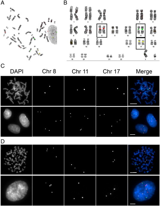
Evaluating the specificity and efficacy of CEP8/11/17. (A) A representative mitotic spread depicting the localization pattern of CEP8 (red), CEP11 (green), and CEP17 (yellow) obtained from diploid, hTERT cells. (B) Karyotypic analysis of the mitotic chromosome spread presented in A demonstrating the specificity of CEP8/11/17 for their respective chromosomes. Each CEP hybridizes with high specificity to the pericentric regions of the corresponding chromosome and presents as two copies (foci) per CEP (one focus per chromosome). (C) Representative images acquired from a mitotic chromosome spread (top) and interphase hTERT cells (bottom) hybridized with CEP8/11/17. Note that each CEP presents as two foci within each interphase nucleus and indicates the presence of two copies of the corresponding chromosome. (D) Representative images acquired from a mitotic chromosome spread (top) and interphase hypotetraploid HeLa cells (bottom) hybridized with CEP8/11/17. Note that four, three, and three copies of CEP8, CEP11, and CEP17, respectively, are observed in both the mitotic spread and interphase cells.