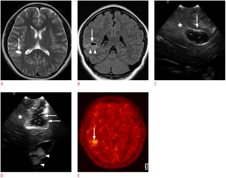
Fig. 7. A 4-year-old girl with right temporal lobe epilepsy.

A, B. Preoperative magnetic resonance images reveal a focal cystic lesion in the right temporal lobe (arrows). Note the ill-defined hyperintensities in the surrounding cortex and the subcortical white matter (arrowheads, B). C. Intraoperative ultrasonography (US) through the right temporal craniostomy shows a well-defined cystic lesion in the periventricular white matter (arrow). Note the illdefined hyperechogenic area at the caudal aspect of the cystic lesion (asterisk). D. After the resection of the cystic lesion in the temporal periventricular white matter with the adjacent cortex and the subcortical white matter, fluid-filled surgical defects (arrows) are clearly seen on US. However, the ill-defined hyperechoic area in the caudal part of the surgical defect remains (asterisk). Anechoic cerebrospinal fluid and hyperechoic choroid plexus (arrowheads) can be used as anatomic indicators. The pathologic diagnosis was a ganglioglioma. E. A methionine positron emission tomography-computed tomography image obtained 1 year later demonstrates residual tumors in the right temporal lobe (arrow).
