Abstract
Primary lymphoepithelioma-like carcinoma of the skin (LE-lCS) is a very rare cutaneous malignancy of uncertain origin. The neoplasm reveals typical morphological similarity to undifferentiated nasopharyngeal carcinoma (lymphoepithelioma). This case report presents a 47-year-old man with a 5 mm erythematous papule on dorsal nose of six months duration. The patient underwent complete surgical excision and is disease free 7 months later.
Keywords: EBV, lymphoepithelioma-like carcinoma, lymphoepithelioma, nasopharyngeal carcinoma, primary skin tumour
What was known?
Lymphoepithelioma-like carcinomas are carcinomas that arise outside of the nasopharynx, but resemble a lymphoepithelioma histologically.
Introduction
The term lymphoepithelioma-like carcinoma refers to a group of nasopharyngeal epithelial tumors histologically characterized by aggregates of malignant undifferentiated cells surrounded by a dense reactive lymphoplasmacellular infiltrate. Clinically, it presents as a flesh-colored firm papulo-plaque and/or papulonodular lesion on the face, scalp, or shoulder of middle aged to elderly individuals. Primary lymphoepithelioma-like carcinoma of the skin (LE-lCS) is a very rare cutaneous malignancy of uncertain origin. Here in, we are reporting a rare case of Lymphoepithelioma – like carcinoma of the skin in a 47 years old male.
Case Report
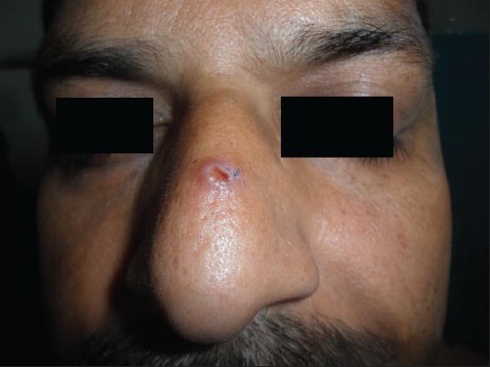
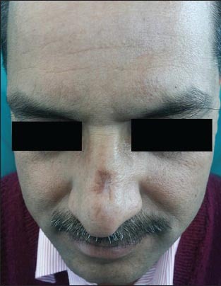
A 47-year-old male presented to our outpatient department with the complaint of a single red raised asymptomatic lesion on the dorsum of nose about 2 cm from the glabella for six months. Initially it was tiny in size and gradually progressed to achieve its present size [Figure 1]. There was no history of insect bite and trauma. On cutaneous examination, there was a single 4-5 mm erythematous papule present over the dorsal aspect of the nose. The overlying skin was thinned out with crusting at the center. No telangiectasia and local or distant lymphadenopathy were noted. The lesion was completely excised with a 2 mm normal margin with a clinical diagnosis of basal cell carcinoma (BCC). There is no recurrence seen after 7 months follow-up [Figure 2].
Figure 1.

Erythematous papule with overlying crust at center
Figure 2.

No recurrence after 7 months follow-up
Histopathological findings
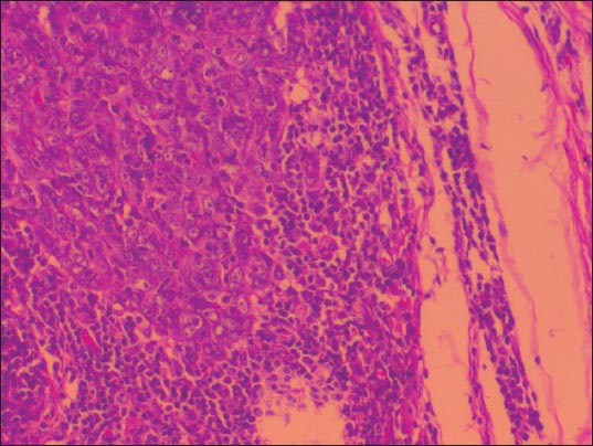
Microscopy revealed a well-defined intradermal tumor without epidermal infiltration. The epidermis showed no ulceration. The tumor consisted of a syncytium of tumor cells showing indistinct cell borders with vesicular nuclei and prominent eosinophilic nucleoli [Figure 3], surrounded by predominantly lymphocytic infiltrate.
Figure 3.

Syncytium of tumor cells surrounded by predominant lymphocytic infiltrate
Immunohistochemical profile
The tumor cells showed expression of cytokeratins (CK) (AE1/AE3), CK-5/6 and P-63 (focal). The tumor was negative for CK-20. Epstein-barr virus (EBV)-associated antibodies were negative.
Discussion
Swanson et al., documented the first case of lymphoepithelioma-like carcinoma of the skin (LE-lCS). Primary LE-lCS is a very rare cutaneous neoplasm of uncertain origin. Some people have proposed epidermal and adnexal origin due to trichilemmal and eccrine differentiation. It rarely presents as epithelial dysplasia with epidermal involvement.[1,2,3]
It usually presents as a single firm papulo-plaque and/or papulonodular lesion of variable color, over the facial or neck region. It affects men and women approximately equally in their seventh decade. Cutaneous lymphoepithelioma-like carcinoma is histologically distinct from other primary skin tumors showing well-demarcated lobules or nests of large, epithelioid cells closely associated with a dense, mixed T and B lymphocytic infiltrates. The epithelioid component has no connection with the epidermis. The cells have poorly defined eosinophilic cytoplasm and vesicular nuclei with prominent nucleoli and increased mitotic activity, including atypical mitotic figures. The main differential diagnosis includes undifferentiated nasopharyngeal carcinoma or metastatic lymphoepithelioma-like carcinoma from other sites, cutaneous lymphoma, and adnexal tumor.
Work-up for EBV and primary lymphoepithelioma-like carcinoma (LE-lC) in other organs may help in differentiating primary skin tumors from metastases. While undifferentiated nasopharyngeal carcinoma and some LE-lC of the other sites are associated with EBV.[4] The primary skin tumors have never been reactive for EBV-encoded RNA.
The association of LE-lC and EBV varies in different organs. EBV is usually associated with the tumors in lung, thymus, salivary glands, and stomach. Treatment of choice for LE-lCS is complete surgical excision.[5] Radiotherapy is also a useful modality whenever complete surgical excision is not possible.[6]
Cutaneous LE-lCS has low metastatic potential and good prognosis. Most of the cases (78%) were free of disease after treatment whereas 10% of the patients had a local recurrence and only 2 % patients developed lymph node metastases with a lethal outcome.[7,8]
In conclusion, LE-lCS is an exceedingly rare, but distinct pathological entity of unclear histogenesis. Immunohistochemistry, and clinical work-up including head and body CT scans and thorough ENT examination, are important for the diagnosis and treatment. Despite its similarity to lymphoepithelioma, a conservative surgical approach is advised.[2,5,9] Furthermore, this case supports the lack of a relationship between EBV and LE-lCS; this is in keeping with the other reported cases of this primary skin tumor.[10]
What is new?
Lymphoepithelioma-like carcinoma of the skin over dorsum of nose is very rare tumor. Complete surgical excision is the treatment of choice with a good outcome.
Footnotes
Source of support: Nil
Conflict of Interest: Nil.
References
- 1.Ko T, Muramatsu T, Shirai T. Lymphoepithelioma-like carcinoma of the skin. J Dermatol. 1997;24:104–9. doi: 10.1111/j.1346-8138.1997.tb02752.x. [DOI] [PubMed] [Google Scholar]
- 2.Robins P, Perez MI. Lymphoepithelioma-like carcinoma of the skin treated by Mohs micrographic surgery. J Am Acad Dermatol. 1995;32:814–6. doi: 10.1016/0190-9622(95)91485-4. [DOI] [PubMed] [Google Scholar]
- 3.Wick MR, Swanson PE, LeBoit PE, Strickler JG, Cooper PH. Lymphoepithelioma-like carcinoma of the skin with adnexal differentiation. J Cutan Pathol. 1991;18:93–102. doi: 10.1111/j.1600-0560.1991.tb00134.x. [DOI] [PubMed] [Google Scholar]
- 4.Iezzoni JC, Gaffey MJ, Weiss LM. The role of Epstein-Barr virus in lymphoepithelioma-like carcinomas. Am J Clin Pathol. 1995;103:308–15. doi: 10.1093/ajcp/103.3.308. [DOI] [PubMed] [Google Scholar]
- 5.Dozier SE, Jones TR, Nelson-Adesokan P, Hruza GJ. Lymphoepithelioma-like carcinoma of the skin treated by Mohs micrographic surgery. Dermatol Surg. 1995;21:690–4. doi: 10.1111/j.1524-4725.1995.tb00271.x. [DOI] [PubMed] [Google Scholar]
- 6.Ortiz-Frutos FJ, Zarco C, Gil R, Ballestin C, Iglesias L. Lymphoepithelioma-like carcinoma of the skin. Clin Exp Dermatol. 1993;18:83–6. doi: 10.1111/j.1365-2230.1993.tb00979.x. [DOI] [PubMed] [Google Scholar]
- 7.Swanson SA, Cooper PH, Mills SE, Wick MR. Lymphoepithelioma- like carcinoma of the skin. Mod Pathol. 1988;1:359–65. [PubMed] [Google Scholar]
- 8.Gillum PS, Morgan MB, Naylor MF, Everett MA. Absence of Epstein-Barr virus in lymphoepithelioma-like carcinoma of the skin. Polymerase chain reaction evidence and review of five cases. Am J Dermatopathol. 1996;18:478–82. doi: 10.1097/00000372-199610000-00006. [DOI] [PubMed] [Google Scholar]
- 9.Jimenez F, Clark RE, Buchanan MD, Kamino H. Lymphoepithelioma-like carcinoma of the skin treated with Mohs micrographic surgery in combination with immune staining for cytokeratins. J Am Acad Dermatol. 1995;32:878–81. doi: 10.1016/0190-9622(95)91552-4. [DOI] [PubMed] [Google Scholar]
- 10.Carr KA, Bulengo-Ransby SM, Weiss LM, Nickoloff BJ. Lymphoepitheliomalike carcinoma of the skin. A case report with immunophenotypic analysis and in situ hybridization for Epstein-Barr viral genome. Am J Surg Pathol. 1992;16:909–13. [PubMed] [Google Scholar]


