Abstract
Morquio's syndrome, also known as mucopolysaccharidosis type IV is an autosomal recessive disorder, caused by deficiency of n-acetylgalactosamine-6-sulphate. Anesthetic management of this syndrome is a great challenge, especially in pediatric age group as “cannot ventilate, cannot intubate” scenario can be encountered by anesthesiologist due to the possibility of total airway collapse. Herewith, we are reporting a case of child with Morquio's syndrome where I-gel assisted fiber-optic intubation was used for safe endotracheal intubation.
Keywords: Difficult airway, fiber-optic bronchoscope, I-gel, Morquio's syndrome
INTRODUCTION
Morquio's syndrome, also known as mucopolysaccharidosis type IV (MPS IV) is an autosomal recessive disorder caused by deficiency of n-acetylgalactosamine-6-sulfate. It was first described by Morquio and Brailsford and was listed as a “rare disease” by the Office of Rare Diseases of National Institutes of Health, with a prevalence of 1/200,000-250,000 people.[1] Patients with MPS IV pose a great challenge for the anesthesiologists because of their inherent problems such as difficult airway, neck instability, restrictive pulmonary disease, and end-organ damage. Herewith, we describe the anesthetic management of a child with Morquio's syndrome posted for fixation of atlanto axial subluxation.
CASE REPORT
A 3-year-old male child presented with a history of weakness of both lower limb and snoring for the past 6 months. Child was weighing 10.5 kg with a height of 78 cm and had clinical features like coarse facies, large head and extremely short neck. He was not able to stand with support but able to sit with support. On inspection pigeon shaped chest, increased anterior-posterior diameter, protuberant belly and cervico-dorsal scoliosis were present. Neurological examination revealed brisk deep tendon reflex with power of 4/5 in both upper limb and 2/5 in both lower limb. Preoperative airway examination revealed mallampatti grade 3, buck teeth, macroglossia and neck fixed in full extension with a full restriction of movements.
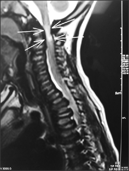
Hemogram, urine and liver function test were within normal limits. Echocardiography showed mild aortic regurgitation with no wall motion abnormality. Chest X-ray revealed wide ribs, foreshortened clavicle and cevico-dorsal scoliosis. Spiral computed tomography cervical-spine revealed atlanto axial subluxation and rotation of C2 over C1. Magnetic resonance imaging spine showed cord compression at C1 with the loss of cerebrospinal fluid space and signal changes in cord below the compression [Figure 1]. Skeletal radiograph findings were as follows: X-ray skull revealed normal calvarium with j-shaped pituitary fossa; X-ray hands showed flexion deformity at interphalangeal joints and wrist joint; X-ray pelvis showed bilateral wide iliac blades with obliquely placed acetabular roof and coxa vera deformity of femur.
Figure 1.

Magnetic resonance imaging cervical spine sagittal section, showing cord compression at C1 vertebrae with loss of cerebrospinal fluid space
He was posted for fixation of atlanto axial subluxation. Difficult intubation cart with the appropriate size of laryngeal mask airway (LMA), I-gel, endotracheal tubes (ETs) and fiberoptic bronchoscope was kept ready. Monitoring included 5 lead electrocardiogram, SpO2, noninvasive blood pressure and end-tidal carbon dioxide. Anesthesia was induced with incremental sevoflurane in 100% oxygen by face mask with Ayres T piece and spontaneous ventilation was maintained. A 22 G intravenous cannula was secured and injection. Fentanyl 20 μg was given intravenously. When adequate depth of anesthesia was attained, 2 size I-gel was inserted with manual in-line stabilization. I-gel position was confirmed with capnography. A 3.6 outer diameter fiber-optic bronchoscope which was loaded with 4.00 mm ET was introduced through the I-gel into the trachea. Once the position of the bronchoscope was confirmed ET tube was railroaded into the trachea under vision, followed by removal of bronchoscope and I-gel. Anesthesia was maintained with isoflurane, air and oxygen. Muscle paralysis was achieved with injection atracurium with neuromuscular monitoring. Cord decompression at C1 and C1-C2 stabilization done in the prone position. Rest of the intraoperative period remained uneventful and postoperatively child was shifted to Intensive Care Unit for elective ventilation in view of airway edema. Child was extubated on 1st postoperative day and discharged on 8th postoperative day.
DISCUSSION
Mucopolysaccharidoses are uncommon genetic diseases related to the metabolism of connective tissue due to deficiency of specific lysosomal enzymes, which leads to the storage of partially degraded glycosaminoglycans causing progressive cellular, organ, and multisystemic damage. Classification of MPS disorders includes 7 major types (I-IX) on the basis of clinical features, age at presentation, and biochemical alterations.[2]
The Morquio-Brailsford syndrome is MPS IV A in which there is accumulation of keratin sulfate and chondroitin-6-sulfate in connective tissue, skeletal system, teeth, brain, heart, liver, spleen, trachea and bronchial tree leading to characteristic physical appearance and resulting in end organ dysfunction.[3] Anesthetic management of such patients require proper preoperative planning and preparation and the main areas of concern in our patient were:
Difficult airway.
Unstable neck.
Restrictive pulmonary disease due to cervico-thoracic kyphoscoliosis and.
Paediatric age group.
Difficult mask ventilation was anticipated because of bulky pharyngeal soft tissue due to the deposition of mucopolysaccharides in orophayrnx, tongue, floor of the mouth, epiglottis, aryepiglottic folds which also mandates the use of smaller endotrachael tube. Presence of prominent maxillae, short neck, restricted mouth opening due to involvement of the temporomandibular joints, atlanto-axial instability and hypoplasia of the odontoid process makes direct laryngoscopy and intubation difficult to perform. All these factors may lead to a “cannot intubate/cannot ventilate” situation.[4]
In a series reporting on occiptio-cervical fusion in 17 patients with Morquio's syndrome (age 3-22) intubation under general anesthesia was described.[5] Awake fibreoptic intubations have been previously described in pediatric[6] and adult[7] cases of Morquio's syndrome. However, the deposition of soft tissue in the neck and oropharynx in Morquio's syndrome may present difficulties for conventional fiberoptic intubation. In our case, awake fiberoptic intubation was not attempted as child was not cooperative. Hence, child was induced with sevoflurane and maintained on spontaneous ventilation till I-gel has been placed.
I-gel has been reported to rescue the airway and facilitate fiberoptic tracheal intubation in patients with difficult intubation and it has been compared with other supraglottic airway devices for ease of insertion in airway training manikins and was found to be very useful compared to other devices.[8] I-gel has also got its own limitations as it needs bigger mouth opening than flexible LMA, but smaller than what is needed for intubating LMA. Michalek et al. has also described the successful intubation using I-gel has a conduit for fiberoptic guided intubation in adult patients.[9] Sharma et al. described difficulties removing the I-gel after intubation[10] whereas in our case we did not encounter any such difficulties during removal.
Thus, I-gel guided fiberoptic intubation can be a simple and useful technique in the airway management that can avoid “cannot ventilate, cannot intubate” situations in children with Morquio's syndrome.
Footnotes
Source of Support: Nil
Conflict of Interest: None declared.
REFERENCES
- 1.Kadic L, Driessen JJ. General anesthesia in an adult patient with Morquio syndrom with emphasis on airway issues. Bosn J Basic Med Sci. 2012;12:130–3. doi: 10.17305/bjbms.2012.2513. [DOI] [PMC free article] [PubMed] [Google Scholar]
- 2.Sam JA, Baluch AR, Niaz RS, Lonadier L, Kaye AD. Mucopolysaccharidoses: Anesthetic considerations and clinical manifestations. Middle East J Anaesthesiol. 2011;21:243–50. [PubMed] [Google Scholar]
- 3.McLaughlin AM, Farooq M, Donnelly MB, Foley K. Anaesthetic considerations of adults with Morquio's syndrome — A case report. BMC Anesthesiol. 2010;10:2. doi: 10.1186/1471-2253-10-2. [DOI] [PMC free article] [PubMed] [Google Scholar]
- 4.Spinello CM, Novello LM, Pitino S, Raiti C, Murabito P, Stimoli F, et al. Anesthetic management in mucopolysaccharidoses. ISRN Anesthesiol. 2013 Article ID 791983: 10 pages. [Google Scholar]
- 5.Ransford AO, Crockard HA, Stevens JM, Modaghegh S. Occipito-atlanto-axial fusion in Morquio-Brailsford syndrome. A ten-year experience. J Bone Joint Surg Br. 1996;78:307–13. [PubMed] [Google Scholar]
- 6.Tzanova I, Schwarz M, Jantzen JP. Securing the airway in children with the Morquio-Brailsford syndrome. Anaesthesist. 1993;42:477–81. [PubMed] [Google Scholar]
- 7.Bartz HJ, Wiesner L, Wappler F. Anaesthetic management of patients with mucopolysaccharidosis IV presenting for major orthopaedic surgery. Acta Anaesthesiol Scand. 1999;43:679–83. doi: 10.1034/j.1399-6576.1999.430614.x. [DOI] [PubMed] [Google Scholar]
- 8.Jackson KM, Cook TM. Evaluation of four airway training manikins as patient simulators for the insertion of eight types of supraglottic airway devices. Anesthesia. 2007;62:388–93. doi: 10.1111/j.1365-2044.2007.04983.x. [DOI] [PubMed] [Google Scholar]
- 9.Michalek P, Hodgkinson P, Donaldson W. Fiberoptic intubation through an I-gel supraglottic airway in two patients with predicted difficult airway and intellectual disability. Anesth Analg. 2008;106:1501–4. doi: 10.1213/ane.0b013e31816f22f6. [DOI] [PubMed] [Google Scholar]
- 10.Sharma S, Scott S, Rogers R, Popat M. The I-gel airway for ventilation and rescue intubation. Anesthesia. 2007;62:419–20. doi: 10.1111/j.1365-2044.2007.05045.x. [DOI] [PubMed] [Google Scholar]


