Abstract
Prosthetic dentistry involves the replacement of missing and contiguous tissues with artificial substitutes to restore and maintain the oral functions, appearance, and health of the patient. The treatment of edentulous areas with ridge defects poses a challenging task for the dentist. Management of such cases involves a wide range of treatment options comprising mainly of surgical interventions and non surgical techniques such as use of removable, fixed or fixed- removable partial dentures. But each treatment plan undertaken should be customized according to patient needs. A variety of factors such as quality and quantity of existing contiguous hard and soft tissues, systemic condition and economic status of the patient play an important role in treatment planning, clinical outcome and prognosis. This case report presents the restoration of a Seibert's Class III ridge defect by an economical modification of Andrews Bridge in a 32 Year old patient.
Keywords: Andrews bridge, fixed-removable partial dentures, modified Andrews bridge, prosthetic dentistry, Siebert's Class III ridge defect
Introduction
The treatment of edentulous areas with ridge defects poses a challenging task for the dentist. A variety of factors such as quality and quantity of existing contiguous hard and soft tissues, systemic condition and economic status of the patient play a significant role in treatment planning, clinical outcome and prognosis. Management of such cases involves a wide range of treatment options comprising mainly of soft tissue augmentation (employed mainly for Siebert's Class I defects),[1,2,3] bone augmentation techniques by Inlay and Onlay grafting (mainly for Siebert's Class II and III defects)[4,5] and combination technique incorporating soft tissue and bone augmentation. The clinical situations in which surgical augmentation cannot be carried out on grounds of the systemic condition of the patients or their reluctance for the surgical procedure mandate the use of alternative treatment protocols.[6,7] Such alternate treatment protocols include removable partial dentures, fixed partial dentures[8,9] with gingival colored porcelain and fixed-removable partial dentures known as Andrews bridge.[8,9] This case report presents the restoration of a Seibert's Class III ridge defect by modified Andrews bridge in a 32-year-old patient.
Case Report
A 32-year-old male reported with a chief complaint of unesthetic appearance of the face due to depression in the lip region on the left side of the face. Extra orally, two scars and a prominent depression in lower lip region were noticeable on the left side of the face. On intraoral examination, mandibular anterior and premolar teeth were missing and a bony defect (12 mm × 19 mm) with flabby tissue at the base was present at the respective region [Figure 1a]. The complete case history of the patient was taken, which revealed that the patient had undergone treatment for comminuted mandibular fracture in symphseal-parasymphyseal region on the left side of face 3 years back. This segment of the mandible along with five teeth (mandibular anteriors and premolars) was removed during open reduction of the fracture followed by placement of titanium reconstruction plate. The intraoral closure of the defect was done by placing the temporal flap over the defect. The relative positions of the two mandibular segments and the reconstruction plate could be clearly appreciated on orthopantogram [Figure 1]. The tissues in the defect region were 12 mm below the cervical margins of the adjacent teeth, attributing it as a Siebert's Class III defect [Figure 1b]. The various treatment modalities were thoroughly explained to the patient. Considering the reluctance for the surgical procedure and economic status of the patient a modification of Andrews bridge was chosen as the treatment option. The modification planned was to replace the costlier prefabricated bar and sleeve attachment of conventional Andrews system with two small disc shaped magnets. The advantages and disadvantages were clearly explained to the patient and informed consent was taken. The step by step procedure is as follows:
Figure 1.

(a) Preoperative orthopantogram (b) Preoperative interocclusal view showing Siebert's Class III defect
Diagnostic impressions were made and casts were prepared
The selected abutment teeth (41, 36) were prepared for metal ceramic restorations. The gingival retraction was done using gingival retraction cord (Ultrapak) and putty wash impression (Express STD Putty 3M, ESPE, USA and Express Light body STD Quick Wash, 3M ESPE, USA) was made. Provisional crowns (Protemp – II, 3M ESPE, USA) were cemented on both the prepared teeth
The maxillary and mandibular casts were mounted, and the wax pattern was fabricated in Blue Inlay Wax (Bego, USA) comprising of a rectangular bar connecting copings on both the prepared teeth. The pattern was casted in Nickel chrome alloy (Wiron 99, Bego, USA)
The cast metal framework was tried in patient and checked for any impingement of the basal tissues
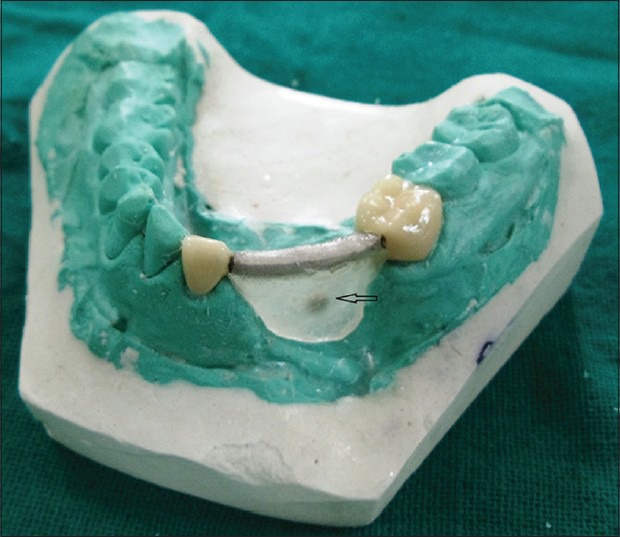
The framework was again tried in after application of Porcelain (Ceramco-3, Dentsply, USA). Afterwards, the framework was reseated on the cast and a self-polymerizing clear acrylic (DPI, India) flange was fabricated from the bar in the framework till the soft tissues on the base of the defect. The tissue surface of the flange simulated the sanitary pontic design [Figure 2]. A magnet (NdFeB magnet, Ni-plated–disc shaped magnet 2 mm × 1.5 mm in size, Techtone Electronics, Mumbai, India) was placed in buccolingual and mesiodistal center of the lingual surface of this flange [Figure 3a]
The framework was cemented in vivo with temporary cement (Rely-X Temp, 3M ESPE, USA). An impression of the mandibular arch after cementation of the framework was made using irreversible hydrocolloid (Algitex, DPI, India). The cast was poured (Kalstone, Kalhabhai, India) and undercuts were blocked. The cast thus obtained was used for the fabrication of removable component of the prosthesis
Mounting of the casts was followed by teeth arrangement and esthetic try in
Acrylization of prosthesis was done with heat cure acrylic resin (DPI, India)
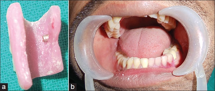
The undercuts in the prosthesis were removed before insertion. A counter magnet was placed on intaglio surface of lingual flange of the removable part of the prosthesis corresponding to the magnet on fixed part of the prosthesis [Figure 3a]. The framework was cemented permanently, and the removable component was placed [Figure 3b]
The technique of insertion and removal of the prosthesis was taught to the patient. Oral hygiene Instructions and recall schedule were explained to the patient.
Figure 2.

Fixed component with acrylic flange fabricated and magnet placed in it
Figure 3.

(a) Counter magnet placed in the removable component (b) Postoperative view showing removable component placed over the cemented fixed component
Discussion
The management of extensive ridge defect (Siebert's Class III) by a fixed-removable partial denture in a mandibular fracture patient has been explained. The Andrews system is basically composed of two components: Fixed component (retainers on abutments joined by bar) and removable component. The conventional Andrews system requires a castable bar (incorporated in the fixed component) and sleeve (incorporated in the removable component) attachment, which provides a precision while seating and retention. In this case, the bar and clip attachment of Andrews bridge system was replaced with magnets.[10,11] Ni-plated, disc shaped, miniature (2 mm × 1.5 mm), open field Neodynium-Iron-Boron magnets were used. The magnetic attraction between the magnets provides the required retention to the appliance. This modification makes the appliance economical still maintaining all the basic requirements of Andrews bridge system such as facilitating good oral hygiene maintenance in addition to providing good functional and esthetic results. Moreover, these magnets do not exert any deleterious effects on the human tissues.[12] The support in this system is mainly derived from the abutment teeth so no pressure was applied to the tissues at the base of the defect which were devoid of any bone support.
There are certain shortcomings of the modification which are worth mentioning. The surfaces of magnets are exposed to oral fluids, which may lead to tarnish and corrosion. This may produce disruption of the protective coating of the magnets leading to some cytotoxic effects.[12,13] It may result in the decreased retentive force upon usage.[14] Hence maintaining constant recalls and replacing the magnets as early as signs of corrosion develop may play a significant role in the durability and functioning of the appliance.
The surgical grafting procedures still remain the best treatment options for the cases with unesthetic ridge defects. However, considering the treatment cost, invasiveness and treatment period in surgical procedures, the fixed removable system provides a rapid and economical substitute for achieving the desired treatment goals in cases with severe ridge defects.
Footnotes
Source of Support: Nil.
Conflict of Interest: None declared.
References
- 1.Seibert JS. Reconstruction of deformed, partially edentulous ridges, using full thickness onlay grafts. Part I. Technique and wound healing. Compend Contin Educ Dent. 1983;4:437–53. [PubMed] [Google Scholar]
- 2.Abrams L. Augmentation of the deformed residual edentulous ridge for fixed prosthesis. Compend Contin Educ Gen Dent. 1980;1:205–13. [PubMed] [Google Scholar]
- 3.Langer B, Calagna L. The subepithelial connective tissue graft. J Prosthet Dent. 1980;44:363–7. doi: 10.1016/0022-3913(80)90090-6. [DOI] [PubMed] [Google Scholar]
- 4.Smidt A, Goldstein M. Augmentation of a deformed residual ridge for the replacement of a missing maxillary central incisor. Pract Periodontics Aesthet Dent. 1999;11:229–32. [PubMed] [Google Scholar]
- 5.van den Bergh JP, ten Bruggenkate CM, Tuinzing DB. Preimplant surgery of the bony tissues. J Prosthet Dent. 1998;80:175–83. doi: 10.1016/s0022-3913(98)70107-6. [DOI] [PubMed] [Google Scholar]
- 6.Mueninghoff LA, Johnson MH. Fixed-removable partial denture. J Prosthet Dent. 1982;48:547–50. doi: 10.1016/0022-3913(82)90360-2. [DOI] [PubMed] [Google Scholar]
- 7.Shankar RY, Raju AV, Raju DS, Babu PJ, Kumar DR, Rao DB. A fixed removable partial denture treatment for severe ridge defects. Int J Dent Case Rep. 2011;1:112–8. [Google Scholar]
- 8.Everhart RJ, Cavazos E., Jr Evaluation of a fixed removable partial denture: Andrews bridge system. J Prosthet Dent. 1983;50:180–4. doi: 10.1016/0022-3913(83)90008-2. [DOI] [PubMed] [Google Scholar]
- 9.Immekus JE, Aramany M. A fixed-removable partial denture for cleft-palate patients. J Prosthet Dent. 1975;34:286–91. doi: 10.1016/0022-3913(75)90105-5. [DOI] [PubMed] [Google Scholar]
- 10.Riley MA, Walmsley AD, Harris IR. Magnets in prosthetic dentistry. J Prosthet Dent. 2001;86:137–42. doi: 10.1067/mpr.2001.115533. [DOI] [PubMed] [Google Scholar]
- 11.Javid N. The use of magnets in a maxillofacial prosthesis. J Prosthet Dent. 1971;25:334–41. doi: 10.1016/0022-3913(71)90196-x. [DOI] [PubMed] [Google Scholar]
- 12.Balu K. An innovative method for magnet retained over denture. J Indian Acad Dent Spec. 2010;1:52–5. [Google Scholar]
- 13.Vrijhoef MM, Mezger PR, Van der Zel JM, Greener EH. Corrosion of ferromagnetic alloys used for magnetic retention of overdentures. J Dent Res. 1987;66:1456–9. doi: 10.1177/00220345870660090901. [DOI] [PubMed] [Google Scholar]
- 14.Riley MA, Williams AJ, Speight JD, Walmsley AD, Harris IR. Investigations into the failure of dental magnets. Int J Prosthodont. 1999;12:249–54. [PubMed] [Google Scholar]


