Figure 7.
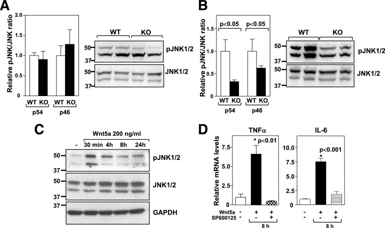
Wnt5a induces JNK signaling and proinflammatory cytokine expression in macrophages. JNK1/2 phosphorylation was evaluated by Western blot analysis in whole epididymal WAT (A) or SVFs (B) obtained from Wnt5a-KO and WT mice fed an HFHS diet for 12 weeks. Four mice per genotype were analyzed. Left: Densitometric quantification of the phospho-JNK/JNK ratio. Right: Representative immunoblots. C: JNK1/2 phosphorylation was evaluated by Western blot analysis in BM-derived macrophages treated with 200 ng/mL recombinant Wnt5a. A representative immunoblot is shown. D: BM-derived macrophages were treated with 200 ng/mL recombinant Wnt5a protein for 8 h in the absence or presence of 10 μmol/L SP600125, and TNF-α and IL-6 expression was evaluated by qRT-PCR. The graphs show the average of three independent experiments. Asterisk indicates the experimental group corresponding to the P value shown in the panel. pJNK, phospho-JNK.

