Abstract
The fly pharyngeal sense organs lie at the transition between external and internal nutrient sensing mechanisms. Here, we investigate the function of pharyngeal sweet gustatory receptor neurons (GRNs), demonstrating that they express a subset of the nine previously identified sweet receptors and respond to stimulation with a panel of sweet compounds. We show that pox-neuro (poxn) mutants lacking taste function in the legs and labial palps have intact pharyngeal sweet taste, which is both necessary and sufficient to drive preferred consumption of sweet compounds by prolonging ingestion. Moreover, flies putatively lacking all sweet taste show little preference for nutritive or non-nutritive sugars in a short-term feeding assay. Together, our data demonstrate that pharyngeal sense organs play an important role in directing sustained consumption of sweet compounds, and suggest that post-ingestive sugar sensing does not effectively drive food choice in a simple short-term feeding paradigm.
Keywords: Chemosensation, nutrient sensing, feeding, sweet taste
Introduction
Sweet taste plays a key role in promoting ingestion of nutritionally rich sources of carbohydrates. Adult Drosophila express sweet taste receptors in gustatory receptor neurons (GRNs) located in the legs, labellum, and a set of three pharyngeal sense organs collectively referred to as the internal mouthparts1. Sweet GRNs in the legs and labellum are broadly tuned to sugar stimuli, and their activation initiates feeding behaviours including the proboscis extension reflex (PER)2-7. However, neither the physiology nor behavioural roles of pharyngeal GRNs have been described.
The pharyngeal sense organs consist of the dorsal and ventral cibarial sense organs (DCSO, VCSO) and the more distal labral sense organ (LSO)1,8. The DCSO has two gustatory sensilla on each side of the midline, each housing three GRNs. Each side of the VCSO and LSO has three gustatory sensilla housing a total of eight and ten GRNs, respectively8,9. Axons from pharyngeal GRNs project via the pharyngeal nerve to the subesophageal zone (SEZ) of the brain, where they target an area that is distinct from the projections of leg and labellar GRNs1,3. Mapping of body parts to different areas of the SEZ raises the possibility that taste detection by the legs, labellum, and pharyngeal sense organs may each have distinct ethological importance.
The Drosophila genome encodes 68 members of the gustatory receptor (Gr) family, with nine classified as sweet receptors2-7,10-13. Eight of these, Gr5a, Gr61a, and Gr64a-64f, are closely related in sequence and are the defining members of a clade of insect sweet taste receptors14. Both expression and functional analyses suggest that sweet GRNs co-express multiple sweet receptors4,10,11,13,15. Furthermore, mapping of Gr promoter-GAL4 expression patterns to identified sensilla in the labellum and tarsi suggests that individual sweet Grs may be expressed in overlapping but distinct subsets of sweet GRNs8,9,16,17. In addition to the sweet clade, a highly conserved receptor, Gr43a, also functions as a sugar receptor5,13,18. Interestingly, Gr43a, which is expressed in a few neurons in the protocerebrum, appears to be restricted to some tarsal and pharyngeal GRNs in the gustatory system13,16,17.
In addition to sweet taste, mounting evidence suggests that the caloric content of sugars can drive feeding preferences in both flies and mammals19-26. To distinguish between the nutritional and gustatory effects of various sugars, it would be beneficial to examine animals that completely lack taste sensory input19,21,22. One proposed means of achieving this effect in flies has been to use pox-neuro (poxn) mutants, in which external taste bristles are transformed into mechanosensory bristles21,22,27,28. However, there is evidence that the pharyngeal sense organs of poxn mutants retain expression of at least some gustatory genes29. If poxn mutants have functional pharyngeal taste sensilla, this could account for their observed preference for caloric sugars21,22.
Here, we attempt to parse out the distinctions between external and internal sugar sensing by examining the role of pharyngeal taste in the fly. We show that the pharyngeal sense organs include several GRNs that express sweet taste receptors and respond to sweet compounds. We also demonstrate that poxn mutants have functional pharyngeal sweet GRNs, and use silencing of these neurons in the poxn mutant background to examine the effects of taste and nutrient sensing on feeding decisions. We find that flies lacking all identified sweet GRN function lose most of their preference for sweet compounds in a simple short-term binary choice assay. Our results suggest that taste is the primary influence on short-term feeding decisions and that post-ingestive sugar sensing plays at most only a minor role in this context.
Results
Pharyngeal GRNs express sweet receptors
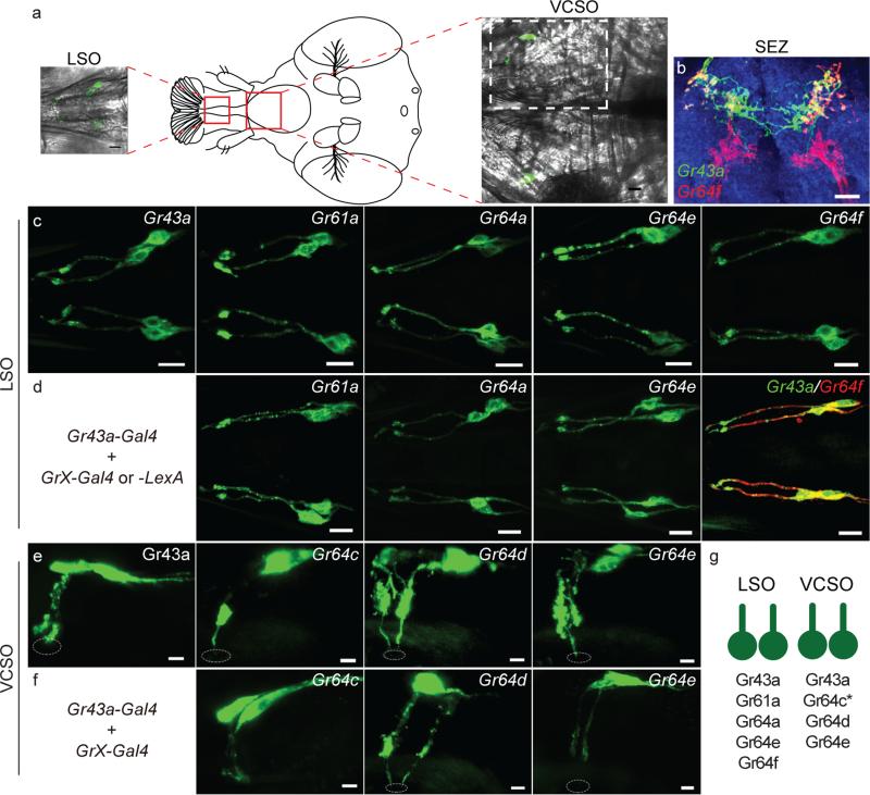
Previous studies have suggested that sweet receptors are expressed in internal pharyngeal GRNs4,13,30. To assign selected sweet receptors to identified pharyngeal GRNs, we used seven previously reported Gr promoter-GAL4 transgenes16,17 and a LexA knock-in allele of Gr64f13. Mapping was based on examination of GFP expression in the LSO, VCSO, and DCSO of flies carrying each Gr-GAL4 or GrLexA driver, as well as analysis of GFP expression in flies carrying two different drivers. Six of the seven sweet Gr-GAL4 drivers, as well as Gr64fLexA, showed expression in the pharynx and suggest that the LSO and VCSO are innervated by sweet GRNs (Figure 1). We did not detect any Gr5a-GAL4 expression in the pharynx, and none of the sweet Gr-GAL4 lines tested showed expression in the DCSO (not shown). The map identifies two candidate sweet GRNs per side of the LSO, which co-express Gr43a and Gr64e with other members of the sweet clade. The VCSO also contains two candidate sweet GRNs per side, both of which express Gr43a-GAL4 and Gr64e-GAL4. Thus, Gr43a- and Gr64e-GAL4 offer two tools to explore the physiological and behavioural roles of the pharyngeal sense organs. Both are expressed in all identified candidate pharyngeal sweet neurons as well as GRNs in the legs; however, Gr64e-GAL4 is also expressed in the taste hairs and taste pegs of the labellum, while Gr43a-GAL4 lacks labellar expression, but is expressed in sugar-sensing neurons in the protocerebrum 13,16,17,30.
Figure 1.
Pharyngeal GRNs express sweet Grs (a) Cartoon showing the positions of the LSO and VCSO, with associated images of each structure from a fly expressing GFP under control of Gr43a-GAL4. Dotted white box indicates area shown in e-f. (b) Axonal projections of Gr43a-GAL4 (green) and Gr64fLexA (red) to the SEZ. Overlapping regions are from LSO projections. (c) Gr-GAL4-driven GFP expression in LSO. Scale bars are 10 μm in c-d. (d) LSO GFP expression from flies carrying Gr43a-GAL4 and indicated second Gr-GAL4 or Gr64fLexA. (e) Gr-GAL4-driven GFP expression in VCSO. (f) VCSO GFP expression from flies carrying Gr43a-GAL4 and indicated second Gr-GAL4. Scale bars are 5 μm in e-f. Dotted circles indicate the cuticular pore of sensilla. (g) Schematic of observed sweet Gr expression in LSO and VCSO GRNs. Asterisk indicates that Gr64c- GAL4 expression is seen in only one neuron per side of the VCSO.
Pharyngeal GRNs detect a variety of sugars
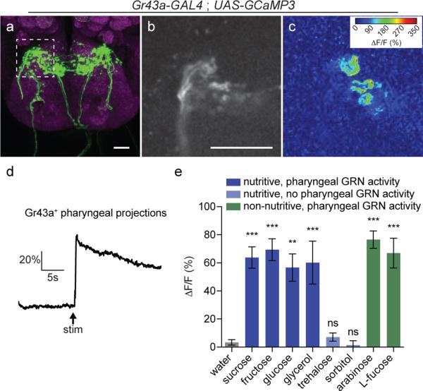
To examine the role of sweet taste detected by the pharyngeal sense organs, we began by measuring the response properties of pharyngeal neurons expressing Gr43a. We expressed GCaMP3 under the control of Gr43a-GAL4 and used an in vivo imaging preparation to measure the calcium responses of GRN axon terminals in the SEZ during consumption of sweet stimuli (Figure 2). Pharyngeal Gr43a+ GRNs exhibited broad tuning to sweet compounds, with responses to both nutritive (sucrose, fructose, glucose) and non-nutritive (arabinose, L-fucose) sugars, as well as the sweet sugar alcohol glycerol (Figure 2e). Consistent with the reported lack of Gr5a expression in the pharyngeal sense organs, we did not observe responses to the Gr5a ligand trehalose3-5. We also saw no response to the nutritive sugar alcohol sorbitol, which is generally considered tasteless to the fly. Taken together these data confirm that, as predicted by their gustatory receptor expression, a subset of pharyngeal GRNs is activated by the ingestion of sweet compounds. Additionally, it is notable that calcium responses in pharyngeal GRNs were sustained much longer than those previously observed from stimulation of labellar GRNs6,31.
Figure 2.
Pharyngeal GRNs respond to sweet compounds (a) Immunofluorescence of anti-GFP (green) and nc82 (magenta) in the SEZ of flies expressing GCaMP3 under control of Gr43a-GAL4. Dotted line shows area imaged in panel (b). (b) Single optical section of baseline GCaMP3 fluorescence in pharyngeal GRN axon terminals. Scale bars are 20 μm in a-b. (c) Heat map showing change in GCaMP3 fluorescence during ingestion of 1 M fructose. (d) Representative trace of fluorescence change of GCaMP3 in Gr43a axon terminals during ingestion of 1 M fructose. Arrow indicates time at which stimulus is applied to the proboscis to initiate feeding. (e) Peak fluorescence changes of GCaMP3 in Gr43a axon terminals during ingestion of 1 M solutions of the indicated compounds. Values represent mean +/− s.e.m. for n = 5 flies per stimulus (n = 4 for sorbitol), with data collected over at least 2 days. Asterisks indicate significant difference from water by one-way ANOVA with Bonferroni correction for multiple comparisons: **p < 0.01, ***p < 0.001, ns = not significant.
poxn mutants retain functional pharyngeal sense organs
To investigate the behavioural role of internal pharyngeal GRNs, we began by examining null mutants for the transcription factor poxn. poxn mutants lack external taste bristles, which are instead transformed into mechanosensory bristles28. This has resulted in the use of poxn mutants as taste-blind flies21,22. However, the pharyngeal sense organs of poxn mutants have been reported to express a reporter for the apparently gustatory-specific odorant binding protein OBP56b, raising the possibility that taste sensilla may be intact in these tissues29. Moreover, many of the neurons in the pharyngeal organs of the adult originate in the embryo and persist through the larval stages and metamorphosis, which contrasts with the general principle that adult sensory structures are born during metamorphosis and suggests that the pharyngeal organs may depend on an entirely distinct developmental program9. We therefore began by asking whether poxn mutants have functional pharyngeal taste sensilla.
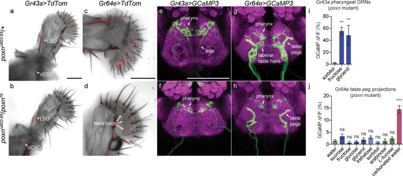
Transheterozygotes for two poxn null alleles (poxn70 and poxnΔM22-B5) showed normal expression of Gr43a-GAL4 in GRNs of the LSO and VCSO (Figure 3a,b). Additionally, brains from poxn null mutants had morphologically normal projections from pharyngeal GRNs, while they lacked the leg projections seen in otherwise wild-type flies (Figure 3e,f). Examining Gr64e-GAL4 expression in the poxn background confirmed these results and additionally demonstrated that labellar taste peg GRNs are also present in poxn mutants (Figure 3c,d,g,h). To ask whether the pharyngeal GRNs of poxn mutants are functional, we expressed GCaMP3 under the control of Gr43a-GAL4 in the poxn null mutant background, and measured calcium responses during ingestion of sweet compounds. We observed robust activation of Gr43a+ pharyngeal GRNs upon ingestion of fructose and glycerol but not sorbitol (Figure 3i). Due to the technical difficulties in stimulating flies lacking external taste sensation to ingest sweet tastants during calcium imaging, we did not expand our analysis to a larger panel of compounds. However, it is very likely that poxn Gr43a+ pharyngeal neurons retain the same receptive fields seen in a wild-type background (Figure 2e). By contrast, Gr64e+ taste peg GRNs did not respond to any of the sweet compounds tested but were activated by carbonated water (Figure 3j), as previously reported for taste pegs in a wild-type background32. Together, these data demonstrate unequivocally that poxn mutants retain functional pharyngeal taste sensilla that are capable of responding to sweet compounds. Moreover, while functional taste peg GRNs also exist in these mutants, they do not respond to sweet compounds and thus are unlikely to affect our subsequent behavioural analyses of sweet taste preferences driven by the pharyngeal sense organs.
Figure 3.
poxn null mutants retain functional pharyngeal sense organs (a,b) Pharyngeal GRNs labeled by Gr43a-GAL4 driving UAS-TdTomato in poxnΔM22-B5/+ heterozygotes (a) and poxnΔM22-B5/poxn70 null mutants (b). Arrows point to GRNs in the LSO and VCSO. (c,d) Labellar GRNs labeled by Gr64e-GAL4 driving UAS-TdTomato in poxnΔM22-B5/+ heterozygotes (a) and poxnΔM22-B5/poxn70 null mutants (b). Arrows point to taste peg GRNs in d. (e-h) Immunofluorescence of anti-GFP (green) and nc82 (magenta) in the brains of poxnΔM22-B5/+ heterozygotes (e,g) and poxnΔM22-B5/poxn70 null mutants (f,h) expressing GCaMP3 under control of Gr43a-GAL4 (e,f) or Gr64e-GAL4 (g,h). Arrows point to GRN projections originating from the various body locations. (i-j) Peak fluorescence changes of GCaMP3 in Gr43a-GAL4 pharyngeal (i) or Gr64e-GAL4 taste peg (j) axon terminals in poxnΔM22-B5/poxn70 null mutants during ingestion of the indicated compounds. Values represent mean +/− s.e.m. for n = 5 flies, with data collected over at least 2 days. Asterisks indicate significant difference from sorbitol (i) or water (j) by one way ANOVA with Bonferroni correction for multiple comparisons: **p < 0.01, ***p < 0.001, ns = not significant. Scale bars are 100 μm.
poxn mutants prefer sweet compounds
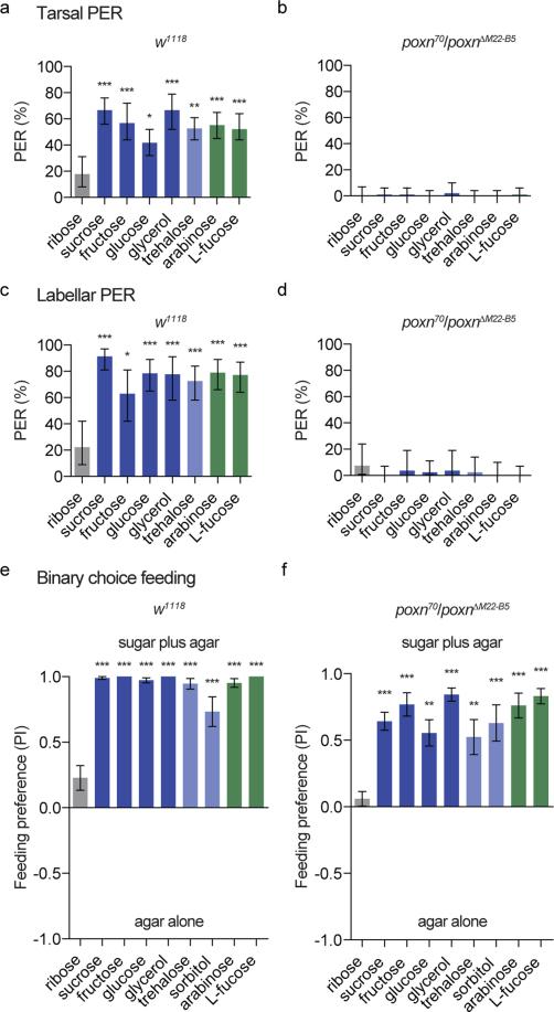
Given that poxn mutants have functional pharyngeal sweet taste, but apparently lack all peripheral sweet taste sensation, we asked whether pharyngeal taste is sufficient to direct consumption of a variety of sweet compounds. First, we verified that poxn null mutants lack peripheral sweet taste responses by performing the proboscis extension reflex (PER)7,33,34. We used ribose as a negative control stimulus, as it evokes no significant response from L-type taste sensilla on the labellum4. However, our observation that ribose elicits a mean PER response of ~20% in control flies suggests that it may stimulate some appetitive taste neurons at a low level (Figure 4). Alternatively, these responses could be the result of osmotic differences between the ribose solution and water used to water satiate the flies prior to testing35,36. Nevertheless, control flies (w1118) showed robust PER to all sweet compounds tested, responding at frequencies significantly higher than those elicited by ribose (Figure 4a,c). By contrast, PER was entirely abolished in poxn mutants following stimulation of either the labellum or the tarsal segment of the legs (Figure 4b,d).
Figure 4.
poxn null mutants lack peripheral taste responses but prefer sweet compounds (a-d) PER responses of w1118 (a,c) and poxnΔM22-B5/poxn70 null mutant (b,d) flies following stimulation of the tarsi (a,b) or labellum (c,d) with the indicated compounds. Values represent percentage of stimulations resulting in a positive response; error bars show 95% binomial confidence interval, and asterisks indicate significant difference from ribose stimulation: * p < 0.05, ** p < 0.01, *** p < 0.001 by Fisher's exact test. n = 17- 50 flies for w1118 tarsal PER, n = 17-33 flies for poxn tarsal PER, n = 9-19 flies for w1118 labellar PER, and n = 9-17 flies for poxn labellar PER. (e,f) Preference of w1118 (e) and poxnΔM22-B5/poxn70 null mutant flies (f) for 100 mM solutions of the indicated compounds in 1% agar versus agar alone. Values represent mean +/− s.e.m. for n = 10 groups of 10 flies each, with independent replicates performed over at least 2 days. Asterisks indicate significant difference from ribose preference by one-way ANOVA with Bonferroni correction for multiple comparisons: **p < 0.01, ***p < 0.001.
Next, we subjected poxn mutants and controls to a variation of the binary feeding choice paradigm in which groups of food-deprived flies were allowed to feed for two hours on drops of 1% agar with or without a given test compound. Again we used ribose as a negative control because it has little to no taste response or nutritional value4,26. Control flies showed a small preference for ribose (PI = 0.23 +/− 0.09) over plain agar, which may be due to weak taste responses or the osmolarity differences discussed above (Figure 4e). This effect was reduced in poxn mutants (PI = 0.06 +/− 0.06).
As expected, poxn mutants and controls displayed robust preference for sucrose, fructose, glucose, and glycerol (Figure 4f). All of these compounds are nutritional23,25,26 and activate Gr43a+ pharyngeal GRNs (Figure 2e). The potential role of nutritional content in guiding feeding decisions is supported by the observation that poxn mutants preferred two compounds, trehalose and sorbitol, that are caloric but did not stimulate pharyngeal GRNs in our experiments (Figure 4f). Importantly, poxn mutants also strongly preferred arabinose and L-fucose, both of which stimulate sugar taste but offer no caloric value4,23,25,26. These data strongly suggest that activation of pharyngeal taste neurons is sufficient to drive consumption behaviour, and that this activation accounts for at least part of the previously reported preference of poxn mutants for caloric sugars21,22.
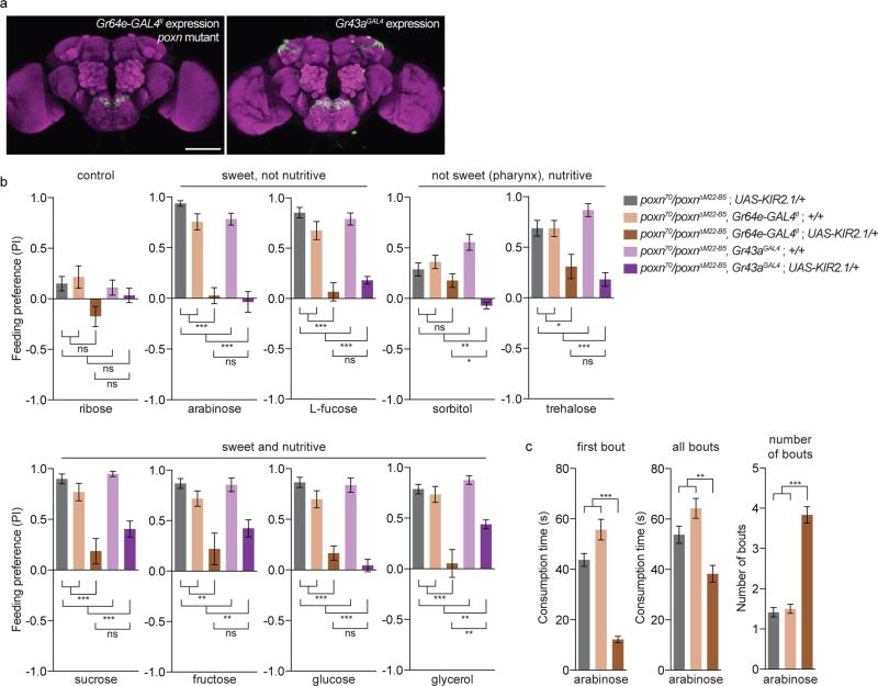
To further examine the role of pharyngeal taste in driving the preference of poxn mutants for sweet compounds, we silenced Gr64e+ pharyngeal GRNs in the poxn mutant background through expression of the inward rectifying potassium channel KIR2.137. Importantly, the insertion of Gr64e-GAL4 used (Gr64e-GAL4II) lacks expression in the taste pegs (Figure 5a), meaning that silencing specifically affects pharyngeal GRNs. Silencing of Gr64e+ GRN function in poxn mutants resulted in a complete loss of preference for arabinose and L-fucose over agar alone, indicating that Gr64e+ pharyngeal GRNs are necessary for the preference for non-caloric sweet sugars in poxn mutants (Figure 5b). This suggests that Gr64e-GAL4 likely labels the complete set of pharyngeal sweet GRNs, and that poxn mutants lacking Gr64e+ pharyngeal GRN function may be sweet-blind.
Figure 5.
Pharyngeal GRNs are necessary for the preference of poxn mutants for sweet compounds (a) Immunofluorescence of anti-GFP (green) and nc82 (magenta), showing expression of the Gr64e-GAL4II and Gr43aGAL4 drivers used in the behavioural experiments shown. Gr64e-GAL4 is shown in a poxn null mutant background, while Gr43aGAL4 is in a poxn/+ heterozygous background. Scale bars are 100 μm. (b) Preference of indicated genotypes for 100 mM solutions of the specified compounds in 1% agar (positive) versus agar alone (negative). (c) Temporal consumption characteristics of the indicated genotypes in response to stimulation with 50 mM arabinose. Values represent mean +/− s.e.m. for n = 10 groups of 10 flies each in b and n = 29-60 flies in c, with independent replicates performed over at least 2 days. Asterisks indicate significant difference by one-way ANOVA with Bonferroni correction for multiple comparisons: *p < 0.05, **p < 0.01, ***p < 0.001, ns = not significant.
Since poxn, Gr64e-silenced flies lack both peripheral and identified pharyngeal sweet taste, we used them to re-evaluate the role of post-ingestive nutrient sensing in driving preference for a number of sugars. Silencing of pharyngeal taste caused only a mild, insignificant decrease in the preference of poxn mutant flies for sorbitol (Figure 5b), consistent with previously reported post-ingestive mechanisms promoting consumption of this compound, and its reported tastelessness13. However, it's worth noting that the preference for sorbitol in these experiments was weak, so it is difficult to confidently ascribe a taste-independent effect. In contrast to sorbitol, the preference for trehalose was significantly reduced following silencing of Gr64e+ pharyngeal GRNs, suggesting that this sugar may stimulate pharyngeal GRNs at a level below the sensitivity of our calcium imaging but enough to affect behaviour.
Strikingly, silencing Gr64e+ pharyngeal GRNs dramatically reduced the preference of poxn flies to all four sweet and nutritive compounds tested (Figure 5b). These data suggest that taste is the dominant driver of feeding preference in our short-term binary choice assay, and, in contrast to previous reports21,22, taste-independent post-ingestive sugar sensing has little, if any, effect on consumption behaviour in this context.
To further probe the factors influencing sugar preference, we repeated the silencing experiment using a GAL4 knock-in allele of Gr43a that is expressed in the same complement of pharyngeal GRNs as Gr64e-GAL4II but also shows additional expression in identified sugar-sensing neurons in the protocerebrum and a population of neurons in the proventricular ganglion13. Consistent with a role for Gr43a+ brain neurons in promoting the ingestion of sorbitol, we observed a complete lack of sorbitol preference in Gr43a-silenced poxn mutants (Figure 5b). Notably, this was a significant reduction compared to both genetic controls and Gr64e-silenced flies. Interestingly, while the behavioural assay used may lack the resolution to tease out small differences between Gr64e and Gr43a silencing, we observed a trend towards increased preference for sweet and nutritive sugars in Gr43a-silenced mutants compared to Gr64e-silenced flies. This trend, which was insignificant for sucrose and fructose but significant for glycerol, could reflect weaker silencing with the Gr43aGAL4 driver, a difference in genetic background, or the previously reported role for Gr43a+ brain neurons in promoting feeding termination of nutritive sweet sugars in some contexts13. Nevertheless, overall our silencing results strongly support the conclusion that short-term sugar preferences are primarily taste-mediated, even in poxn mutants lacking external gustatory sensilla.
Pharyngeal sweet GRNs sustain ingestion
Based on the anatomical position of pharyngeal GRNs, we wondered whether they might affect food preference by preferentially sustaining the ingestion of sweet compounds. To test this we subjected poxn, Gr64e-silenced flies to a temporal consumption assay38 and compared their behaviour to that of poxn controls with intact pharyngeal GRN function (Figure 5c). We found that poxn flies with silenced pharyngeal GRNs displayed a dramatic reduction in the duration of consumption during the first bout of feeding on a solution of sweet, non-nutritive arabinose compared to non-silenced controls, as well as a reduction in total feeding time over multiple bouts and an elevated number of feeding bouts prior to “satiety” (defined here as the refusal to initiate further feeding). These data support the notion that pharyngeal GRNs indeed function to sustain ingestion of sweet, appetitive food sources.
Discussion
Much is known about sugar sensing through peripheral sweet taste neurons in the fly's legs and labellum, and there is growing interest in mechanisms that sense dietary sugars following ingestion39,40. Sugar sensing by the pharyngeal sense organs, however, has remained virtually unexplored due to their inaccessibility to electrophysiology and the lack of genetic tools to specifically manipulate their function. This represents an important gap in our understanding of how sugar feeding is regulated, since the pharyngeal sense organs must operate following the initiation of feeding, but prior to ingestion. They are therefore poised to provide feedback during feeding, contributing to the ongoing decision of whether to maintain or terminate ingestion.
Our calcium imaging data demonstrated that the receptive fields of pharyngeal sweet GRNs are in line with predictions based on receptor expression and specificities4,5,10,11,13,15,41. All putative pharyngeal sweet GRNs co-express Gr43a and Gr64e, which are receptors tuned to fructose and glycerol, respectively, and we observed robust calcium responses to those two compounds. Expression of Gr61a in the LSO likely accounts for the response to glucose, since Gr61a-dependent glucose responses are seen in tarsal GRNs lacking Gr5a41. Likewise, sucrose and L-fucose responses can be accounted for by the expression of Gr64a in the LSO4,5,10. Notably, we did not observe responses to trehalose, consistent with the lack of Gr5a expression, which is necessary for trehalose responses in the labellum4. However, our behavioural data suggested that Gr64e+ pharyngeal GRNs mediate some attraction to trehalose. This apparent discrepancy could be explained if trehalose excites sweet pharyngeal GRNs at a level too low to observe significant calcium responses in our preparation, a possibility supported by previous reports that overexpression of Gr43a or Gr64e confers trehalose sensitivity to tarsal GRNs or ab1C olfactory receptor neurons, respectively5,13.
Using poxn mutants lacking peripheral taste, we demonstrated that pharyngeal sweet GRNs are sufficient to drive preference for sweet compounds in binary choice feeding assays. These results indicate that appetitive taste is not absolutely necessary for feeding initiation, and that flies must “sample” potential food sources in the absence of sweet taste input from the legs and labellum. Moreover, we provide evidence that activation of pharyngeal sweet GRNs prolongs feeding by providing a positive feedback signal to sustain ingestion. This proposed role for pharyngeal GRN function is also consistent with their physiology, which exhibits prolonged activation during consumption of sweet compounds compared to previously reported responses in the labellum6,31. It is likely that this prolonged activation functions to maintain ingestion during feeding bouts lasting up to several seconds. In the future, it will be interesting to examine whether pharyngeal sweet taste plays any specific roles outside of feeding. For example, pharyngeal bitter GRNs appear to be important in selection of egg laying substrates42.
A key component of our work is the demonstration that poxn mutants have a functional pharyngeal taste system that is critical in guiding their preference for sweet compounds, which therefore precludes their experimental use as “taste-blind” flies. The behavioural preference we observed of poxn mutant flies for the non-nutritional sweetners L-fucose and arabinose contrasts with the conclusion reached by Dus and colleagues (2011), who performed a similar experiment with the artificial sweetner sucralose. One possible explanation for this difference is that arabinose and L-fucose may activate pharyngeal sweet GRNs more potently than sucralose. Another possible source of the discrepancy is that Dus and colleagues analyzed their feeding data by plotting the total proportion of flies to eat each option in the binary choice assay, while we analyzed relative preference between the options. Nevertheless, the strong dependence of poxn mutants on Gr64e+ GRNs for preferred consumption of both nutritive and non-nutritive sweeteners demonstrates that the majority of the preference of these mutants for sweet compounds is mediated by pharyngeal taste sensitivity.
By silencing Gr64e+ GRNs in a poxn mutant background, we created for the first time a fly that may completely lack sweet taste, allowing one to reevaluate the taste-independent role for nutrient sensing mechanisms in behaviour. While we did observe some evidence for taste-independent selection of nutritive carbohydrates, the observed effect was much weaker than previously suggested21,22. Although ample additional evidence supports the existence of taste-independent carbohydrate sensing13,23-26, its specific role in regulating feeding remains unclear. Why do we observe such weak preference of putatively sweet-blind flies for nutritive sugars? One possibility is the particular behavioural assay used, which operates over only two hours. We have previously reported increasing effects of caloric content on feeding preferences over the course of a 16-hour assay26. It will be instructive to reevaluate the feeding behaviour of poxn, sweet GRN-silenced flies over longer time periods using different behavioural paradigms.
Our newly established putatively sweet-blind flies also afforded the opportunity to examine potential nutrient sensing mechanisms in a taste-independent context. For example, by comparing Gr43a+-silenced to Gr64e+-silenced flies in a poxn mutant background, we were able to observe effects for non-taste Gr43a+ populations that are largely consistent with those previously reported13. Further evaluation of these and other putative nutrient-sensing cell populations in a sweet taste-blind background will continue to shed light on the apparently complex interactions between the taste-dependent and -independent mechanisms that ultimately guide critical feeding decisions.
Methods
Fly stocks
Flies were raised on standard cornmeal fly food at 25°C and 70% relative humidity. The following fly lines were used: Gr5a-GAL4, Gr43a-GAL4, Gr64a-GAL4, Gr61a-GAL4, Gr64c-GAL4, Gr64d-GAL4, and Gr64e-GAL4 (ref16); Gr64fLexA and Gr43aGAL4 (ref13); UAS-GCaMP3 (ref43); UAS-KIR2.1 (ref37); UAS-TdTomato and UAS-GFP (Bloomington stock center); poxnΔM22-B5 (ref44); poxn70 (ref 28).
Immunohistochemistry
Immunohistochemistry was carried out as previously described33. The primary antibodies used were rabbit anti-GFP (1:1000; Invitrogen) and mouse nc82 (1:50, DSHB). The secondary antibodies used were goat anti-rabbit Alexa-488 and goat anti-mouse Alexa-568 (Invitrogen). Images are maximum intensity projections of confocal z-stacks acquired using a Leica SP5 II confocal microscope with 25x water immersion objective.
Gr expression mapping
Expression of sweet Gr promoter-GAL4 lines was mapped in the three pharyngeal sense organs by visualizing UAS-mCD8::GFP fluorescence in live tissue. Proboscis tissue was dissected and mounted in 80% glycerol in 1X PBS before imaging with a Leica SP5 laser scanning confocal microscope. For double driver analysis, the UAS-mCD8::GFP transgene was under the control of two different Gr promoter-Gal4 transgenes and the number of GFP-labeled neurons were compared to those in flies containing a single Gal4 driver alone.
GCaMP imaging
Female flies aged 2-12 days were used for calcium imaging. To prepare flies for imaging, they were briefly anesthetized and all legs were removed to allow unobstructed access to the proboscis. Using a customized chamber, each fly was mounted by inserting the cervix into individual collars. To further immobilize the head, nail polish was applied in a thin layer to seal the head to the chamber. Melted wax was applied using a modified dental waxer to adhere the fully extended proboscis to the chamber rim. The antennae and associated cuticle covering the SEZ were removed and AHL buffer with ribose45 was immediately injected into the preparation to cover the exposed brain. A coverslip was inserted into the chamber to keep the proboscis dry and separated from the bath solution.
GCaMP3 fluorescence was viewed with a Leica SP5 II laser scanning confocal microscope equipped with a tandem scanner and HyD detector. The relevant area of the SEZ was visualized using the 25x water objective with an electronic zoom of 8. Images were acquired at a speed of 8000 lines per second with a line average of 4, resulting in collection time of 60 ms per frame at a resolution of 512 × 200 pixels. The pinhole was opened to 2.68 AU. Stimuli were applied to the proboscis using a pulled glass pipette, and flies were allowed to ingest solutions during imaging. The maximum change in fluorescence (ΔF/F) was calculated as the peak intensity change divided by the average intensity over 10 frames prior to stimulation.
Behavioural assays
For PER, adult female flies were aged 3-10 days and starved on 1% agar at room temperature (~22°C) for 24 hours before testing. For tarsal PER, flies were mounted on glass slides using nail polish. For labellar PER, flies were placed inside a pipette tip cut to size so that only the head was exposed. Flies were then sealed into the tube with tape, and then adhered to a glass slide with double-sided tape. Flies were allowed 1-2 hours to recover before testing began. Flies were stimulated with water on their front tarsi or labella for tarsal and labellar PER, respectively, and allowed to drink until satiated. Each fly was then stimulated with a tastant on either the tarsi or labella, and responses to each of three trials were recorded. Flies were provided with water between each tastant. All stimuli were delivered with a 1 mL syringe attached to a 20 μL pipette tip. For statistical purposes, each trial was treated as an independent unit of analysis.
Binary choice preference tests were performed similarly to previous descriptions 16,21,46. Female flies aged 3-8 days were sorted into groups of 10 at least two days prior to the experiment, and starved on 1% agar at room temperature (~22°C) for 24 hours before testing. For the assay, flies were transferred into standard vials containing six 10 μL dots of agar that alternated in color. The agar substrates were 1% agar with or without the test stimulus at a concentration of 100 mM. Each choice contained either 0.125 mg/mL blue (Erioglaucine, FD&C Blue#1) or 0.5 mg/mL red (Amaranth, FD&C Red#2) dye, and half the replicates for each stimulus were done with the dyes swapped to control for any dye preference. Flies were allowed to feed for 2 hours in the dark at 25°C and then frozen and scored for abdomen color. Preference index (PI) for sugar was calculated as ((# of flies labeled with the stimulus color) – (# of flies labeled with the plain agar color))/(total number of flies that fed).
The temporal consumption assay was performed on flies deprived of food for 24 hours38. As described above for PER, flies were mounted on glass slides using nail polish and allowed to recover for 1-2 hours in a humidified chamber. Water satiated flies were then offered 50 mM arabinose on their labella. Once they initiated feeding, the time between starting and stopping their first feeding bout was recorded (first bout length). The fly was then offered arabinose again and if they failed to reinitiate feeding for three consecutive stimulations, the assay was terminated for that fly. If flies began a new bout of feeding upon stimulation, the time of the subsequent bouts was added to the first bout to determine total consumption time, and number of bouts was recorded.
Statistical analyses
For PER analyses, the 95% binomial confidence interval was calculated using JavaStat (http://statpages.org/confint.html). Fisher's exact tests were calculated using Graphpad QuickCalcs (http://www.graphpad.com/quickcalcs/). All other statistical tests performed using GraphPad Prism 6 or SPSS software. No statistical test was used to predetermine sample size. Sample size ranges were chosen based on previously published examples of the same assays.
Acknowledgements
We thank Adriana Lomeli for assistance with fly crosses; Kristin Scott, Greg Suh, and the Bloomington stock center for fly strains; and Makoto Hiroi for designing and building the calcium imaging chamber. This work was funded by the Canadian Institutes of Health Research (CIHR) operating grant MOP-114934 (M.D.G), Natural Sciences and Engineering Research Council (NSERC) Discovery grant RGPIN 402184-11 (M.D.G.), Canadian Foundation for Innovation (CFI) grant 27290 (M.D.G.), Whitehall Foundation research grant 2010-12-42 (A.D.), National Institutes of Health research project grant R01DC013587 (A.D.), a CIHR New Investigator Award (M.D.G.), an NSERC PGS-D3 scholarship (E.E.L.), and a UBC Dean of Science Summer Research Position (A.Y.J.).
Footnotes
Author contributions
Y-C.C. performed the wild-type Gr-GAL4 expression analysis. E.E.L. and M.D.G. performed the experiments to examine Gr-GAL4 expression in poxn mutants and the GCaMP imaging. E.E.L. performed proboscis extension experiments and the majority of binary feeding assays. A.Y.J. performed initial binary feeding assays. E.E.L. and Y-C.C. performed temporal consumption assays. E.E.L., Y-C.C., A.Y.J., A.D., and M.D.G. planned experiments and analyzed the data. M.D.G. and A.D. conceived of and supervised the project. M.D.G. wrote the paper with A.D., E.E.L., and Y-C.C.
Competing financial interests
The authors declare no competing financial interests.
References
- 1.Stocker R. The organization of the chemosensory system in Drosophila melanogaster: a review. Cell Tissue Res. 1994;275:3–26. doi: 10.1007/BF00305372. [DOI] [PubMed] [Google Scholar]
- 2.Hiroi M, Meunier N, Marion-Poll F, Tanimura T. Two antagonistic gustatory receptor neurons responding to sweet-salty and bitter taste in Drosophila. J Neurobiol. 2004;61:333–342. doi: 10.1002/neu.20063. [DOI] [PubMed] [Google Scholar]
- 3.Wang Z, Singhvi A, Kong P, Scott K. Taste representations in the Drosophila brain. Cell. 2004;117:981–991. doi: 10.1016/j.cell.2004.06.011. [DOI] [PubMed] [Google Scholar]
- 4.Dahanukar A, Lei Y, Kwon J, Carlson J. Two Gr genes underlie sugar reception in Drosophila. Neuron. 2007;56:503–516. doi: 10.1016/j.neuron.2007.10.024. [DOI] [PMC free article] [PubMed] [Google Scholar]
- 5.Freeman EG, Wisotsky Z, Dahanukar A. Detection of sweet tastants by a conserved group of insect gustatory receptors. Proc Natl Acad Sci USA. 2014;111:1598–1603. doi: 10.1073/pnas.1311724111. [DOI] [PMC free article] [PubMed] [Google Scholar]
- 6.Marella S, et al. Imaging taste responses in the fly brain reveals a functional map of taste category and behavior. Neuron. 2006;49:285–295. doi: 10.1016/j.neuron.2005.11.037. [DOI] [PubMed] [Google Scholar]
- 7.Dethier VG. The hungry fly: A physiological study of the behavior associated with feeding. Harvard University Press; 1976. [Google Scholar]
- 8.Nayak SV, Singh RN. Sensilla on the tarsal segments and mouthparts of adult Drosophila melanogaster meigen (Diptera: Drosophilidae). International Journal of Insect Morphology and Embryology. 1983;12:273–291. [Google Scholar]
- 9.Gendre N. Integration of complex larval chemosensory organs into the adult nervous system of Drosophila. Development. 2004;131:83–92. doi: 10.1242/dev.00879. [DOI] [PubMed] [Google Scholar]
- 10.Jiao Y, Moon S, Montell C. A Drosophila gustatory receptor required for the responses to sucrose, glucose, and maltose identified by mRNA tagging. Proc Natl Acad Sci U S A. 2007;104:14110–14115. doi: 10.1073/pnas.0702421104. [DOI] [PMC free article] [PubMed] [Google Scholar]
- 11.Slone J, Daniels J, Amrein H. Sugar receptors in Drosophila. Curr Biol. 2007;17:1809–1816. doi: 10.1016/j.cub.2007.09.027. [DOI] [PMC free article] [PubMed] [Google Scholar]
- 12.Scott K, et al. A chemosensory gene family encoding candidate gustatory and olfactory receptors in Drosophila. Cell. 2001;104:661–673. doi: 10.1016/s0092-8674(01)00263-x. [DOI] [PubMed] [Google Scholar]
- 13.Miyamoto T, Slone J, Song X, Amrein H. A Fructose Receptor Functions as a Nutrient Sensor in the Drosophila Brain. Cell. 2012;151:1113–1125. doi: 10.1016/j.cell.2012.10.024. [DOI] [PMC free article] [PubMed] [Google Scholar]
- 14.Kent LB, Robertson HM. Evolution of the sugar receptors in insects. BMC Evol Biol. 2009;9:41. doi: 10.1186/1471-2148-9-41. [DOI] [PMC free article] [PubMed] [Google Scholar]
- 15.Jiao Y, Moon SJ, Wang X, Ren Q, Montell C. Gr64f Is Required in Combination with Other Gustatory Receptors for Sugar Detection in Drosophila. Current Biology. 2008;18:1797–1801. doi: 10.1016/j.cub.2008.10.009. [DOI] [PMC free article] [PubMed] [Google Scholar]
- 16.Weiss LA, Dahanukar A, Kwon JY, Banerjee D, Carlson JR. The Molecular and Cellular Basis of Bitter Taste in Drosophila. Neuron. 2011;69:258–272. doi: 10.1016/j.neuron.2011.01.001. [DOI] [PMC free article] [PubMed] [Google Scholar]
- 17.Ling F, Dahanukar A, Weiss LA, Kwon JY, Carlson JR. The molecular and cellular basis of taste coding in the legs of Drosophila. J Neurosci. 2014;34:7148–7164. doi: 10.1523/JNEUROSCI.0649-14.2014. [DOI] [PMC free article] [PubMed] [Google Scholar]
- 18.Sato K, Tanaka K, Touhara K. Sugar-regulated cation channel formed by an insect gustatory receptor. Proc Natl Acad Sci USA. 2011;108:11680–11685. doi: 10.1073/pnas.1019622108. [DOI] [PMC free article] [PubMed] [Google Scholar]
- 19.De Araujo IE, et al. Food Reward in the Absence of Taste Receptor Signaling. Neuron. 2008;57:930–941. doi: 10.1016/j.neuron.2008.01.032. [DOI] [PubMed] [Google Scholar]
- 20.Sclafani A. Oral, post-oral and genetic interactions in sweet appetite. Physiology & Behavior. 2006;89:525–530. doi: 10.1016/j.physbeh.2006.03.021. [DOI] [PMC free article] [PubMed] [Google Scholar]
- 21.Dus M, Min S, Keene AC, Lee GY, Suh GSB. Taste-independent detection of the caloric content of sugar in Drosophila. Proc Natl Acad Sci USA. 2011;108:11644–11649. doi: 10.1073/pnas.1017096108. [DOI] [PMC free article] [PubMed] [Google Scholar]
- 22.Dus M, Ai M, Suh GSB. Taste-independent nutrient selection is mediated by a brain-specific Na+ /solute co-transporter in Drosophila. Nat Neurosci. 2013;16:526–528. doi: 10.1038/nn.3372. [DOI] [PMC free article] [PubMed] [Google Scholar]
- 23.Burke CJ, Waddell S. Remembering nutrient quality of sugar in Drosophila. Curr Biol. 2011;21:746–750. doi: 10.1016/j.cub.2011.03.032. [DOI] [PMC free article] [PubMed] [Google Scholar]
- 24.Burke CJ, et al. Layered reward signalling through octopamine and dopamine in Drosophila. Nature. 2012;492:433–437. doi: 10.1038/nature11614. [DOI] [PMC free article] [PubMed] [Google Scholar]
- 25.Fujita M, Tanimura T. Drosophila evaluates and learns the nutritional value of sugars. Curr Biol. 2011;21:751–755. doi: 10.1016/j.cub.2011.03.058. [DOI] [PubMed] [Google Scholar]
- 26.Stafford JW, Lynd KM, Jung AY, Gordon MD. Integration of Taste and Calorie Sensing in Drosophila. J Neurosci. 2012;32:14767–14774. doi: 10.1523/JNEUROSCI.1887-12.2012. [DOI] [PMC free article] [PubMed] [Google Scholar]
- 27.Nottebohm E, et al. The gene poxn controls different steps of the formation of chemosensory organs in Drosophila. Neuron. 1994;12:25–34. doi: 10.1016/0896-6273(94)90149-x. [DOI] [PubMed] [Google Scholar]
- 28.Awasaki T, Kimura K. pox-neuro is required for development of chemosensory bristles in Drosophila. J Neurobiol. 1997;32:707–721. doi: 10.1002/(sici)1097-4695(19970620)32:7<707::aid-neu6>3.0.co;2-8. [DOI] [PubMed] [Google Scholar]
- 29.Galindo K, Smith DP. A large family of divergent Drosophila odorant- binding proteins expressed in gustatory and olfactory sensilla. Genetics. 2001;159:1059–1072. doi: 10.1093/genetics/159.3.1059. [DOI] [PMC free article] [PubMed] [Google Scholar]
- 30.Wisotsky Z, Medina A, Freeman E, Dahanukar A. Evolutionary differences in food preference rely on Gr64e, a receptor for glycerol. Nat Neurosci. 2011;14:1534–1541. doi: 10.1038/nn.2944. [DOI] [PubMed] [Google Scholar]
- 31.Chu B, Chui V, Mann K, Gordon MD. Presynaptic gain control drives sweet and bitter taste integration in Drosophila. Curr Biol. 2014;24:1978–1984. doi: 10.1016/j.cub.2014.07.020. [DOI] [PubMed] [Google Scholar]
- 32.Fischler W, Kong P, Marella S, Scott K. The detection of carbonation by the Drosophila gustatory system. Nature. 2007;448:1054–1057. doi: 10.1038/nature06101. [DOI] [PubMed] [Google Scholar]
- 33.Gordon MD, Scott K. Motor Control in a Drosophila Taste Circuit. Neuron. 2009;61:373–384. doi: 10.1016/j.neuron.2008.12.033. [DOI] [PMC free article] [PubMed] [Google Scholar]
- 34.Rajashekhar KP, Singh RN. Organization of motor neurons innervating the proboscis musculature in Drosophila melanogaster meigen (diptera: Drosophilidae). International Journal of Insect Morphology and Embryology. 1994;23:225–242. [Google Scholar]
- 35.Cameron P, Hiroi M, Ngai J, Scott K. The molecular basis for water taste in Drosophila. Nature. 2010;465:91–95. doi: 10.1038/nature09011. [DOI] [PMC free article] [PubMed] [Google Scholar]
- 36.Chen Z, Wang Q, Wang Z. The Amiloride-Sensitive Epithelial Na+ Channel PPK28 Is Essential for Drosophila Gustatory Water Reception. J Neurosci. 2010;30:6247–6252. doi: 10.1523/JNEUROSCI.0627-10.2010. [DOI] [PMC free article] [PubMed] [Google Scholar]
- 37.Baines R, Uhler J, Thompson A, Sweeney S, Bate M. Altered electrical properties in Drosophila neurons developing without synaptic transmission. J Neurosci. 2001;21:1523–1531. doi: 10.1523/JNEUROSCI.21-05-01523.2001. [DOI] [PMC free article] [PubMed] [Google Scholar]
- 38.Pool A-H, et al. Four GABAergic InterneuronsImpose Feeding Restraint in Drosophila. Neuron. 2014;83:164–177. doi: 10.1016/j.neuron.2014.05.006. [DOI] [PMC free article] [PubMed] [Google Scholar]
- 39.Itskov PM, Ribeiro C. The dilemmas of the gourmet fly: the molecular and neuronal mechanisms of feeding and nutrient decision making in Drosophila. Front Neurosci. 2013;7:12. doi: 10.3389/fnins.2013.00012. [DOI] [PMC free article] [PubMed] [Google Scholar]
- 40.Miyamoto T, Wright G, Amrein H. Nutrient sensors. Current Biology. 2013;23:R369–R373. doi: 10.1016/j.cub.2013.04.002. [DOI] [PMC free article] [PubMed] [Google Scholar]
- 41.Miyamoto T, Chen Y, Slone J, Amrein H. Identification of a Drosophila Glucose Receptor Using Ca2+ Imaging of Single Chemosensory Neurons. PLoS ONE. 2013;8:e56304. doi: 10.1371/journal.pone.0056304. [DOI] [PMC free article] [PubMed] [Google Scholar]
- 42.Joseph RM, Heberlein U. Tissue-Specific Activation of a Single Gustatory Receptor Produces Opposing Behavioral Responses in Drosophila. Genetics. 2012;192:521–532. doi: 10.1534/genetics.112.142455. [DOI] [PMC free article] [PubMed] [Google Scholar]
- 43.Tian L, et al. Imaging neural activity in worms, flies and mice with improved GCaMP calcium indicators. Nat Meth. 2009;6:875–881. doi: 10.1038/nmeth.1398. [DOI] [PMC free article] [PubMed] [Google Scholar]
- 44.Boll W. The Drosophila Pox neuro gene: control of male courtship behavior and fertility as revealed by a complete dissection of all enhancers. Development. 2002;129:5667–5681. doi: 10.1242/dev.00157. [DOI] [PubMed] [Google Scholar]
- 45.Liu C, et al. A subset of dopamine neurons signals reward for odour memory in Drosophila. Nature. 2012;488:512–516. doi: 10.1038/nature11304. [DOI] [PubMed] [Google Scholar]
- 46.Ribeiro C, Dickson BJ. Sex Peptide Receptor and Neuronal TOR/S6K Signaling Modulate Nutrient Balancing in Drosophila. Current Biology. 2010;20:1000–1005. doi: 10.1016/j.cub.2010.03.061. [DOI] [PubMed] [Google Scholar]


