



Abstract
Reported herein is the use of proximity-induced non-covalent energy transfer for the detection of medium-sized polycyclic aromatic hydrocarbons (PAHs). This energy transfer occurs within the cavity of γ-cyclodextrin in various aqueous environments, including human plasma and coconut water. Highly efficient energy transfer was observed, and the efficiency of the energy transfer is independent of the concentration of γ-cyclodextrin used, demonstrating the importance of hydrophobic binding in facilitating such energy transfer. Low limits of detection were also observed for many of the PAHs investigated, which is promising for the development of fluorescence-based detection schemes.
INTRODUCTION
The accurate, sensitive, and selective detection of polycyclic aromatic hydrocarbons (PAHs) remains a crucial research objective, as many of these compounds are known or suspected carcinogens (1), environmental pollutants (2), and endocrine disruptors (3). They are known components of crude and processed oil (4), and have been found in the blood (5) and breast milk (6) of populations living in oil spill affected areas, and in seafood from the Gulf of Mexico following the Deepwater Horizon oil spill (7).
Despite the widespread prevalence and known toxicity of these compounds, current detection methods often have significant limitations, including the requirement for tedious sample preparation prior to analysis (8), or the inability to accurately distinguish structurally similar PAHs with widely disparate toxicities (9). Previous research in our group introduced a fundamentally new approach for the detection of PAHs, which relies on using the PAHs as energy donors in combination with high quantum yield fluorophore acceptors including BODIPY (compound 10) and Rhodamine 6G (compound 11) (Figure 1) (10–14). Energy transfer from the PAH to the fluorophore occurs when both are bound in the cavity of commercially available, non-toxic γ-cyclodextrin, leading to a new, brightly fluorescent signal in the presence of the PAH of interest (Figure 2). This energy transfer based detection has been used in complex biological fluids (14) and with PAHs that have been extracted from crude oil samples (13).
Figure 1.
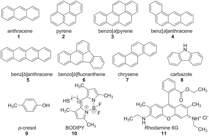
Structures of all analytes and fluorophores targeted for investigation.
Figure 2.

Schematic illustration of cyclodextrin-promoted energy transfer.
Previous research in our group focused predominantly on lower molecular weight PAHs, including anthracene, pyrene, and benzo[a]pyrene, with exceptionally efficient energy transfer observed from pyrene 2 and benzo[a]pyrene 3 to BODIPY 10. While benzo[a]pyrene is highly toxic (15), many of the lower molecular weight homologs (naphthalene, anthracene, and pyrene) have greatly reduced toxicities (16). Higher molecular weight PAHs, including benz[a]anthracene, benz[b]anthracene, benzo(b)fluoranthene, and chrysene (compounds 4–7), are significantly more toxic to a wide variety of organisms (17,18). The detection of higher molecular weight PAHs via cyclodextrin-promoted energy transfer would have significant potential utility in PAH detection in complex environments and in environmental remediation efforts. Other known toxicants include heterocyclic aromatic compounds such as carbazole (compound 8) (19) and environmental toxicants such as p-cresol (compound 9) (20), and the ability to use cyclodextrin-based energy transfer for the detection of these classes of toxicants would provide significant operational advantages. Reported herein are the results of our efforts towards achieving these goals: detecting higher molecular weight PAHs, heteroaromatic toxicants, and low molecular weight environmental toxicants via cyclodextrin-promoted, non-covalent energy transfer.
EXPERIMENTAL
Materials and Methods
All chemicals were obtained from Sigma Aldrich Chemical Company and were used as received. Compound 10 was synthesized following literature-reported procedures (21). Human plasma was obtained from Innovative Technologies. Coconut water was obtained from CVS Pharmacy in Kingston, Rhode Island. 1H NMR spectra were recorded on a Bruker 300 MHz spectrometer. UV-Visible spectra were recorded on an Agilent 8453 spectrometer equipped with a photodiode array detector. Fluorescence spectra were recorded on a Shimadzu RF-5301PC spectrophotofluorimeter.
Energy Transfer Experimental Procedures
For full experimental details and copies of all spectra, see the Electronic Supporting Information.
Energy transfer experiments were conducted as follows.
All analytes (4–9) and fluorophores (10, 11) were dissolved in tetrahydrofuran (THF). Small amounts of these THF solutions were added to a 10 mM aqueous solution of γ-cyclodextrin in phosphate buffered saline (PBS) buffered at pH 7.4. Following thorough mixing, the fluorescence emission of the solution was recorded from excitation at the analyte’s excitation wavelength and from excitation at the fluorophore’s excitation wavelength, and the percent energy transfer efficiencies were calculated according to Equation (1):
| (1) |
where IDA is the integrated emission of the fluorophore from analyte excitation and IA is the integrated fluorophore emission from direct fluorophore excitation.
Two control experiments were conducted for each analyte-fluorophore combination.
The 10 mM γ-cyclodextrin solution was replaced with a 0 mM solution, and the same procedures were followed, to elucidate the beneficial effect of γ-cyclodextrin on promoting efficient energy transfer.
- The fluorophore emission via excitation at the analyte’s excitation wavelength (but in the absence of the analyte) was measured, and compared to the fluorophore emission via analyte excitation in the presence of the analyte. The analyte comparison ratio was calculated according to Equation (2):
where FA is the fluorophore emission via analyte excitation in the absence of an analyte, and FDA is the fluorophore emission via analyte excitation in the presence of the analyte. Ratios close to 1 indicate a limited contribution of the analyte, which means that the observed energy transfer peaks are likely due to the fluorophore itself having a nonzero absorbance at the analyte’s excitation wavelength, rather than a result of legitimate energy transfer.(2)
Analogous experiments were conducted in phosphate buffer (buffered at pH 7.4 but with less total salt content), coconut water, and human plasma.
Limit of Detection Experimental Procedures
Limits of detection for all analyte-fluorophore combinations were calculated by constructing calibration curves for each combination, with the analyte concentration on the X-axis and the integrated fluorophore emission on the Y-axis. The minimum analyte concentration necessary to elucidate a fluorescence response, defined as the limit of detection, was calculated from these calibration curves following literature-reported procedures (22). The limit of quantification was calculated for each combination as well, defined as the minimum concentration of analyte necessary to elucidate a quantifiable fluorescence signal response.
RESULTS AND DISCUSSION
Previous results from our research group demonstrated highly efficient cyclodextrin-promoted energy transfer for a wide range of toxicants and fluorophores, with greater than 100% energy transfer efficiencies observed in several cases (10–14). These energy transfer efficiencies were particularly efficient for pyrene and benzo[a]pyrene, as well as for highly toxic aromatic amines such as 2-aminofluorene (23), 2-acetylaminofluorene (23), and benzidine (24), in combination with fluorophores 10 and 11. Attempts to extend this cyclodextrin-promoted energy transfer to analytes 4–9 resulted in highly efficient energy transfer in several cases (Table 1), in particular for analytes 4, 6, and 8 in combination with fluorophore 10 (Figure 3). The analyte comparison ratios for each of these cases were substantially less than 1, indicating that the observed energy transfer peaks were due to legitimate energy transfer from the analyte to the fluorophore.
Table 1.
Percent energy transfer efficiencies for analytes 4–9 with fluorophores 10 and 11 in PBSa
| Analyte | Fluorophore | 10 mM γ-CD | 0 mM γ-CD |
|---|---|---|---|
| 4 | 10 | 140.2 | 122.0 |
| 5 | 10 | b | b |
| 6 | 10 | 274.3 | 526.6 |
| 7 | 10 | 25.1 | 22.4 |
| 8 | 10 | 44.8 | 87.5 |
| 9 | 10 | 30.2 | 29.3 |
| 4 | 11 | 6.4 | 6.6 |
| 5 | 11 | b | b |
| 6 | 11 | 25.8 | 34.1 |
| 7 | 11 | 19.4 | 21.1 |
| 8 | 11 | 12.0 | 12.9 |
| 9 | 11 | 24.2 | 27.9 |
All reported results are an average of at least three trials.
Excessive overlap between the analyte and fluorophore emissions prevented efficient integration.
Figure 3.
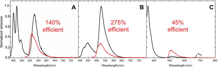
Energy transfer in PBS observed for (A) 4–10; (B) 6–10; and (C) 8–10. The black line represents analyte excitation and the grey line (red in online version) represents direct fluorophore excitation.
A closer examination of the control experiments done in the absence of cyclodextrin indicated that for many of these analyte-fluorophore pairs, the observed energy transfer efficiencies in 10 mM cyclodextrin were unchanged or even lower than energy transfer efficiencies observed in 0 mM cyclodextrin. These surprising results point to the existence of more complex intermolecular phenomena that are occurring, rather than the previously observed ternary complex formation in γ-cyclodextrin (Figure 2).
Cases where the energy transfer efficiencies are unchanged in the presence and absence of cyclodextrin are likely a result of the donor and acceptor interacting via non-covalent, non-cyclodextrin promoted hydrophobic binding, leading to energy transfer even in the absence of cyclodextrin. For such cases, the addition of 10 mM cyclodextrin has no effect on this intermolecular hydrophobic bonding, and no changes in the energy transfer efficiencies are observed. This non-cyclodextrin promoted energy transfer requires exceptionally strong hydrophobic binding to ensure that the donor and acceptor are bound in close proximity without an external host to enforce such proximity. This strong binding is enabled by the larger size of the aromatic PAHs compared to previously studied smaller analogs (25), which increases their hydrophobic surface areas and the strength of the hydrophobic binding observed.
Cases where the energy transfer efficiencies are lower in 10 mM cyclodextrin compared to the cyclodextrin-free solution can be explained by the same non-cyclodextrin dependent hydrophobic binding and energy transfer. In these cases, however, the addition of cyclodextrin causes one of the two energy transfer partners to bind in the cyclodextrin cavity, which disrupts the close proximity of the donor and acceptor. This cyclodextrin-enforced separation of the donor and acceptor leads to a decrease in the observed energy transfer efficiencies.
Experimental evidence for the importance of strong hydrophobic binding in promoting cyclodextrin-free energy transfer comes from a direct comparison of energy transfer efficiencies in phosphate buffered saline and in phosphate buffer (without saline) (Table 2). Energy transfer efficiencies observed in phosphate buffer for compound 6 were markedly decreased compared to the energy transfer efficiencies observed in PBS. Sodium is a known kosmotropic agent (26,27), meaning that it stabilizes hydrophobic interactions such as those required for cyclodextrin-independent energy transfer (28). Because the sodium content of PBS is substantially higher than that of the phosphate buffer, hydrophobic binding is strengthened under those conditions.
Table 2.
A comparison of the energy transfer efficiencies in 10 mM γ-cyclodextrin dissolved in PBS and in phosphate buffera
| Analyte | Fluorophore | ET in PBS | ET in phosphate buffer |
|---|---|---|---|
| 6 | 10 | 274.3 | 169.6 |
| 6 | 11 | 25.8 | 8.2 |
| 8 | 10 | 44.8 | 41.8 |
All reported results are an average of at least three trials.
Moreover, the addition of cyclodextrin had either no effect (for 6–11) or a negative effect (for 6–10) on the observed energy transfer efficiencies in phosphate buffer, again pointing to the significant contribution of non-cyclodextrin dependent intermolecular hydrophobic binding. Even in the absence of added sodium, the strong donor-acceptor hydrophobic binding leads to high energy transfer efficiencies.
Similar behavior was also observed in more complex fluids, including coconut water (which has been used as a plasma surrogate in emergencies) (29,30) and human plasma (31). Coconut water is known to contain a wide variety of small molecule nutrients, including vitamins, sugars, and amino acids (32), as well as macromolecules such as proteins and lipids (33). Many of these analytes can potentially interfere with efficient non-covalent energy transfer. Plasma contains numerous lipids and proteins (34), including proteins that are known to bind highly lipophilic molecules (35). These analytes also have the potential to interfere with the desired energy transfer. Energy transfer from the PAH to the fluorophore was observed in the presence of 10 mM γ-cyclodextrin (Figure 4), but was even more pronounced in the absence of the cyclodextrin (Table 3), pointing to a scenario in which the introduction of the cyclodextrin leads to a disruption of hydrophobic binding between the donor and acceptor.
Figure 4.

Energy transfer from analyte 6 to fluorophore 10 in (A) coconut water with cyclodextrin; (B) coconut water without cyclodextrin; (C) plasma with cyclodextrin; and (D) plasma without cyclodextrin. The black line represents analyte excitation and the grey line (red in online version) represents direct fluorophore excitation.
Table 3.
A comparison of energy transfer efficiencies in the presence and absence of 10 mM γ-cyclodextrin in coconut water and plasmaa
| Analyte | Fluorophore | coconut water
|
plasma
|
||
|---|---|---|---|---|---|
| 10 mM γ-CD | 0 mM γ-CD | 10 mM γ-CD | 0 mM γ-CD | ||
| 6 | 10 | 45.8 | 260.6 | 26.0 | 36.8 |
| 6 | 11 | 5.5 | 8.6 | 8.3 | 17.0 |
| 8 | 10 | 31.7 | 115.5 | 26.3 | 29.1 |
All reported results are an average of at least three trials.
Overall, the medium-sized polycyclic aromatic hydrocarbons demonstrate efficient cyclodextrin-independent energy transfer, but demonstrate a reluctance to form ternary complexes in the γ-cyclodextrin cavity. This reluctance is likely due to the larger dimensions of these analytes (Table 4), which can preclude efficient binding in the cyclodextrin cavity. The interior diameter of γ-cyclodextrin has been reported to be between 7.5 and 8.3 Å, with a cavity height of 7.9 Å (36). Analytes 4–7 have dimensions that are comparable to or larger than the cyclodextrin cavity size, which explains their reluctance to form ternary complexes.
Table 4.
Calculated cavity dimensions for analytes 4–9a
| Analyte | Height | Width |
|---|---|---|
| 4 | 10.7 Å | 7.2 Å |
| 5 | 5.0 Å | 11.6 Å |
| 6 | 7.2 Å | 11.4 Å |
| 7 | 7.2 Å | 10.5 Å |
| 8 | 5.3 Å | 9.0 Å |
| 9 | 5.0 Å | 6.6 Å |
Calculations were performed using Spartan software, semi-empirical PM3-level calculations.
The ability of the analytes to undergo highly efficient cyclodextrin-independent energy transfer means that the development of detection methods based on this energy transfer is possible. The practicality of these detection methods will be greatly enhanced by the ability to detect toxicants at low concentrations (at or below recommended exposure limits). The limits of detection for each analyte-fluorophore pair were calculated, and micromolar concentration limits were determined for a number of the analytes (see ESI for more details). Most interestingly, low detection limits were also calculated for energy transfer in complex fluids such as plasma and coconut water (Table 5), highlighting the potential utility of this method for toxicant detection in complex environments.
Table 5.
Limits of detection for selected donor-acceptor combinations
| Analyte | Fluorophore | PBS | Phosphate Buffer | Coconut Water | Plasma |
|---|---|---|---|---|---|
| 6 | 10 | 18.6 μM | a | 24.7 μM | 44.4 μM |
| 6 | 11 | a | 12.6 μM | 20.6 μM | 44.0 μM |
| 8 | 10 | 54.2 μM | 9.6 μM | a | 46.8 μM |
Attempts to calculate limits of detection for these analyte-fluorophore combinations led to nonsensical values; current efforts are focused on trying to understand these results.
CONCLUSION
In summary, highly efficient energy transfer has been demonstrated from a variety of medium sized polycyclic aromatic hydrocarbons to high quantum yield fluorophores. This energy transfer is independent of the concentration of cyclodextrin, and likely occurs due to the strong hydrophobic binding between the PAH energy donors and fluorophore acceptors. The hydrophobic binding is stronger in PBS than in saline-free phosphate buffer, and also occurs with moderate to high efficiencies in coconut water and human plasma. The robustness of the hydrophobic binding and the cyclodextrin-independent energy transfer leads to low detection limits for a wide variety of the analytes under investigation, highlighting the practical applications of this energy transfer in the development of turn-on detection schemes. Efforts towards the development of such detectors are in progress, and results will be reported in due course.
Footnotes
Color versions of one or more of the figures in the article can be found online at www.tandfonline.com/gpol.
References
- 1.Irigaray P, Belpomme D. Basic Properties and Molecular Mechanisms of Exogenous Chemical Carcinogens. Carcinogenesis. 2010;31:135–48. doi: 10.1093/carcin/bgp252. [DOI] [PubMed] [Google Scholar]
- 2.Craig ZR, Wang W, Flaws JA. Endocrine-Disrupting Chemicals in Ovarian Function: Effects on Steroidogenesis, Metabolism and Nuclear Receptor Signaling. Reproduction. 2011;142:633–46. doi: 10.1530/REP-11-0136. [DOI] [PubMed] [Google Scholar]
- 3.Manzetti S. Polycyclic Aromatic Hydrocarbons in the Environment: Environmental Fate and Transformation. Polycyclic Aromatic Compounds. 2013;33:311–30. [Google Scholar]
- 4.Yim UH, Kim M, Ha SY, Kim S, Shim WJ. Oil Spill Environmental Forensics: the Hebei Spirit Oil Spill Case. Environmental Science & Technology. 2012;46:6431–7. doi: 10.1021/es3004156. [DOI] [PubMed] [Google Scholar]
- 5.Lemiere S, Cossu-Leguille C, Bispo A, Jourdain M-J, Lanhers M-C, Burnel D, Vasseur P. DNA Damage Measured by the Single-Cell Gel Electrophoresis (Comet) Assay in Mammals Fed With Mussels Contaminated by the Erika’ Oil-Spill. Mutation Research - Genetic Toxicology and Environmental Mutagenesis. 2005;581:11–21. doi: 10.1016/j.mrgentox.2004.10.015. [DOI] [PubMed] [Google Scholar]
- 6.Crepeaux G, Bouillaud-Kremarik P, Sikhayeva N, Rychen G, Soulimani R, Schroeder H. Exclusive Prenatal Exposure to a 16 PAH Mixture Does Not Impact Anxiety-Related Behaviours and Regional Brain Metabolism in Adult Male Rats: A Role for the Period of Exposure in the Modulation of PAH Neurotoxicity. Toxicology Letters. 2013;221:40–6. doi: 10.1016/j.toxlet.2013.05.014. [DOI] [PubMed] [Google Scholar]
- 7.Xia K, Hagood G, Childers C, Atkins J, Rogers B, Ware L, Armbrust K, Jewell J, Diaz D, Gatian N, Folmer H. Polycyclic Aromatic Hydrocarbons (PAHs) in Mississippi Seafood from Areas Affected by the Deepwater Horizon Oil Spill. Environmental Science & Technology. 2012;46:5310–8. doi: 10.1021/es2042433. [DOI] [PubMed] [Google Scholar]
- 8.Lieberzeit PA, Dickert FL. Sensor Technology and its Application in Environmental Analysis. Analytical and Bioanalytical Chemistry. 2007;387:237–47. doi: 10.1007/s00216-006-0926-z. [DOI] [PubMed] [Google Scholar]
- 9.Gratz S, Mohrhaus A, Gamble B, Gracie J, Jackson D, Roetting J, Ciolino L, McCauley H, Schneider G, Crockett D, Krol W, Arsenault T, White J, Flottmeyer M, Johnson Y, Heitkemper D, Fricke F. Screen for the Presence of Polycyclic Aromatic Hydrocarbons in Select Seafoods Using LC-Fluorescence. Laboratory Information Bulletin. 2010;4475:1–39. [Google Scholar]
- 10.Mako T, Marks P, Cook N, Levine M. Fluorescent Detection of Polycyclic Aromatic Hydrocarbons in Ternary Cyclodextrin Complexes. Supramolecular Chemistry. 2012;24:743–7. [Google Scholar]
- 11.Serio N, Miller K, Levine M. Efficient Detection of Polycyclic Aromatic Hydrocarbons and Polychlorinated Biphenyls via Three-Component Energy Transfer. Chemical Communications. 2013;49:4821–3. doi: 10.1039/c3cc40534f. [DOI] [PubMed] [Google Scholar]
- 12.Radaram B, Potvin J, Levine M. Highly Efficient Non-Covalent Energy Transfer in All-Organic Macrocycles. Chemical Communications. 2013;49:8259–61. doi: 10.1039/c3cc45128c. [DOI] [PubMed] [Google Scholar]
- 13.Serio N, Chanthalyma C, Prignano L, Levine M. Cyclodextrin-Enhanced Extraction and Energy Transfer of Carcinogens in Complex Oil Environments. ACS Applied Materials & Interfaces. 2013;5:11951–7. doi: 10.1021/am403702n. [DOI] [PubMed] [Google Scholar]
- 14.Serio N, Chanthalyma C, Prignano L, Levine M. Cyclodextrin-Promoted Energy Transfer for Broadly Applicable Small-Molecule Detection. Supramolecular Chemistry. 2014 doi: 10.1080/10610278.2013.860226. [DOI] [PMC free article] [PubMed] [Google Scholar]
- 15.Kaefferlein HU, Marczynski B, Mensing T, Bruening T. Albumin and Hemoglobin Adducts of Benzo[a]pyrene in Humans-Analytical Methods, Exposure Assessment, and Recommendations for Future Directions. Critical Reviews in Toxicology. 2010;40:126–50. doi: 10.3109/10408440903283633. [DOI] [PubMed] [Google Scholar]
- 16.Nisbet ICT, LaGoy PK. Toxic Equivalency Factors (TEFs) for Polycyclic Aromatic Hydrocarbons (PAHs) Regulatory Toxicology and Pharmacology. 1992;16:290–300. doi: 10.1016/0273-2300(92)90009-x. [DOI] [PubMed] [Google Scholar]
- 17.Pickering RW. Toxicity of Polyaromatic Hydrocarbons Other Than Benzo[a]pyrene: A Review. Journal of Toxicology, Cutaneous and Ocular Toxicology. 2000;19:55–67. [Google Scholar]
- 18.Engraff M, Solere C, Smith KEC, Mayer P, Dahlloef I. Aquatic Toxicity of PAHs and PAH Mixtures at Saturation to Benthic Amphipods: Linking Toxic Effects to Chemical Activity. Aquatic Toxicology. 2011;102:142–9. doi: 10.1016/j.aquatox.2011.01.009. [DOI] [PubMed] [Google Scholar]
- 19.Bradshaw TD, Bell DR. Relevance of the Aryl Hydrocarbon Receptor (AhR) for Clinical Toxicology. Clinical Toxicology. 2009;47:632–42. doi: 10.1080/15563650903140423. [DOI] [PubMed] [Google Scholar]
- 20.Niwa T. Recent Progress in the Analysis of Uremic Toxins by Mass Spectrometry. Journal of Chromatography B. 2009;877:2600–6. doi: 10.1016/j.jchromb.2008.11.032. [DOI] [PubMed] [Google Scholar]
- 21.Shepherd JL, Kell A, Chung E, Sinclar CW, Workentin MS, Bizzotto D. Selective Reductive Desorption of a SAM-Coated Gold Electrode Revealed Using Fluorescence Microscopy. Journal of the American Chemical Society. 2004;126:8329–35. doi: 10.1021/ja0494095. [DOI] [PubMed] [Google Scholar]
- 22.Saute B, Premasiri R, Ziegler L, Narayanan R. Gold Nanorods as Surface Enhanced Raman Spectroscopy Substrates for Sensitive and Selective Detection of Ultra-Low Levels of Dithiocarbamate Pesticides. Analyst. 2012;137:5082–7. doi: 10.1039/c2an36047k. [DOI] [PubMed] [Google Scholar]
- 23.Dutta S, Li Y, Johnson D, Dzantiev L, Richardson CC, Romano LJ, Ellenberger T. Crystal Structures of 2-Acetylaminofluorene and 2-Aminofluorene in Complex with T7 DNA Polymerase Reveal Mechanisms of Mutagenesis. Proc of the National Academy of Science USA. 2004;101:16186–91. doi: 10.1073/pnas.0406516101. [DOI] [PMC free article] [PubMed] [Google Scholar]
- 24.Chen MH-C, Hsu L-C, Wu J-L, Yeh C-W, Tsai J-N, Hseu Y-C, Hsu L-S. Exposure to Benzidine Caused Apoptosis and Malformation of Telencephalon Region in Zebrafish. Environmental Toxicology. 2013 doi: 10.1002/tox.21873. [DOI] [PubMed] [Google Scholar]
- 25.Gouliarmou V, Smith KEC, de Jonge LW, Mayer P. Measuring Binding and Speciation of Hydrophobic Organic Chemicals at Controlled Freely Dissolved Concentrations and without Phase Separation. Analytical Chemistry. 2012;84:1601–8. doi: 10.1021/ac2028497. [DOI] [PubMed] [Google Scholar]
- 26.Chaladze R, Gvaramia M, Kurtanidze M, Bezarashvili G, Rukhadze M. Study of Influence of Salt Additives on Mixed Reverse Micelles via Reverse Microemulsion Chromatography and UV-Visible Spectroscopy. Chemistry Letters. 2012;41:1035–7. [Google Scholar]
- 27.Cray JA, Russell JT, Timson DJ, Singhal RS, Hallsworth JE. A Universal Measure of Chaotropicity and Kosmotropicity. Environmental Microbiology. 2013;15:287–96. doi: 10.1111/1462-2920.12018. [DOI] [PubMed] [Google Scholar]
- 28.Wei F, Li H, Ye S. Specific Ion Interaction Dominates over Hydrophobic Matching Effects in Peptide-Lipid Bilayer Interactions: The Case of Short Peptide. Journal of Physical Chemisty C. 2013;117:26190–6. [Google Scholar]
- 29.Campbell-Falck D, Thomas T, Falck TM, Tutuo N, Clem K. The Intravenous Use of Coconut Water. American Journal of Emergency Medicine. 2000;18:108–11. doi: 10.1016/s0735-6757(00)90062-7. [DOI] [PubMed] [Google Scholar]
- 30.Prathapan A, Rajamohan T. Antioxidant and Antithrombotic Activity of Tender Coconut Water in Experimental Myocardial Infarction. Journal of Food Biochemistry. 2011;35:1501–7. [Google Scholar]
- 31.Tremblay A, Joanisse DR. Calcium Intake, Body Composition and Plasma Lipid-Lipoprotein Concentrations in Adults. Australian Journal of Dairy Technology. 2005;60:66–9. [Google Scholar]
- 32.Sandhya VG, Rajamohan T. Comparative Evaluation of the Hypolipidemic Effects of Coconut Water and Lovastatin in Rats Fed Fat-Cholesterol Enriched Diet. Food and Chemical Toxicology. 2008;46:3586–92. doi: 10.1016/j.fct.2008.08.030. [DOI] [PubMed] [Google Scholar]
- 33.Das Purkayastha M, Kalita D, Mahnot NK, Mahanta CL, Mandal M, Chaudhuri MK. Effect of L-Ascorbic Acid Addition on the Quality Attributes of Micro-Filtered Coconut Water Stored at 4°C. Innovative Food Science and Emerging Technologies. 2012;16:69–79. [Google Scholar]
- 34.Li M, Hagerman AE. Interactions between Plasma Proteins and Naturally Occurring Polyphenols. Current Drug Metabolism. 2013;14:432–45. doi: 10.2174/1389200211314040006. [DOI] [PubMed] [Google Scholar]
- 35.Lucek RW, Coutinho CB. The Role of Substituents in the Hydrophobic Binding of the 1,4-Benzodiazepines by Human Plasma Proteins. Molecular Pharmacology. 1976;12:612–9. [PubMed] [Google Scholar]
- 36.DelValle EMM. Cyclodextrins and Their Uses: A Review. Process Biochemistry. 2004;39:1033–46. [Google Scholar]