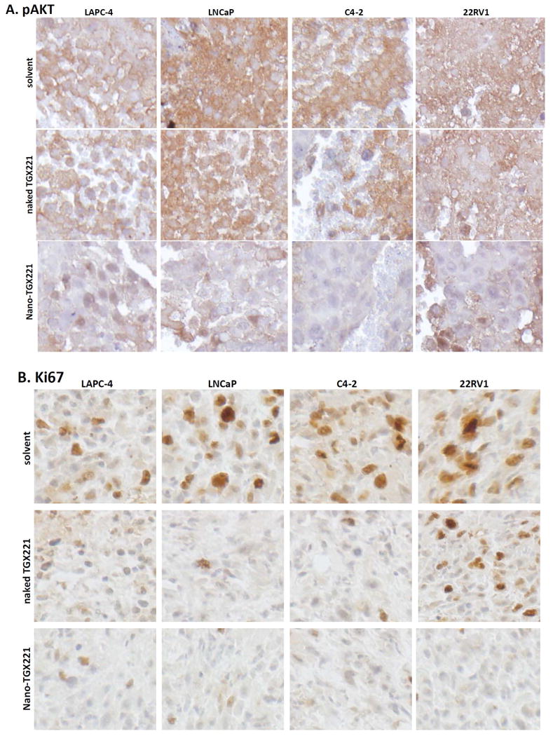
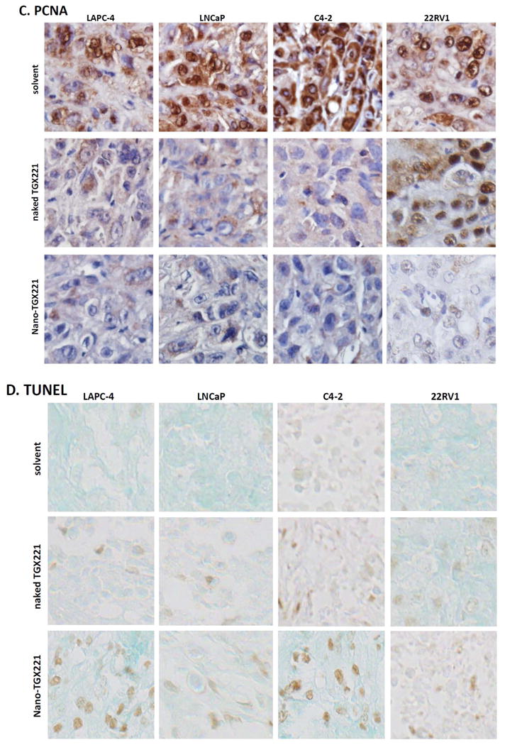
Fig 3.


A-D TGX221 suppresses AKT phosphorylation (A), reduces cell proliferation (B, Ki67; C, PCNA) and induces apoptosis (D, TUNEL). Paraffin-embedded tissue sections from harvested xenografts after treatment indicated on the left side of the panel (as described in Fig 2) were used for the immunostaining with antibodies listed on each panels. Representative microscopic images were shown for the immunostaining results. Magnification, × 200.
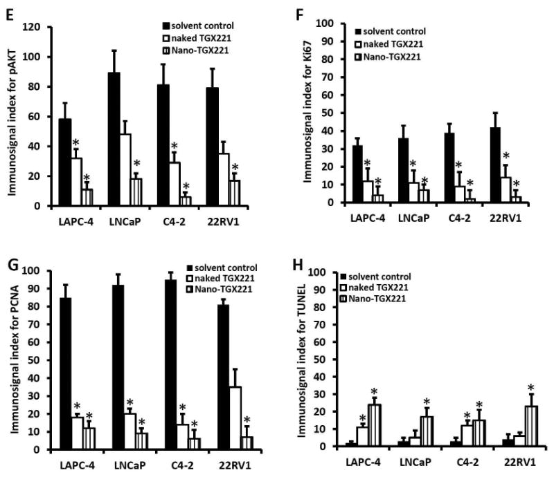
E-H Quantitative data from each immunostaining assays were summarized as average value (MEAN) of the immnosignal indexes as described (24). Error bars indicate the SEM of the MEAN. The asterisks indicate significant differences compared to the solvent control (P < 0.05, Student t-test).
