Abstract
Background:
To compare the variation in size of left and right maxillary central incisors for male patients (using digital calipers of 0.01 mm accuracy). To compare the variation in size of left and right maxillary central incisors for female patients (using digital calipers of 0.01 mm accuracy). To find out the difference between the maxillary central incisors of men and women. Its clinical applicability if difference exists.
Materials and Methods:
A total of 70 dental students of PMNM Dental College and Hospital were selected. Of 70 dental students, 40 male and 30 female were selected. Impressions were made for all subjects, using irreversible hydrocolloid (Algitex, manufacturer DPI, Batch-T-8804) using perforated stock metal trays. The mesiodistal crown width and cervical width were measured for each incisor and recorded separately for left and right teeth. The length was measured for each incisor and recorded separately for left and right maxillary central incisor using digitec height caliper.
Results:
The mean value of maximum crown length of maxillary left central incisor of male was greater in length compared with maxillary right central incisor. Mean value of maximum crown length for male patient right and left side was greater compared with maximum crown length of female patient.
Conclusion:
When compared the dimensions of teeth between two sex, male group shows larger values to female group.
Keywords: Central incisor, digital vernier caliper, digitec height caliper
Introduction
The subject of esthetics has always been a challenging area in dentistry. Esthetics is a derivative of the Greek word “Aiesthetikos” meaning perceptive. It is not a totally scientific or objective discipline, nor is it totally an art form. Denture esthetics is a blending or combination of art and science of prosthodontics. Esthetics in complete denture prosthesis covers a spectrum of restoration of facial contours to an understanding of tooth size and shape, gingival margin contour and color.1 With respect to esthetic appearance of the face, maxillary central Incisors are considered to be the key teeth when treating edentulous patients. Currently, several available methods and techniques for tooth selection are based exclusively on central incisors since they are the most visible teeth during facial activity.2 When no pre-extraction records are available, selecting the proper anterior teeth size for edentulous patients can be difficult. Because a systematic approach is needed in such situation several anatomic measurements have been suggested, including the bizygomatic width, interpupillary distance, interalar width and intercommisural width. Lombardi (1973) proposed that dental and facial esthetics were optimized if features, such as the central to lateral width and lateral to canine width, were repeated in proportion when the patient is viewed from the front. He considered the use of the “Golden proportion.”3 Previous workers have suggested normal ranges and average values for the size of all permanent teeth, but these are mostly based on plaster cast measurements. An additional effort should be made by the manufactures of artificial teeth to ensure that the molds of artificial teeth correspond to these measurements of natural teeth. Therefore, a study was conducted to collect data on the size of permanent maxillary central incisor teeth. With view to improve denture esthetics by guiding the manufacturers to adhere to these norms in artificial teeth.
Materials and Methods
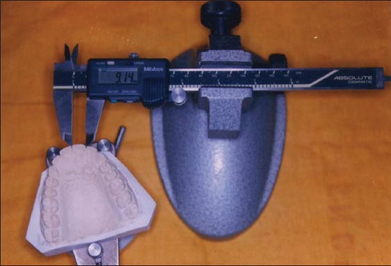
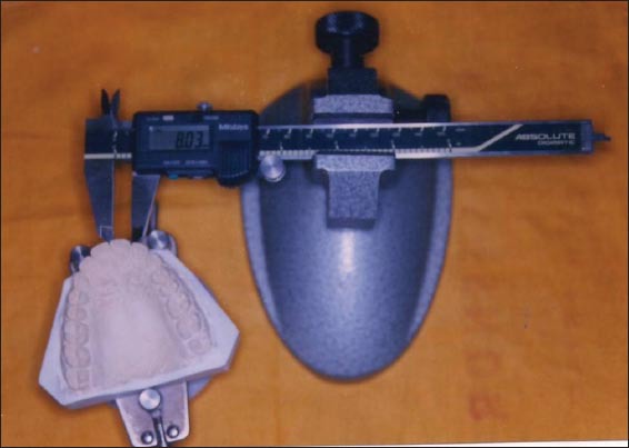
Dental students of PMNM Dental College and Hospital were selected who fulfilled the following criteria in their natural dentition. Both maxillary central incisors were present and in reasonably good alignment. Both incisors exhibited no abrasion, restoration, caries or obvious deformities. Gingival inflammation or hypertrophy that would impede the accurate measurement of the crown length and cervical width of the central incisor teeth was not present. Of 70 dental students, 40 male and 30 female were selected. Impressions were made for all subjects, using irreversible hydrocolloid (Algitex, manufacturer DPI, Batch-T-8804) using perforated stock metal trays. The criteria in selection of stock metal tray. The impression trays were selected with proper extension. The tray is placed first in the mouth over the labial frenum. The posterior extent of the tray relative to the posterior palatal seal area is maintained and the handle is dropped downward to permit visual inspection posteriorly. The dental stone (Kalstone type 3 by Kalabhai) was used for cast. Thus, the cast of all subjects were made, numbered and stored. Measurements were made on the casts using digital Vernier caliper (for width of central incisor [Figures 1 and 2]) to record the mesiodistal crown width and cervical width. The digital Vernier caliper was fixed in position with screw thread and having finally pointed ends that would fit interdentally. The recorded distance was measured to an accuracy of 0.01 mm.
Figure 1.

Digital vernier caliper showing mesiodistal crown width.
Figure 2.

Digital vernier caliper showing cervical crown width.
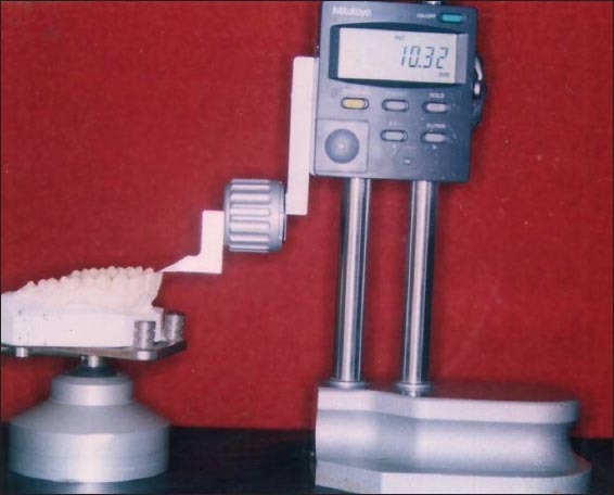
The divider was used to confirm the bulkiest mesiodistal portion of the tooth. Digitec height caliper (for length of central incisor [Figure 3]) used to record the height. Digitec height caliper was fixed in position with screw thread and having finally pointed end. Initially, the pointed end was placed at the lowest attachment of gingival tissue (gingival zenith) to the labial surface of tooth and the scale of the digitec height caliper was adjusted to 0.00 mm and digitec height calipers screw was raised and the pointed end was placed on the incisal edge of the maxillary central incisor. The length was measured for each incisor and recorded separately for left and right maxillary central incisor. SPSS version 16 was used for analysis. Descriptive data which included mean, standard deviation, minimum and maximum values were calculated for each group (side-wise and sex-wise). Student’s t-test was used to compare the mean values between two groups.
Figure 3.

Digitec height caliper pointed tip placed at the lowest attachment of gingival tissue.
Results
The present study was undertaken to assess the variation in size, between maxillary right and left central incisors for male and female patients and to compare the differences between sex groups. Table 1 represents the measurement of 140 central incisors of 40 male and 30 female patients for crown length in millimeter, mesiodistal crown width in millimeter and cervical crown width for right and left maxillary central incisor and combination of both the groups. Mean mesio-distal width of female male right central incisor 8.23 mm and left central incisor 8.16 mm. Mean mesio-distal width of male right central incisor 8.47 mm and left central incisor 8.43 mm. Maximum crown length of female right central incisor 9.14 mm and left central incisor 9.26 mm. Maximum crown length of female left central incisor 10.17 mm and left central incisor 10.32 mm. Table 2 shows descriptive statistics on various dimensions of right central incisor teeth mean value of maximum crown length (mm) for including male and female is 9.73 mm and for left central incisor teeth 9.86 mm. Mesio-distal crown diameter including male and female right central incisor 8.37 mm and for left central incisor 8.31 mm. Cervical crown width including male and female for right central incisor 7.90 mm and for left incisor 7.81 mm. Table 3 represents the comparison of tooth size between male and female for maximum crown length in millimeter mesiodistal crown width cervical crown width for right and left incisor. Mean value of maximum crown length for male patient right and left side was greater compared to maximum crown length of female patient. Right and left side by 10.17-9.14 (Right side) and 10.32-9.26 (Left side). mean value of mesiodistal crown width of male patient right and left side was greater compared with mesiodistal crown width of female patient right and left side by 8.47-8.23 mm (Right side) and 8.43-8.16 mm (Left side). In this study it was observed that crown length of maxillary left central incisor of male and female are of greater in length compared with right central incisor by 0.12 mm for female and 0.15 mm for male group. The mesiodistal crown width of maxillary right central incisor was greater when compared with maxillary left central incisor by 0.07 mm for female group and 0.04 mm for male group. The cervical crown width of right central incisor was greater compared to left central incisor by 0.1 mm for female group and 0.08 mm for male group. Mavroskoufis and Ritchie (1980) said when all the three measurements are measured between right and left was found to correspond to each other, they are termed as identical teeth. If one or two dimensions had a difference not exceeding 0.2 mm, then the teeth were classified as similar. Teeth with differences in all three dimensions, one of which in the excess of 0.2 mm, were classified as dissimilar. In the present study after statistical analysis, I found 13% identical (9 patient), 27% similar (19 patient) and 60% dissimilar (42 patient).
Table 1.
Descriptive statistics on various dimensions of central incisor teeth.

Table 2.
Descriptive statistics on various dimensions of central incisor teeth.

Table 3.
Comparison of tooth size and form between males and females.

Discussion
Esthetics may be defined as the branch of philosophy dealing with beauty. In dentistry, the theory and philosophy that deal with beauty and the beautiful, especially with respect to the appearance of a dental restoration, as achieved through its form and/or color. Those subjective or objective elements and principles underlying the beauty and attractiveness of an object, design or principle. Esthetics is the combination of qualities, such as shape, proportion, color of human face or form, or in other objects that delights the sight. Artificial denture esthetic is generally considered to be naturalness in the appearance of the orofacial regions, in the function of the mandible and lips, and the using esthetically appropriate tooth forms and alignments with composition and colors. The relative dimensions of teeth seem to be among the most objective dental criteria within the esthetic requirement (Magne et al., 2003).4 LaVere et al. (1992)5-7 measured the crown of right and left maxillary central incisor. The width was measured from mesial to distal contact points. The length was measured from the gingival margin to the incisal edge or where gingival recession was present then the cementoenamel junction to the incisal edge was considered. Tooth size ratios represent a valid diagnostic tool that allow for an educated prediction of treatment outcomes and may also limit the necessity for diagnostic set ups and the size of the maxillary central and lateral incisors also presented high variability. This suggests that they could be responsible for incongruity in the anterior ratio and should therefore be examined clinically at the beginning of treatment to detect any major size and shape variation (Santoro et al., 2000).8 The teeth can be modified by grinding or other personalizing features incorporated to change the outline form. Frush and Fisher9-12 have suggested a simple method of modifying replacement anterior teeth according to sex, personality and age of the patient. In this study, 70 (40 male and 30 female) dental students of PMNM Dental College and Hospital were selected and the measurements of crown length, mesiodistal crown width and cervical crown width for male and female students are noted separately. The mean value of cervical crown width for male subject’s right and left side was greater compared to mean value of cervical crown width for female subjects right and left side (7.99-7.77 mm – right side 7.91-7.67 mm – left side). Moorrees and Reed (1964)13 using Americans, found a difference of 0.38mm between the mean values of 87 men and 87 women subjects (8.78 mm and 8.40 mm respectively) and noted that this difference existed in the whole range of values from 7.9 to 10.1mm for men and from 7.1 to 9.8 mm for women subjects using Boley Gauge of 0.1 mm accuracy. Mavroskoufis and Ritchie (1980)1 measured 140 central incisors (70 from each side) in London dental students and provided a mean value for the mesiodistal crown width of 8.90 mm for the right side and 8.87 mm for the left side. Measurements of the mean cervical width were similarly slightly larger on the right side (8.36 mm) as compared with the left side (8.27 mm). In the present study, the percentage of 13% (9 patients) teeth showed identical, 27% (19 patient) similar to each other and 60% (42 patients) are dissimilar in size represented by Pai diagram. These findings are in agreement with the result based on Mavroskoufis and Ritchie 1980. In 10 patients (14%) the central incisors were identical. In 16 patients (23%) they were similar. In 44 patients (63%) they were dissimilar. It is apparent that in most of the subjects, both the maxillary central incisors are not identical to each other in respect to crown length, mesiodistal crown width and cervical width in the selected group. The change in size of these central incisors plays a key role in establishing natural smile of denture patient. Hence, it is suggesting our study values to mold makers to incorporate these changes in their mold (as these values having an important role in the esthetic requirement) to make the artificial teeth to look more natural.
Conclusion
Within the limitation of this study, the following conclusions were drawn; the crown length of maxillary left central incisor for male and female was greater compared to maxillary right central incisor. The mesiodistal crown width of maxillary right central incisor for male and female was greater compared to maxillary left central incisor. The cervical crown width of maxillary right central incisor for male and female was greater compared to maxillary left central incisor. The crown length of right and left side for male patient shows more difference compared to right and left side of female patient and the mesiodistal crown width, cervical width of female patient right and left side shows more difference compared to male patient right and left side. When compared the dimensions of teeth between two sex, male group shows larger values to female group.
Footnotes
Conflicts of Interest: None
Source of Support: Nil
References
- 1.Mavroskoufis F, Ritchie GM. Variation in size and form between left and right maxillary central incisor teeth. J Prosthet Dent. 1980;43(3):254–7.1. doi: 10.1016/0022-3913(80)90398-4. [DOI] [PubMed] [Google Scholar]
- 2.Alvesalo L, Tammisalo E, Townsend G. Upper central incisor and canine tooth crown size in 47, XXY males. J Dent Res. 1991;70(7):1057–60. doi: 10.1177/00220345910700070801. [DOI] [PubMed] [Google Scholar]
- 3.Rosenstiel SF, Ward DH, Rashid RG. Dentists’ preferences of anterior tooth proportion – A web-based study. J Prosthodont. 2000;9(3):123–36. [PubMed] [Google Scholar]
- 4.Magne P, Gallucci GO, Belser UC. Anatomic crown width/length ratios of unworn and worn maxillary teeth in white subjects. J Prosthet Dent. 2003;89(5):453–61. doi: 10.1016/S0022-3913(03)00125-2. [DOI] [PubMed] [Google Scholar]
- 5.LaVere AM, Marcroft KR, Smith RC, Sarka RJ. Denture tooth selection: An analysis of the natural maxillary central incisor compared to the length and width of the face. Part I. J Prosthet Dent. 1992;67(5):661–3. doi: 10.1016/0022-3913(92)90166-8. [DOI] [PubMed] [Google Scholar]
- 6.LaVere AM, Marcroft KR, Smith RC, Sarka RJ. Denture tooth selection: An analysis of the natural maxillary central incisor compared to the length and width of the face: Part II. J Prosthet Dent. 1992;67(6):810–12. doi: 10.1016/0022-3913(92)90589-3. [DOI] [PubMed] [Google Scholar]
- 7.LaVere AM, Marcroft KR, Smith RC, Sarka RJ. Denture tooth selection: Size matching of natural anterior tooth width with artificial denture teeth. J Prosthet Dent. 1994;72(4):381–4. doi: 10.1016/0022-3913(94)90557-6. [DOI] [PubMed] [Google Scholar]
- 8.Santoro M, Ayoub ME, Pardi VA, Cangialosi TJ. Mesiodistal crown dimensions and tooth size discrepancy of the permanent dentition of Dominican Americans. Angle Orthod. 2000;70(4):303–7. doi: 10.1043/0003-3219(2000)070<0303:MCDATS>2.0.CO;2. [DOI] [PubMed] [Google Scholar]
- 9.Frush JP, Fisher RD. How dentogenic restorations interpret the sex factor. J Prosthet Dent. 1956;6:160–72. [Google Scholar]
- 10.Frush JP, Fisher RD. Age factor in dentogenics. J Prosthet Dent. 1957;7:5–13. [Google Scholar]
- 11.Frush JP, Fisher RD. The dynesthetic interpretation on the dentogenic concept. J Prosthet Dent. 1958;8:558–81. [Google Scholar]
- 12.Frush JP, Fisher RD. Dentogenics: Its practical application. J Prosthe Dent. 1959;9:914–21. [Google Scholar]
- 13.Moorrees CF, Reed RB. Correlations among crown diameters of human teeth. Arch Oral Biol. 1964;9:685–97. doi: 10.1016/0003-9969(64)90080-9. [DOI] [PubMed] [Google Scholar]


