

Abstract
Brown tumors are giant cell focal lesion that arises as a result of abnormal bone metabolism in patients with hyperparathyroidism (HPT). The lesions localize in areas of extensive bone resorption, which is replaced by fibrovascular tissue and giant cells with abundant deposits hemorrhage and hemosiderin. A rare case of brown tumor of mandible in a 22-year-old woman is reported here. This case emphasizes the importance of a detailed systemic investigation for all lesions in the maxillofacial region and also discusses the diverse presentations associated with primary HPT.
Keywords: Brown tumor, giant cell lesion, mandible, primary hyperparathyroidism
Introduction
Hyperparathyroidism (HPT) is a disease in which there may be a complex of biochemical, anatomic and clinical abnormalities resulting from increased secretion of parathyroid hormone (PTH). It may occur in primary, secondary and tertiary forms. Primary HPT is characterized by excess secretion PTH due to an abnormality in one or more of the parathyroid glands.1 Parathyroid adenomas were considered to be the main etiology in about 85% of primary HPT cases. The name “brown tumor” derives from the color, which is caused by the vascularity, hemorrhage and deposits of hemosiderin.2 It is generally accepted that bone involvement is a late manifestation of primary hyperparathyroidism (PHPT). Patients presenting in this manner were once more common. However, such a presentation is rare because most cases of PHPT are detected early and before symptomatic bone lesions appear due to improved blood screening techniques.3
Case Report
A female patient aged 22 years presented with a swelling on the left side of the face since past 4 months, which gradually enlarged up to the present size. Patient gives history of generalized weakness, lethargy and weight loss noticed since past few months. Her family history and past medical history was non-significant.
On extra-oral examination a diffuse swelling was noticed on left side of the face, measuring approximately 6 cm × 7 cm, extending superiorly from the infraorbital margin to inferiorly 3 cm below the lower border of mandible, medially 2 cm away from the ala of nose to tragus of the ear laterally (Figure 1). The skin over the swelling was shiny and stretched. There was no ulceration or erythema noticed over the swelling. On palpation, swelling was firm to hard and tender.
Figure 1.

A diffuse swelling was on left side of the face.
On intraoral examination, an ulceroproliferative growth in the vestibule on the left side, extending from the mesial aspect of lower left canine to the retromolar region posteriorly. Obliteration of buccal and lingual vestibule was observed. The growth was covered with pseudomembranous slough, and the indentations of upper molars were seen over the swelling. The swelling has displaced lower first premolar buccally and lower second premolar lingually, lower first molar anteriorly and lower second and third molars posteriorly (Figure 2).
Figure 2.
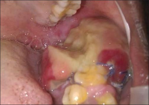
An ulceroproliferative growth in the vestibule on left side, extending from the mesial aspect of lower left canine to the retromolar region.
On palpation swelling was soft to firm in consistency and non-tender. No associated discharge or bleeding noticed. Left submandibular lymph nodes were palpable which was tender and firm in consistency. Provisional diagnosis of ameloblastoma and a differential diagnosis of aneurysmal bone cyst, odontogenic cyst were given. Radiographic examination with orthopantamogram showed large unilocular radiolucency on the left side, extending from mandibular first premolar to mandibular third molar without involving the lower border of mandible. There was generalized decrease in density of mandible, and there was resorption of roots of teeth from second premolar to the second molar (Figure 3).
Figure 3.

Orthopantamogram with large unilocular radiolucency on left side, extending from mandibular first premolar to mandibular third molar.
Patient was advised blood investigations that revealed alkaline phosphatase and PTH levels of 762 IU/L (Normal: 36-14 IU/L) and 452.5 pg/ml (Normal: 12-65 pg/ml) respectively. Serum calcium level was 12.5 mg/dl (Normal: 8.5-10.5 ml/dl).
Patient underwent high-resolution sonography of neck, which revealed a well-defined lesion below the lower pole of the thyroid on the right side between the carotid and trachea measuring about 2.25 cm × 0.46 cm. The lesion had hyperechoic solid component and cystic component with septations. A parathyroid technetium scintiscan (99Tcm SESTAMIBI; Technetium-99 MIBI; methoxy-isobutyl-isonitrile) findings were showing abnormally high uptake is observed at the lower pole of the thyroid lobe interpreted as hyperplasia of right inferior parathyroid with possible brown tumor of mandible (Figure 4).
Figure 4.

Parathyroid technetium scintiscan showing abnormally high uptake observed at the lower pole of the right thyroid lobe.
An incisional biopsy of the mandibular lesion was done which revealed hemorrhagic fibrovascular connective tissue with multinucleated giant cells consistent with the diagnosis of a giant cell lesion. Parathyroid adenoma excision was done under general anesthesia. Recovery was uneventful and patient discharged after 1 week. The resected gland was histologically suggestive of hyperplasia. The surgical removal of the bony lesion was done by curettage. Patient was given oral calcium supplementation in addition to vitamin D3 for possible post-operative hypocalcemia. Considerable reduction in the size of the oral lesion was noted after 6 months.
Discussion
Brown tumor also known as osteitis fibrosa, cystica generalisata or Von Recklinghausen’s disease of bone, is a metabolic bone disease that develops in primary, secondary or tertiary HPT. It should be differentiated from other true giant cell tumors of bone, and it represents reparative granuloma rather than a true neoplastic process.2,4 Brown tumors can occur in any location but are most common in the ribs, clavicle and pelvis. Although the reported occurrence in mandible is 4.5% of subjects in a 220-case HPT study, it is rare to find brown tumor as the initial clinical manifestation in primary HPT.5
Our patient exhibited none of the classical symptoms of hypercalcemia that include “bony pain/bone fractures, renal stones, abdominal groans, and psychic moans.” Patients with hypercalcemia also present with associated symptoms such as paraesthesia, headaches, recent fractures, constipation, polyuria, and polydipsia. However, most of the patients are asymptomatic and are usually identified as part of routine investigations.6
The reported prevalence of brown tumor is 0.1% and can occur in mandible, maxilla, clavicle, ribs and pelvic bones. Furthermore the frequency of occurrence is more among persons older than 50 years of age with a male to female ratio of 1:3.7,8 In the present case, the patient was 22-year-old female and mandibular brown tumor was found to be the first clinical manifestation of PHPT.
Clinically, brown tumors may present as small, asymptomatic swelling in the jaw bone or as a painful exophytic mass. The radiographic appearance is usually as a well-demarcated monolocular or multilocular osteolytic lesion infrequently associated with root resorption and loss of the lamina dura. The diagnosis has to be confirmed by establishing elevated serum calcium and PTH levels because histological features alone are insufficient as it may resemble any giant cell tumor. The parathyroid technetium scintiscan is one of the most preferred imaging modality to localize diseased parathyroid glands prior to surgery.9
Histologically, brown tumors are characterized by vascular fibroblastic stroma and several osteoclast-like multinucleated giant cells often interspersed with hemorrhagic infiltrates and hemosiderin deposits.10 The initial step in the management of primary HPT involves control of HPT and a partial parathyroidectomy is considered effective in spontaneous regression of small osteolytic jaw lesions. However, surgical excision may be indicated in large symptomatic lesion usually done after parathyroid surgery as was done in the present case.5,11 Post-operative hypocalcemia may occur in patients who undergo a partial parathyroidectomy. Therefore, calcium supplements could be required as was done with this patient.2,12,13
Conclusion
Even though the improvement of various diagnostic process and biochemical tests results in early diagnosis of HPT, the dentists should be alert of the possible occurrence of brown tumors in the jaws of the previously diagnosed patients. In addition, it is essential that the dental surgeon be aware of oral manifestations associated with systemic diseases, hence the importance of careful physical examination and thorough investigation for the diagnosis and treatment success.
Footnotes
Conflicts of Interest: None
Source of Support: Nil
References
- 1.Movahedian B, Razavi SM, Hasheminia D, Rezaei M. Simultaneous maxillary and mandibular brown tumors in secondary hyperparathyroidism: A case report. Dent Res J. 2008;5(1):41–5. [Google Scholar]
- 2.Selvi F, Cakarer S, Tanakol R, Guler SD, Keskin C. Brown tumour of the maxilla and mandible: A rare complication of tertiary hyperparathyroidism. Dentomaxillofac Radiol. 2009;38(1):53–8. doi: 10.1259/dmfr/81694583. [DOI] [PubMed] [Google Scholar]
- 3.Jebasingh F, Jacob JJ, Shah A, Paul TV, Seshadri MS. Bilateral maxillary brown tumours as the first presentation of primary hyperparathyroidism. Oral Maxillofac Surg. 2008;12(2):97–100. doi: 10.1007/s10006-008-0105-9. [DOI] [PubMed] [Google Scholar]
- 4.Soundarya N, Sharada P, Prakash N, Pradeep G. Bilateral maxillary brown tumors in a patient with primary hyperparathyroidism: Report of a rare entity and review of literature. J Oral Maxillofac Pathol. 2011;15:56–9. doi: 10.4103/0973-029X.80027. [DOI] [PMC free article] [PubMed] [Google Scholar]
- 5.Suarez-Cunqueiro MM, Schoen R, Kersten A, Klisch J, Schmelzeisen R. Brown tumor of the mandible as first manifestation of atypical parathyroid adenoma. J Oral Maxillofac Surg. 2004;62(8):1024–8. doi: 10.1016/j.joms.2004.02.011. [DOI] [PubMed] [Google Scholar]
- 6.Carr ER, Contractor K, Remedios D, Burke M. Can parathyroidectomy for primary hyperparathyroidism be carried out as a day-case procedure? J Laryngol Otol. 2006;120(11):939–41. doi: 10.1017/S0022215106002350. [DOI] [PubMed] [Google Scholar]
- 7.Keyser JS, Postma GN. Brown tumor of the mandible. Am J Otolaryngol. 1996;17(6):407–10. doi: 10.1016/s0196-0709(96)90075-7. [DOI] [PubMed] [Google Scholar]
- 8.Queiroz SM, Vasconcelos RG, Andrade AL, Amorim AG, Gordón-Nuñes MA, Freitas R, et al. Maxillary brown tumor associated with chronic kidney failure: A case report. J Bras Patol Med Lab. 2013;49(6):424–8. [Google Scholar]
- 9.Triantafillidou K, Zouloumis L, Karakinaris G, Kalimeras E, Iordanidis F. Brown tumors of the jaws associated with primary or secondary hyperparathyroidism. A clinical study and review of the literature. Otolaryngol Head Neck Surg. 2006;27:281–6. doi: 10.1016/j.amjoto.2005.11.004. [DOI] [PubMed] [Google Scholar]
- 10.Martínez-Gavidia EM, Bagán JV, Milián-Masanet MA, Lloria de Miguel E, Pérez-Vallés A. Highly aggressive brown tumour of the maxilla as first manifestation of primary hyperparathyroidism. Int J Oral Maxillofac Surg. 2000;29:447–9. [PubMed] [Google Scholar]
- 11.Alhusban M, Baqain ZH. Mandibular brown tumor as the first manifestation of primary hyperparathyroidism: A case report. Saudi Dent J. 2011;23(2):107–9. doi: 10.1016/j.sdentj.2010.11.003. [DOI] [PMC free article] [PubMed] [Google Scholar]
- 12.Phitayakorn R, McHenry CR. Jaw tumor: An uncommon presenting manifestation of primary hyperparathyroidism. World J Endocr Surg. 2010;2(1):45–50. [Google Scholar]
- 13.Yildiz A, Kirdak T, Ersoy A, Ersoy C, Saraydaroglu O, Ayyildiz A, et al. A rare cause of primary hyperparathyroidism presented with giant adenoma, multiple brown tumors and end stage renal failure. Med Sci Case Rep. 2013;2:1–6. Dent Assoc 1965; 70: 1162-5. [Google Scholar]