Abstract
Background:
Sesame oil from the seeds of Sesamum indicum Linn. (Pedaliaceae) has been used traditionally in Indian medical practice of Ayurveda in the treatment of central nervous system disorders and insomnia. A few published reports favor the anti-dementia effect of sesamol (SML), an active constituent of sesame oil.
Objective:
Thus, the present study was aimed to explore the anti-dementia effect and possible mechanism (s) of SML in aluminium chloride (AlCl3)-induced cognitive dysfunction model in rodents with special emphasis on memory centers viz., hippocampus and frontal cortex.
Methods:
Male Wistar rats were exposed to AlCl3 (175 mg/kg p.o.) for 60 days. SML (10 and 20 mg/kg) and rivastigmine (1 mg/kg) were administered orally 45 min before administration of AlCl3 for 60 days. Spatial memory was assessed using Morris water maze test. After 60 days of treatment animals were sacrificed, hippocampus and frontal cortex were collected and analyzed for acetylcholinesterase (AChE) activity, tumor necrosis factor (TNF-α) level, antioxidant enzymes (Glutathione, catalase), lipid peroxidation, and nitrite level. The circulating triglycerides, total cholesterol, low-density lipoprotein (LDL) and high-density lipoprotein (HDL) levels were also analyzed.
Results:
SML significantly prevented behavioral impairments in aluminium-exposed rats. Treatment with SML reversed the increased cholesterol, triglycerides and LDL while raised the HDL levels. SML significantly corrected the effect of AlCl3 on AChE activity. Further, SML reversed the elevated nitric oxide, TNF-α and reduced antioxidant enzymes in hippocampus and frontal cortex.
Conclusion:
The present study suggests the neuro-protection by SML against cognitive dysfunction induced by environmental toxin (AlCl3) in hippocampus and frontal cortex.
Keywords: Aluminium, dementia, hypolipidemia, memory, sesamol, tumor necrosis factor-α
INTRODUCTION
Aluminium, a highly neurotoxic metal, is considered to be involved in the pathogenesis of neurodegenerative disorders like Alzheimer's disease (AD)[1,2,3] and Parkinson's disease.[4] Experimental animals, exposed to aluminium have developed AD-like conditions, characterized by elevated levels of amyloid beta (Aβ) protein and amyloid precursor protein (APP),[5,6] mitochondrial dysfunction, depletion of ATP,[7,8] induction of lipid peroxidation and lipid dystrophy,[9,10] accelerated production of phosphorylated tau,[11] impairment of cholinergic projections[12] and promotion of apoptotic neuronal death.[13,14] Thus, aluminium chloride (AlCl3)-induced cognitive dysfunction model has been widely used for testing drugs against AD.[15,16,17]
Currently approved treatments for AD target neurotransmitter systems and only provide modest improvement in cognitive impairment. Thus, it is necessary to develop effective medications that go beyond acetylcholinesterase (AChE) inhibitors and N-methyl-D-aspartate antagonist. Several studies have demonstrated that hypercholesterolemia could cause dementia and Aβ42 deposition in hippocampal region.[18] Hypercholesterolemia is an outcome of sedentary life-style resulting in obesity and lipid dystrophy. Traditionally, in Indian medical practice of Ayurveda, sesame oil from the seeds of Sesamum indicum Linn. (Pedaliaceae) has been used to correct central nervous system disorders and insomnia.[19] Sesamol (SML), an agent obtained from sesame oil, edible oil, is found to reduce cholesterol and triglyceride levels in acute and chronic models of hyperlipidemia.[20] It is also reported to have antioxidant, neuro-protective,[21] anti-inflammatory[22] and hepatoprotective[23] activities. Although, all these actions of SML are beneficial to overcome the condition of dementia, SML has not been investigated for its behavioral effects in chronic models of dementia. In view of this, the present study was designed to investigate the effect of SML, a lipid lowering agent in AlCl3-mediated behavioral and biochemical changes in rats.
MATERIALS AND METHODS
Animals
Male Wistar rats, weighing 200–250 g (90 days old) procured from Central Animal Research Facility of Manipal University, Manipal were used. Animals were acclimatized to laboratory conditions for 7 days before the experiment and they were maintained under controlled conditions of temperature (23°C ± 2°C), humidity (50% ±5%). The animals were kept under standard conditions of 12 h light/dark cycle in sanitized polypropylene cages containing sterile paddy husk as bedding with free access to food and water ad libitum. The experimental protocol was approved by the Institutional Animal Ethics Committee, Kasturba Medical College, Manipal [IAEC/KMC/73/2012] and was carried out in accordance with the guidelines provided by the Committee for the Purpose of Control and Supervision of Experiments on Animals, Government of India.
Drugs and treatment schedule
Aluminium chloride (Spectrochem Pvt Limited, India), SML (Sigma-Aldrich Co, St. Louis, MO, USA) and rivastigmine (RIV) (Dr. Reddy's Laboratories, Hyderabad, India) solutions were made freshly on each day for administration. AlCl3 was dissolved in distilled water and administered orally once daily at a dose of 175 mg/kg from day 6 onwards (24 h after the completion of retention trial on day 5) for 60 days. This dosing regimen for inducing dementia using AlCl3 was determined according to the previous reports and the high rate of induction and low mortality, which was evident in the pilot study conducted. SML and RIV, at various doses, were administered 45 min before administration of AlCl3 orally after suspending them in 0.5% sodium carboxy methyl cellulose (CMC) in distilled water for 60 days from day 6. On the basis of escape latency time (ELT) on day 5, animals were divided into six groups (n = 8). The groups were as follows:
Group 1: Normal control - Received distilled water (5 ml/kg p.o.).
Group 2: Vehicle control - Receives 0.5% CMC (5 ml/kg p.o.).
Group 3: AlCl3 (175 mg/kg p.o.).
Group 4: RIV (1 mg/kg p.o.) + AlCl3 (175 mg/kg p.o.).
Group 5: SML (10 mg/kg p.o.) + AlCl3 (175 mg/kg p.o.).
Group 6: SML (20 mg/kg p.o.) + AlCl3 (175 mg/kg p.o.).
The doses of the standard drug RIV (1 mg/kg) and the test drug SML (10 mg/kg and 20 mg/kg) were chosen based on the previous literature reports.[24,25,26,27] Body weight of the animals was taken on a daily basis before the treatments.
Spatial memory assessment using Morris water maze
To investigate the spatial learning and memory abilities of the experimental rats, Morris water maze task was performed as described by Morris[28] with minor modifications.[29] It consisted of a circular tank of 150 cm diameter and 40 cm height. The pool was divided into North-East, South-East, South-West and North-West (NW) quadrants. In the NW quadrant a hidden escape platform (10 cm diameter), was placed 2 cm below the water surface.
All rats were trained to find the escape platform. Animals were given four trials per day for 4 consecutive days. Animals were kept on the platform for 30 s and then removed. The rats that could not reach the platform in 20 s on the 4th trial-day were excluded from the study. On the probe day (day 5), the hidden platform was removed, and probe trial was performed with a cut off time of 60 s. All the animals were exposed to one retention trial on day 25, 45 and 65 to evaluate the memory consolidation. Data were acquired through a video camera connected to a computerized tracking system (Any Maze, Ugo Basile, Italy) fixed above the centre of the pool.
Dissection and tissue preparation
On day 65, immediately after the retention trial, the animals were sacrificed by decapitation. Brains were rapidly removed, hippocampus and frontal cortex were dissected according to the method described by Glowinski and Iverson.[30] A 10% w/v homogenate of samples was prepared by homogenizing with ice-cooled 0.1 M phosphate buffer potential of Hydrogen (pH) 7.4 using an ultra Turrax T25 homogenizer at a speed of 9500 rpm thrice at an interval of few seconds. The homogenates were then centrifuged at 15,000 rpm at 4°C for 15 min. Supernatant was collected and used for biochemical estimations.
Estimation of acetylcholinesterase activity
In the supernatant, AChE activity was measured by Ellman method using acetylthiocholine iodide as a substrate.[31] To a reaction mixture containing phosphate buffer (2.8 ml, pH 8), acetylthiocholine iodide (0.05 ml) and 0.05 ml of 5,5’-dithio-bis-2-nitrobenzoic acid (DTNB) (Ellman reagent), 0.1 ml of the supernatant was added. The change in absorbance was measured for 4 min at 60 s interval at 412 nm using ultraviolet–visible spectrophotometer and the change in absorbance per minute was calculated. The results were expressed as micromoles of acetylthiocholine iodide hydrolyzed per min per mg protein.
BIOCHEMICAL EVALUATION
At the end of the experimental period, animals were mildly anaesthetized with diethyl ether and the blood samples were collected by retro-orbital sinus puncture into microcentrifuge tubes. The tubes were then centrifuged at 10,000 rpm for 10 min at 20°C. After centrifugation, the serum was separated at once, divided into aliquots and stored at −20°C until they were used for biochemical analysis.
Collected serum samples were analyzed colorimetrically for triglycerides (glycerophosphate-oxidase-peroxidase (POD) method), total cholesterol (cholesterol oxidase-POD method), low-density lipoprotein (LDL) and high-density lipoprotein (HDL) levels by end point method as per the manufacturer's instructions with the help of diagnostic kits (Aspen laboratories, Mumbai) using Enzyme Linked Immuno Sorbent Assay (ELISA) plate reader.
Estimation of lipid peroxidation and nitrite level
Estimation of nitrite level
Nitrite level in hippocampus and frontal cortex homogenate was measured by Griess reaction.[32] The extent of lipid peroxidation in hippocampus and frontal cortex was quantitatively determined by the method described by Konings and Drijver.[33]
Estimation of antioxidant enzymes
The catalase activity was determined by the method of Aebi et al., 1984[34] and glutathione (GSH) activity based upon the reaction between DTNB and sulfhydryl groups of GSH.[35]
Tumor necrosis factor-α Estimation
Level of tumor necrosis factor (TNF-α) in the supernatant was estimated by rat TNF-α kit as per the experimental protocol given by Invitrogen Corporation, USA. It involves a solid phase sandwich ELISA. The level of TNF-α was expressed as pg/mg of protein.
Estimation of total protein
Total protein was estimated in all tissue samples using Pierce® BCA Protein Assay Kit as per the experimental protocol given by Thermo Scientific, USA. Bovine serum albumin was used as a standard.
Statistical analysis
All the data are expressed as mean ± standard error of the mean. Results were analyzed by one-way analysis of variance, followed by Tukey's post-hoc test using Graph Pad Prism version 5.0 software. P <0.05 was considered as statistically significant.
RESULTS
Body weight
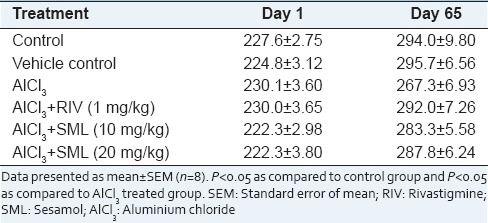
After 60 days, AlCl3 exposure and other treatment groups did not show a significant effect on body weight [Table 1].
Table 1.
Effect of AlCl3 and AlCl3+ treatments (RIV, SML) on the body weight of animals before and after treatment
Spatial memory assessment using Morris water maze
Time to reach hidden platform (Escape latency)
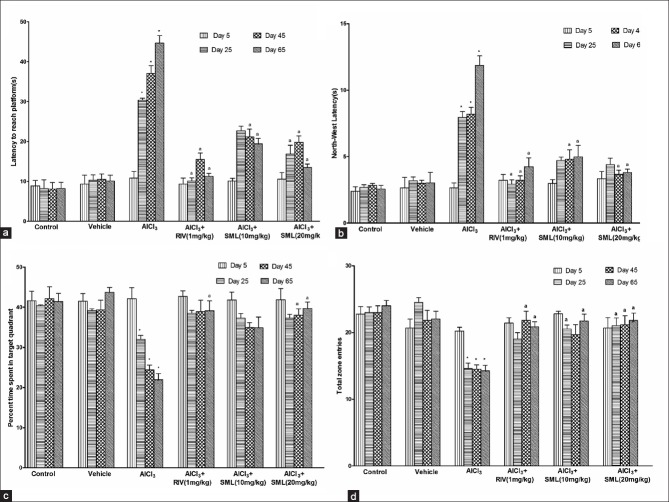
Aluminium chloride intoxication resulted in cognitive impairment as evidenced by significant increase in ELT during probe trials. On day 25, RIV and SML (20 mg/kg) produced a significant decrease in ELT when compared to AlCl3-treated group. On day, 45 and day 65, all the treatment groups significantly improved the cognitive performance (i.e. decreased ELT) of animals relative to AlCl3-treated group [Figure 1a].
Figure 1.
Effect of aluminium chloride (AlCl3) and AlCl3 + treatments (Rivastigmine, Sesamol) on (a) ELT time (latency to reach platform) (b) North-West (NW) latency (c) Percent time spent in target quadrant (NW). (d) Total zone entries and during retention trials before (day 5) and after (day 25, 45 and 65) treatment. Data presented as mean ± standard error of the mean (n = 8). *P < 0.05 as compared to control group and aP < 0.05 as compared to AlCl3 treated group
North-West latency
Following 60 days of AlCl3 administration, dementia was observed in rats as shown by the significant (P < 0.05) increase in the latency to find the target quadrant (NW). In RIV and SML (10 mg/kg and 20 mg/kg) groups, the latency to find the target quadrant was shortened significantly [Figure 1b].
Percentage time spent in target quadrant (north-west)
During the probe trials on day 25, 45 and 65, aluminium treated animals were found to spent significantly less time in the target quadrant (NW) as compared to control group. All the treatment groups significantly increased the time spent in the target quadrant relative to aluminium treated group during probe trial. SML (20 mg/kg) was found to have an activity comparable to RIV [Figure 1c].
Total zone entries
Aluminium treated group showed a significant decrease in total zone transitions on subsequent days of testing as compared to control group. RIV and SML significantly increased total zone transitions of animals as compared to AlCl3 treated group [Figure 1d].
Acetylcholinesterase activity
Chronic AlCl3 exposure significantly decreased AChE activity in the frontal cortex (P < 0.05) and hippocampus (P < 0.05) of rats as compared to normal control group [Figure 2a and b]. RIV also significantly (P < 0.05) decreased cholinesterase activity compared to control group in both hippocampus and frontal cortex. SML (10 mg/kg and 20 mg/kg) significantly reversed the effect of AlCl3 on AChE activity.
Figure 2.
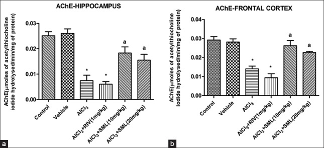
Effect of aluminium chloride (AlCl3) and AlCl3 + treatments (Rivastigmine, Sesamol) on acetylcholinesterase activity in (a) hippocampus (b) frontal cortex. Data presented as mean ± standard error of the mean (n = 8). *P < 0.05 as compared to control group and aP < 0.05 as compared to AlCl3 treated group
Effect of treatments on lipid profile
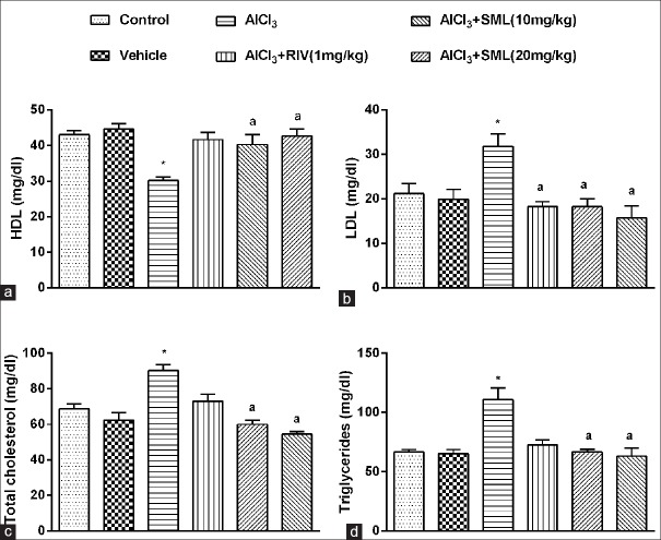
Chronic administration of AlCl3 for 60 days caused significant (P < 0.05) reduction in serum HDL levels [Figure 3a], increase in LDL levels [Figure 3b], total cholesterol [Figure 3c], triglycerides [Figure 3d] as compared to control group. Interestingly RIV significantly (P < 0.05) prevented the rise in LDL levels. Treatment with SML prevented the rise in total cholesterol, triglycerides and LDL levels and increased HDL levels as compared to AlCl3 treated animals.
Figure 3.
Effect of aluminium chloride (AlCl3) and AlCl3 + treatments (Rivastigmine, Sesamol) on (a) High-density lipoprotein (b) Low-density lipoprotein (c) Total cholesterol (d) Triglycerides. Data presented as mean ± standard error of the mean (n = 8). *P < 0.05 as compared to control group and aP < 0.05 as compared to AlCl3 treated group
Estimation of lipid peroxidation and nitrite level
Estimation of malondialdehyde level
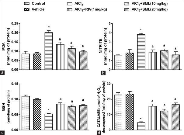
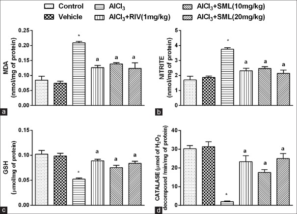
The malondialdehyde (MDA) levels in the hippocampus [Figure 4a] and frontal cortex [Figure 5a] of aluminium treated rats showed a threefold increase as compared to control group. The elevated MDA levels were significantly reversed by RIV and SML. SML at 10 mg/kg and 20 mg/kg dose level showed a better reduction of MDA levels than RIV in the hippocampus region.
Figure 4.
Effect of aluminium chloride (AlCl3) and AlCl3 + treatments (Rivastigmine, Sesamol) on hippocampus (a) malondialdehyde (b) nitrite level (c) glutathione level (d) catalase activity
Figure 5.
Effect of aluminium chloride (AlCl3) and AlCl3 + treatments (Rivastigmine, Sesamol) on frontal cortex (a) Malondialdehyde (b) Nitrite level (c) Glutathione level (d) Catalase activity. Data presented as mean ± standard error of the mean (n = 8). *P < 0.05 as compared to control group and aP < 0.05 as compared to AlCl3 treated group
Estimation of nitrite level
Chronic exposure of animals to AlCl3 caused a significant elevation in nitrite levels in the hippocampus [Figure 4b] and frontal cortex [Figure 5b] as compared to control group of animals. RIV and SML (10 and 20 mg/kg) treatments significantly prevented the rise in levels of nitrite in both frontal cortex and hippocampus. In this case, SML (20 mg/kg) was found to reduce nitrite levels comparable to that seen in the control group.
Estimation of antioxidant enzymes
Catalase and glutathione activity
The hippocampus and frontal cortex of the AlCl3-treated rats were observed to have significant (P < 0.05) reduction in catalase [Figures 4d and 5d] and reduced GSH activity as compared to control animals [Figures 4c and 5c]. RIV (1 mg/kg) and SML (10 mg/kg, 20 mg/kg) enhanced the catalase, and GSH levels significantly as compared to AlCl3 treated group.
Estimation of tumor necrosis factor-α in hippocampus
Tumor necrosis factor-α levels were significantly (P < 0.05) increased (threefold) in the hippocampus of AlCl3 treated animals as compared to vehicle group. Treatment with RIV (P < 0.05), SML (20 mg/kg) significantly (P < 0.05) inhibited this rise in TNF-α levels [Figure 6].
Figure 6.
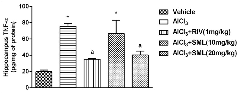
Effect of aluminium chloride (AlCl3) and AlCl3 + treatments (Rivastigmine, Sesamol) on tumor necrosis factor-α level in the hippocampus of rats. Data presented as mean ± standard error of the mean (n = 8). *P < 0.05 as compared to control group and aP < 0.05 as compared to AlCl3 treated group
DISCUSSION
The study investigates the ameliorative effect of the lipid-lowering drug, SML, on AlCl3-induced behavioral and biochemical changes in rodents. Aluminium was shown to accumulate in higher quantities in hippocampal and cortex regions, which are the sites of memory.[36] Spatial memory tasks are highly sensitive to hippocampus and frontal cortex[37] which is severely affected in neurodegenerative conditions such as AD.
Chronic aluminium exposure in animals was reported to cause cognitive decline.[38,39] Cognitive dysfunction is evident from decreased activity of experimental animals in Morris water maze,[24] radial arm maze[38] and passive avoidance task.[40] In the present study, the behavioral changes showed by aluminium exposed rats were inconsistent with previous reports. In Morris water maze test, aluminium exposure resulted in a significant decrease in spatial memory as indicated by increased ELT (time required to reach platform), NW latency (time required to reach target quadrant) and decreased percentage time in the NW zone and total zone entries during the probe trial. The treatment with SML and RIV reversed the memory deficit caused by AlCl3. This suggests the beneficial effects of SML in correcting memory deficit associated with aluminium exposure.
Cholinergic system in the brain plays a major role in modulating learning and memory. Reduction in AChE activity and acetylcholine levels in hippocampus and cortex have been correlated with loss of cognitive function in AD patients.[41] Long-term potentiation in the hippocampal CA1 pyramidal neurons is modulated by AChE.[15] Moreover, it is essential for survival and growth of cells.[42] In experimental animals, aluminium has been shown to decrease AChE activity.[43] It shows a biphasic response on AChE activity, with an initial increase in the activity of the enzyme followed by a marked decrease. Formation of irreversible aluminium complex with high affinity toward the anionic site of enzyme and slow accumulation of aluminium in the brain has been attributed for such biphasic response.[44,45] This explains the significant reduction in AChE activity in both hippocampus and cortex observed in our study after chronic AlCl3 treatment. The toxic effect of aluminium may be attributed to reduced choline uptake,[46] erosion of cholinergic terminals in cortex and hippocampus,[47] and reduced choline acetyl transferase.[48] RIV, the standard AchE inhibitor showed further decrease in AChE activity thereby sustaining the action of the remaining acetylcholine from cholinergic neurons. SML was found to increase the AChE levels in aluminium-exposed rats; this may be attributed to the ability of SML to re-establish the acetylcholine release, thus protecting cholinergic neurons.
Apart from cholinergic deficit leading to memory impairment, effect of dyslipidemia on behavioral changes has been studied. In AD patients, elevation in the levels of total serum cholesterol and LDL-associated cholesterol has been implicated.[49] An increase in the membrane cholesterol enhances the lipid raft area, and the APP present in the rafts gets into contact with β-secretase very easily leading to increased Aβ production.[18] Aluminium through its dyslipidemic property could have contributed to a strong lipid membrane rafts in the brain neuronal membrane leading to AD like syndrome in rats. The dyslipidemia due to aluminium treatment (elevated levels of total cholesterol, LDL, triglycerides and decreased HDL levels) is largely attributed to the accumulation of aluminium in liver causing alteration in lipid metabolism.[50] In our previous study, SML was found to reduce both serum triacylglycerol and cholesterol levels.[19] In the present study, chronic treatment using SML was able to bring down the raised cholesterol, LDL, and triglycerides levels due to aluminium exposure. It also increased the HDL (good cholesterol) level comparable to control.
Oxidative stress and neuro-inflammation are involved in the pathology of neurodegenerative disorders.[51] Lipids are highly vulnerable to oxidative stress. The polyunsaturated fatty acids present in brain get attacked by the free radicals leading to the production of toxic aldehydes as 4-hydroxynonenal and acrolein which in turn, lead to conformational changes of proteins. Studies have suggested the possible involvement of Aβ induced lipid peroxidation in brain generating free radicals and reactive aldehydes resulting in neurodegeneration.[52,53] Nitric oxide (NO), a signaling molecule regulates many physiological functions in the body. It also acts as a free radical to induce nitrergic stress. The nitrergic stress in turn activates the mitochondrial pathway of apoptosis through up regulation of p53,[54] cytochrome c release[55] and through p38 mitogen-activated protein kinase pathway[56] leading to neuronal death. In our study, we observed a significant increase in nitrite and MDA levels in aluminium treated group in accordance with the previous reports.[57] Further, aluminium treated rats showed a decrease in antioxidant system viz., catalase and GSH levels indicating considerable oxidative stress caused by the toxicant. Both RIV and SML treatment normalized the altered levels of nitrite, MDA levels and antioxidant enzyme like catalase, GSH. This may be due to the antioxidant effect of SML[58] and inhibition of NO synthase.[21,22] Ameliorative effect of SML on oxidative stress may be considered as one of the approaches to correct aluminium mediated neurotoxicity.
Accumulation of abnormal protein aggregates like Aβ42 and free radicals (viz., nitrite, reactive oxygen species, reactive nitrogen species) may trigger cellular stress and neuroinflammation by activation of the brain's innate immune system involving microglia and astrocytes. Activation of these immune cells results in the release of inflammatory mediators such as TNF-α, interferon-α, Interleukin-6 resulting in neurodegeneration.[59,60] It has been observed that aluminium exposure has resulted in elevated TNF-α, a key cytokine which stimulates microglia to release glutamate causing excitotoxicity.[61,62] Similar to this we also observed a significant increase in TNF-α level in hippocampus following chronic aluminium exposure. This rise in TNF-α was counteracted by SML indicating its role in preventing neuroinflammation.
CONCLUSION
Sesamol treatment demonstrates a protective effect against AlCl3-induced cognitive dysfunction in rats. The aluminium mediated biochemical changes were reversed, where SML enhanced AChE level in hippocampus and cortex regions through correcting hyperlipidemia, reducing oxidative stress, NO and TNF-α level. Further studies are awaited to establish the role of SML as a potential candidate to control neuronal disturbances.
ACKNOWLEDGMENT
We thank Manipal College of Pharmaceutical Sciences, Manipal and Manipal University for providing research facility and Department of Science (FIST-Scheme) for providing infrastructural support.
Footnotes
Source of Support: Nil
Conflict of Interest: None declared.
REFERENCES
- 1.Campbell A. The potential role of aluminium in Alzheimer's disease. Nephrol Dial Transplant. 2002;17(Suppl 2):17–20. doi: 10.1093/ndt/17.suppl_2.17. [DOI] [PubMed] [Google Scholar]
- 2.Flaten TP. Aluminium as a risk factor in Alzheimer's disease, with emphasis on drinking water. Brain Res Bull. 2001;55:187–96. doi: 10.1016/s0361-9230(01)00459-2. [DOI] [PubMed] [Google Scholar]
- 3.McLachlan D. Aluminium and the risk for Alzheimer's disease. Environmetrics. 1995;6:233–75. [Google Scholar]
- 4.McLachlan DR, Bergeron C, Smith JE, Boomer D, Rifat SL. Risk for neuropathologically confirmed Alzheimer's disease and residual aluminium in municipal drinking water employing weighted residential histories. Neurology. 1996;46:401–5. doi: 10.1212/wnl.46.2.401. [DOI] [PubMed] [Google Scholar]
- 5.Kawahara M, Kato M, Kuroda Y. Effects of aluminium on the neurotoxicity of primary cultured neurons and on the aggregation of beta-amyloid protein. Brain Res Bull. 2001;55:211–7. doi: 10.1016/s0361-9230(01)00475-0. [DOI] [PubMed] [Google Scholar]
- 6.Campbell A, Kumar A, La Rosa FG, Prasad KN, Bondy SC. Aluminium increases levels of beta-amyloid and ubiquitin in neuroblastoma but not in glioma cells. Proc Soc Exp Biol Med. 2000;223:397–402. doi: 10.1046/j.1525-1373.2000.22356.x. [DOI] [PubMed] [Google Scholar]
- 7.Kumar V, Bal A, Gill KD. Impairment of mitochondrial energy metabolism in different regions of rat brain following chronic exposure to aluminium. Brain Res. 2008;1232:94–103. doi: 10.1016/j.brainres.2008.07.028. [DOI] [PubMed] [Google Scholar]
- 8.Lemire J, Mailloux R, Puiseux-Dao S, Appanna VD. Aluminium-induced defective mitochondrial metabolism perturbs cytoskeletal dynamics in human astrocytoma cells. J Neurosci Res. 2009;87:1474–83. doi: 10.1002/jnr.21965. [DOI] [PubMed] [Google Scholar]
- 9.Oteiza PI. A mechanism for the stimulatory effect of aluminium on iron-induced lipid peroxidation. Arch Biochem Biophys. 1994;308:374–9. doi: 10.1006/abbi.1994.1053. [DOI] [PubMed] [Google Scholar]
- 10.Verstraeten SV, Oteiza PI. Al (3+)-mediated changes in membrane physical properties participate in the inhibition of polyphosphoinositide hydrolysis. Arch Biochem Biophys. 2002;408:263–71. doi: 10.1016/s0003-9861(02)00557-x. [DOI] [PubMed] [Google Scholar]
- 11.el-Sebae AH, Abdel-Ghany ME, Shalloway D, Abou Zeid MM, Blancato J, Saleh MA. Aluminium interaction with human brain tau protein phosphorylation by various kinases. J Environ Sci Health B. 1993;28:763–77. doi: 10.1080/03601239309372852. [DOI] [PubMed] [Google Scholar]
- 12.Gulya K, Rakonczay Z, Kása P. Cholinotoxic effects of aluminium in rat brain. J Neurochem. 1990;54:1020–6. doi: 10.1111/j.1471-4159.1990.tb02352.x. [DOI] [PubMed] [Google Scholar]
- 13.Ghribi O, Herman MM, Forbes MS, DeWitt DA, Savory J. GDNF protects against aluminium-induced apoptosis in rabbits by upregulating Bcl-2 and Bcl-XL and inhibiting mitochondrial Bax translocation. Neurobiol Dis. 2001;8:764–73. doi: 10.1006/nbdi.2001.0429. [DOI] [PubMed] [Google Scholar]
- 14.Kawahara M, Kato-Negishi M, Hosoda R, Imamura L, Tsuda M, Kuroda Y. Brain-derived neurotrophic factor protects cultured rat hippocampal neurons from aluminium maltolate neurotoxicity. J Inorg Biochem. 2003;97:124–31. doi: 10.1016/s0162-0134(03)00255-1. [DOI] [PubMed] [Google Scholar]
- 15.Wang B, Xing W, Zhao Y, Deng X. Effects of chronic aluminium exposure on memory through multiple signal transduction pathways. Environ Toxicol Pharmacol. 2010;29:308–13. doi: 10.1016/j.etap.2010.03.007. [DOI] [PubMed] [Google Scholar]
- 16.Sethi P, Jyoti A, Singh R, Hussain E, Sharma D. Aluminium-induced electrophysiological, biochemical and cognitive modifications in the hippocampus of aging rats. Neurotoxicology. 2008;29:1069–79. doi: 10.1016/j.neuro.2008.08.005. [DOI] [PubMed] [Google Scholar]
- 17.Ribes D, Colomina MT, Vicens P, Domingo JL. Impaired spatial learning and unaltered neurogenesis in a transgenic model of Alzheimer's disease after oral aluminium exposure. Curr Alzheimer Res. 2010;7:401–8. doi: 10.2174/156720510791383840. [DOI] [PubMed] [Google Scholar]
- 18.Fonseca AC, Resende R, Oliveira CR, Pereira CM. Cholesterol and statins in Alzheimer's disease: Current controversies. Exp Neurol. 2010;223:282–93. doi: 10.1016/j.expneurol.2009.09.013. [DOI] [PubMed] [Google Scholar]
- 19.Swathy SS, Indira M. The Ayurvedic drug, Ksheerabala, ameliorates quinolinic acid-induced oxidative stress in rat brain. Int J Ayurveda Res. 2010;1:4–9. doi: 10.4103/0974-7788.59936. [DOI] [PMC free article] [PubMed] [Google Scholar]
- 20.Kumar N, Mudgal J, Parihar VK, Nayak PG, Kutty NG, Rao CM. Sesamol treatment reduces plasma cholesterol and triacylglycerol levels in mouse models of acute and chronic hyperlipidemia. Lipids. 2013;48:633–8. doi: 10.1007/s11745-013-3778-2. [DOI] [PubMed] [Google Scholar]
- 21.Kumar P, Kalonia H, Kumar A. Sesamol attenuate 3-nitropropionic acid-induced Huntington-like behavioral, biochemical, and cellular alterations in rats. J Asian Nat Prod Res. 2009;11:439–50. doi: 10.1080/10286020902862194. [DOI] [PubMed] [Google Scholar]
- 22.Chu PY, Chien SP, Hsu DZ, Liu MY. Protective effect of sesamol on the pulmonary inflammatory response and lung injury in endotoxemic rats. Food Chem Toxicol. 2010;48:1821–6. doi: 10.1016/j.fct.2010.04.014. [DOI] [PubMed] [Google Scholar]
- 23.Hsu DZ, Chen KT, Li YH, Chuang YC, Liu MY. Sesamol delays mortality and attenuates hepatic injury after cecal ligation and puncture in rats: Role of oxidative stress. Shock. 2006;25:528–32. doi: 10.1097/01.shk.0000209552.95839.43. [DOI] [PubMed] [Google Scholar]
- 24.Bihaqi SW, Sharma M, Singh AP, Tiwari M. Neuroprotective role of Convolvulus pluricaulis on aluminium induced neurotoxicity in rat brain. J Ethnopharmacol. 2009;124:409–15. doi: 10.1016/j.jep.2009.05.038. [DOI] [PubMed] [Google Scholar]
- 25.Kumar P, Kumar A. Protective effect of rivastigmine against 3-nitropropionic acid-induced Huntington's disease like symptoms: Possible behavioural, biochemical and cellular alterations. Eur J Pharmacol. 2009;615:91–101. doi: 10.1016/j.ejphar.2009.04.058. [DOI] [PubMed] [Google Scholar]
- 26.Kumar P, Kalonia H, Kumar A. Protective effect of sesamol against 3-nitropropionic acid-induced cognitive dysfunction and altered glutathione redox balance in rats. Basic Clin Pharmacol Toxicol. 2010;107:577–82. doi: 10.1111/j.1742-7843.2010.00537.x. [DOI] [PubMed] [Google Scholar]
- 27.Chopra K, Tiwari V, Arora V, Kuhad A. Sesamol suppresses neuro-inflammatory cascade in experimental model of diabetic neuropathy. J Pain. 2010;11:950–7. doi: 10.1016/j.jpain.2010.01.006. [DOI] [PubMed] [Google Scholar]
- 28.Morris R. Developments of a water-maze procedure for studying spatial learning in the rat. J Neurosci Methods. 1984;11:47–60. doi: 10.1016/0165-0270(84)90007-4. [DOI] [PubMed] [Google Scholar]
- 29.Vorhees CV, Williams MT. Morris water maze: Procedures for assessing spatial and related forms of learning and memory. Nat Protoc. 2006;1:848–58. doi: 10.1038/nprot.2006.116. [DOI] [PMC free article] [PubMed] [Google Scholar]
- 30.Glowinski J, Iversen LL. Regional studies of catecholamines in the rat brain. I. The disposition of [3H] norepinephrine, [3H] dopamine and [3H] dopa in various regions of the brain. J Neurochem. 1966;13:655–69. doi: 10.1111/j.1471-4159.1966.tb09873.x. [DOI] [PubMed] [Google Scholar]
- 31.Ellman GL, Courtney KD, Andres V, Jr, Feather-Stone RM. A new and rapid colorimetric determination of acetylcholinesterase activity. Biochem Pharmacol. 1961;7:88–95. doi: 10.1016/0006-2952(61)90145-9. [DOI] [PubMed] [Google Scholar]
- 32.Bredt DS, Snyder SH. Nitric oxide: A physiologic messenger molecule. Annu Rev Biochem. 1994;63:175–95. doi: 10.1146/annurev.bi.63.070194.001135. [DOI] [PubMed] [Google Scholar]
- 33.Konings AW, Drijver EB. Radiation effects on membranes. I. Vitamin E deficiency and lipid peroxidation. Radiat Res. 1979;80:494–501. [PubMed] [Google Scholar]
- 34.Aebi H. Catalase in vitro. Methods Enzymol. 1984;105:121–6. doi: 10.1016/s0076-6879(84)05016-3. [DOI] [PubMed] [Google Scholar]
- 35.Moron MS, Depierre JW, Mannervik B. Levels of glutathione, glutathione reductase and glutathione S-transferase activities in rat lung and liver. Biochim Biophys Acta. 1979;582:67–78. doi: 10.1016/0304-4165(79)90289-7. [DOI] [PubMed] [Google Scholar]
- 36.Poucet B. Spatial cognitive maps in animals: New hypotheses on their structure and neural mechanisms. Psychol Rev. 1993;100:163–82. doi: 10.1037/0033-295x.100.2.163. [DOI] [PubMed] [Google Scholar]
- 37.Macphail EM. Newyork: Columbia University Press; 1993. The Neuroscience of Animal Intelligence: From the Seahare to the Seahorse. [Google Scholar]
- 38.Abdel-Aal RA, Assi AA, Kostandy BB. Rivastigmine reverses aluminium-induced behavioral changes in rats. Eur J Pharmacol. 2011;659:169–76. doi: 10.1016/j.ejphar.2011.03.011. [DOI] [PubMed] [Google Scholar]
- 39.Khan KA, Kumar N, Nayak PG, Nampoothiri M, Shenoy RR, Krishnadas N, et al. Impact of caffeic acid on aluminium chloride-induced dementia in rats. J Pharm Pharmacol. 2013;65:1745–52. doi: 10.1111/jphp.12126. [DOI] [PubMed] [Google Scholar]
- 40.Bhalla P, Garg ML, Dhawan DK. Protective role of lithium during aluminium-induced neurotoxicity. Neurochem Int. 2010;56:256–62. doi: 10.1016/j.neuint.2009.10.009. [DOI] [PubMed] [Google Scholar]
- 41.Hammond P, Brimijoin S. Acetylcholinesterase in Huntington's and Alzheimer's diseases: Simultaneous enzyme assay and immunoassay of multiple brain regions. J Neurochem. 1988;50:1111–6. doi: 10.1111/j.1471-4159.1988.tb10580.x. [DOI] [PubMed] [Google Scholar]
- 42.Appleyard ME. Secreted acetylcholinesterase: Non-classical aspects of a classical enzyme. Trends Neurosci. 1992;15:485–90. doi: 10.1016/0166-2236(92)90100-m. [DOI] [PubMed] [Google Scholar]
- 43.Kumar A, Dogra S, Prakash A. Protective effect of curcumin (Curcuma longa), against aluminium toxicity: Possible behavioral and biochemical alterations in rats. Behav Brain Res. 2009;205:384–90. doi: 10.1016/j.bbr.2009.07.012. [DOI] [PubMed] [Google Scholar]
- 44.Kumar S. Aluminium-induced biphasic effect. Med Hypotheses. 1999;52:557–9. doi: 10.1054/mehy.1997.0693. [DOI] [PubMed] [Google Scholar]
- 45.Kaizer RR, Corrêa MC, Gris LR, da Rosa CS, Bohrer D, Morsch VM, et al. Effect of long-term exposure to aluminium on the acetylcholinesterase activity in the central nervous system and erythrocytes. Neurochem Res. 2008;33:2294–301. doi: 10.1007/s11064-008-9725-6. [DOI] [PubMed] [Google Scholar]
- 46.Julka D, Sandhir R, Gill KD. Altered cholinergic metabolism in rat CNS following aluminium exposure: Implications on learning performance. J Neurochem. 1995;65:2157–64. doi: 10.1046/j.1471-4159.1995.65052157.x. [DOI] [PubMed] [Google Scholar]
- 47.Platt B, Fiddler G, Riedel G, Henderson Z. Aluminium toxicity in the rat brain: Histochemical and immunocytochemical evidence. Brain Res Bull. 2001;55:257–67. doi: 10.1016/s0361-9230(01)00511-1. [DOI] [PubMed] [Google Scholar]
- 48.Hofstetter JR, Vincent I, Bugiani O, Ghetti B, Richter JA. Aluminium-induced decreases in choline acetyltransferase, tyrosine hydroxylase, and glutamate decarboxylase in selected regions of rabbit brain. Neurochem Pathol. 1987;6:177–93. doi: 10.1007/BF02834199. [DOI] [PubMed] [Google Scholar]
- 49.Jarvik GP, Wijsman EM, Kukull WA, Schellenberg GD, Yu C, Larson EB. Interactions of apolipoprotein E genotype, total cholesterol level, age, and sex in prediction of Alzheimer's disease: A case-control study. Neurology. 1995;45:1092–6. doi: 10.1212/wnl.45.6.1092. [DOI] [PubMed] [Google Scholar]
- 50.Fyiad AA. Aluminium toxicity and oxidative damage reduction by melatonin in rats. J Trace Elem Med Biol. 2007;3:1210–7. [Google Scholar]
- 51.Nampoothiri M, Reddy ND, John J, Kumar N, Kutty Nampurath G, Rao Chamallamudi M. Insulin blocks glutamate-induced neurotoxicity in differentiated SH-SY5Y neuronal cells. Behav Neurol 2014. 2014 doi: 10.1155/2014/674164. 674164. [DOI] [PMC free article] [PubMed] [Google Scholar]
- 52.Mark RJ, Lovell MA, Markesbery WR, Uchida K, Mattson MP. A role for 4-hydroxynonenal, an aldehydic product of lipid peroxidation, in disruption of ion homeostasis and neuronal death induced by amyloid beta-peptide. J Neurochem. 1997;68:255–64. doi: 10.1046/j.1471-4159.1997.68010255.x. [DOI] [PubMed] [Google Scholar]
- 53.Mark RJ, Pang Z, Geddes JW, Uchida K, Mattson MP. Amyloid beta-peptide impairs glucose transport in hippocampal and cortical neurons: Involvement of membrane lipid peroxidation. J Neurosci. 1997;17:1046–54. doi: 10.1523/JNEUROSCI.17-03-01046.1997. [DOI] [PMC free article] [PubMed] [Google Scholar]
- 54.Thomas DD, Espey MG, Ridnour LA, Hofseth LJ, Mancardi D, Harris CC, et al. Hypoxic inducible factor 1alpha, extracellular signal-regulated kinase, and p53 are regulated by distinct threshold concentrations of nitric oxide. Proc Natl Acad Sci U S A. 2004;101:8894–9. doi: 10.1073/pnas.0400453101. [DOI] [PMC free article] [PubMed] [Google Scholar]
- 55.Takuma K, Phuagphong P, Lee E, Mori K, Baba A, Matsuda T. Anti-apoptotic effect of cGMP in cultured astrocytes: Inhibition by cGMP-dependent protein kinase of mitochondrial permeable transition pore. J Biol Chem. 2001;276:48093–9. doi: 10.1074/jbc.M108622200. [DOI] [PubMed] [Google Scholar]
- 56.Guner YS, Ochoa CJ, Wang J, Zhang X, Steinhauser S, Stephenson L, et al. Peroxynitrite-induced p38 MAPK pro-apoptotic signaling in enterocytes. Biochem Biophys Res Commun. 2009;384:221–5. doi: 10.1016/j.bbrc.2009.04.091. [DOI] [PMC free article] [PubMed] [Google Scholar]
- 57.Sharma P, Ahmad Shah Z, Kumar A, Islam F, Mishra KP. Role of combined administration of Tiron and glutathione against aluminium-induced oxidative stress in rat brain. J Trace Elem Med Biol. 2007;21:63–70. doi: 10.1016/j.jtemb.2006.12.001. [DOI] [PubMed] [Google Scholar]
- 58.Prasad NR, Mahesh T, Menon VP, Jeevanram RK, Pugalendi KV. Photoprotective effect of sesamol on UVB-radiation induced oxidative stress in human blood lymphocytes in vitro. Environ Toxicol Pharmacol. 2005;20:1–5. doi: 10.1016/j.etap.2004.09.009. [DOI] [PubMed] [Google Scholar]
- 59.Block ML, Zecca L, Hong JS. Microglia-mediated neurotoxicity: Uncovering the molecular mechanisms. Nat Rev Neurosci. 2007;8:57–69. doi: 10.1038/nrn2038. [DOI] [PubMed] [Google Scholar]
- 60.Harry GJ, Kraft AD. Neuroinflammation and microglia: Considerations and approaches for neurotoxicity assessment. Expert Opin Drug Metab Toxicol. 2008;4:1265–77. doi: 10.1517/17425255.4.10.1265. [DOI] [PMC free article] [PubMed] [Google Scholar]
- 61.Tsunoda M, Sharma RP. Modulation of tumor necrosis factor alpha expression in mouse brain after exposure to aluminium in drinking water. Arch Toxicol. 1999;73:419–26. doi: 10.1007/s002040050630. [DOI] [PubMed] [Google Scholar]
- 62.Takeuchi H, Jin S, Suzuki H, Doi Y, Liang J, Kawanokuchi J, et al. Blockade of microglial glutamate release protects against ischemic brain injury. Exp Neurol. 2008;214:144–6. doi: 10.1016/j.expneurol.2008.08.001. [DOI] [PubMed] [Google Scholar]


