Fig. 3.
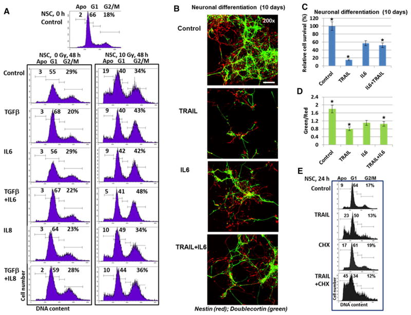
Cell cycle and neuronal differentiation of NSC: effects of cytokines and TGFβ1. a Effects of growth factor/cytokines on the cell cycle-apoptosis of control and irradiated NSC. Recombinant TGFβ1 (20 ng/ml), IL6 (50 ng/ml) and IL8 (50 ng/ml) alone or in combination were added to serum-free NSC media. Cell cycle-apoptosis analysis was performed using PI staining and FACS analysis 48 h after culture initiation. Results of a typical experiment (one from four) are shown. b, c, d Neuronal differentiation of NSC was initiated by serum-free differentiation media in the presence or in the absence of TRAIL (50 mg/ml), IL6 (50 ng/ml) or TRAIL + IL6. After 10 days of growth cells were fixed and used for immunostaining. Confocal analysis of immunofluorescent images was performed using monoclonal Ab against an early neuroprogenitor marker, Nestin (red), and polyclonal Ab against a neuronal marker, Doublecortin (green). Bar = 50 μm. Typical images are shown in b. A ratio of the number of green cells to the number of red cells and relative cell survival 10 days after initiation of differentiation were determined. Pooled results of three independent experiments are shown in c, d. Error bars represent means ± SD (p <0.05, Student’s t test); star indicates a significant difference. e Effects of TRAIL (50 ng/ml), CHX (1 μg/ ml), and TRAIL + CHX on induction of apoptosis in NSC 24 h after treatment

