

Abstract
Peripheral ossifying fibroma (POF) is a reactive gingival overgrowth occurring frequently in the maxillary anterior region of teenagers and young adults. The pink to red growth may be firm to hard in consistency depending on the amount of bone it contains and may become ulcerated as its size increases. POF is commonly associated with poor oral hygiene and early periodontal disease with majority of cases showing no marked underlying bone involvement radiographically. We report an unusual case of a large POF overlying the edentulous mandibular alveolar ridge along with histopathological correlations. This case could be one of the first to demonstrate that there may be a variant of POF affecting edentulous mandibular mucosa that has not been previously recognized.
Keywords: Peripheral ossifying fibroma, Edentulous, Mandible
Introduction
Peripheral ossifying fibroma (POF) is a non neoplastic enlargement of the gingiva that is thought to be reactive in nature. It represents up to 2% of all oral lesions [1]. Other terms used in the reference to POF are peripheral cementifying fibroma, peripheral fibroma with cementogenesis, peripheral fibroma with osteogenesis, peripheral fibroma with calcification, calcified or ossified fibrous epulis, and calcified fibroblastic granuloma [2, 3]. POF affects females more than males, with the anterior maxilla being the most common location of involvement. The lesion occurs in any age group, predominating in the second decade of life [2]. POF is typically a solitary, slow growing nodular mass that is either pedunculated or sessile. The outer surface of POF is usually smooth or ulcerated and pink to red in colour.
The definitive diagnosis is based on histological examination, with identification of cellular connective tissue and the focal presence of bone or other calcifications. Surgery is the treatment of choice, though it has a recurrence rate of 20% [1–3]. POF shows a clinically benign behaviour.
Case Report
A 55-year-old Asian female presented to Terna Dental College with a growth in the oral cavity that had enlarged gradually over the past 12 months. The patient gave history of multiple extractions over the past 2 years. The growth was painless and her main concern was aesthetic and prosthetic rehabilitation.
Clinical examination revealed a round, sessile, non-tender, pink mass with intermittent erythematous islands on the right anterior alveolar mucosa of her edentulous mandible (Fig. 1). It measured 2.5 cm in diameter. The growth had a broad based attachment to the surrounding mucosa, was nonfluctuant and had a firm, rubbery consistency. It did not blanch with pressure.
Fig. 1.
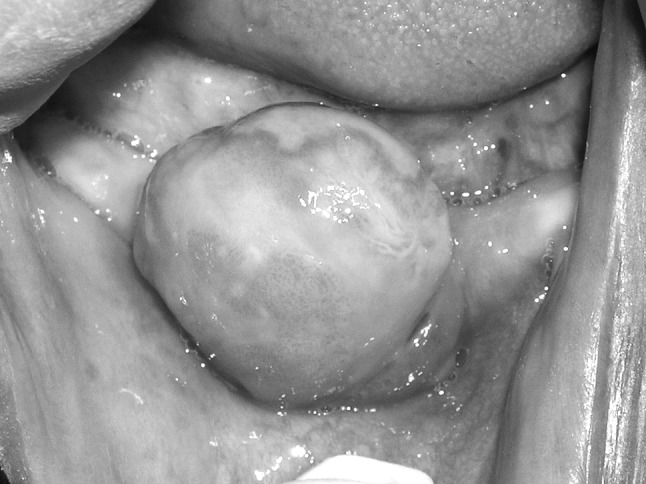
Growth in right edentulous mandibular alveolar ridge
The patient’s medical history was significant for measles, typhoid and urinary tract infections some years ago and malaria in year 2008. She gave no history of smoking, tobacco and betelnut consumption. She was not on any medication. Panoramic radiograph was obtained and was within normal limits with no findings pertaining to the mandibular growth. The differential diagnosis consisted of fibroma, pyogenic granuloma and peripheral giant cell granuloma. The same was discussed with the patient and a surgical excision was planned. Routine blood chemistry and complete blood count levels were within normal limits. Under local anaesthesia, the mass was excised completely along with adjacent mucosa and periosteum (Fig. 2). Haemostasis was achieved and wound sutured with black silk. The mass was submitted to Oral Pathology department for histopathology. The histopathology diagnosis was POF (Fig. 3). The tumour revealed peripheral calcification without adjacent bone changes. The patient presented for follow up on the 3rd, 10th and 21st day postoperatively. Her recovery was uneventful.
Fig. 2.

Photograph showing surgically excised growth
Fig. 3.

Photomicrograph (H & E stain, ×40) fibrous connective tissue stroma showing eosinophilic globules of osteoid tissue
Discussion
Yih et al. [4] and Sciubba and Younai [5] attributed the first description of this disorder to Menzel in 1872. In 1927, Montgomery [6] first used the term ossifying fibroma, by which the lesion is currently known. Ossifying fibroma is a benign bone neoplasm of the jaws composed of fibrous connective tissue with variable amounts of mineralization. The lesion is generally encapsulated—a fact that serves to distinguish it from fibrous dysplasia, which may exhibit similar clinicopathological features. Although the lesion is preferentially located in the jaws, it can also be found elsewhere—including the frontal, ethmoid, sphenoid and temporal bones or orbit, as well as in the anterior cranial fossa [7, 8].
POF may present as a pedunculated nodule, or it may have a broad attachment base [7]. These lesions can be red to pink with areas of ulceration, and their surface may be smooth or irregular. Although they are generally <2 cm in diameter, size can vary; reports range from 0.2 to 3.0 cm [9] and 2 mm to 8 cm [10] and some lesions may be as large as 9 cm in diameter [11].
The female to male ratio reported in the literature varies from 1.22:1 [12] to 4.3:1. POF predominantly occurs in the second decade, with a declining incidence in later years. There are 2 reported cases of POF present at birth, presenting clinically as congenital epuli. In a study, Cuisia and Brannon [9] reported that only 134 out of 657 diagnosed POFs (20%) were in the pediatric population (0–19 years), with 8% in the first decade. In a retrospective study of 431 cases in the Chinese population by Zhang and others [12], the mean age of incidence of POF was found to be 44 years, which is contradictory to previously published literature. POF appears to be more common among people with lighter skin colour [9] and slightly less common among those of Hispanic origin [12].
Histologically, the POF appears to be a nonencapsulated mass of cellular fibroblastic connective tissue of mesenchymal origin, covered with stratified squamous epithelium, which is ulcerated in 23–66% of cases [13]. Most ulcerated lesions occur in patients in the second decade [13]. POFs contain areas of fibrous connective tissue, endothelial proliferation and mineralization. Endothelial proliferation can be profuse in the areas of ulceration, which can be misleading in clinical diagnosis, as the lesion may appear to be a pyogenic granuloma. The mineralized component of POF varies, occurring in approximately 23–35% [12] and 50–75% [13] of cases according to published reports. Mineralization can vary between cementum-like material, bone (woven and lamellar) and dystrophic calcification. The POF lesion is generally small and does not require imaging beyond radiographs. Treatment consists of conservative surgical excision and scaling of adjacent teeth. The rate of recurrence has been reported at 8.9% [14], 9% [9], and 16% [13]. Therefore, regular follow-up is required.
Contributor Information
Sanjay Joshi, Phone: +919869057337, Email: sanjoshi99@gmail.com.
Sima Mazumdar, Email: simaaz@rediffmail.com.
Manish Kumar Pandit, Email: drmanish23@gmail.com.
References
- 1.Batsakis JG. Non-odontogenic tumors: clinical evaluation and pathology. In: Thawley SE, Panje WR, Batsakis JG, Lindberg RD, editors. Comprehensive management of head and neck tumors. 2. Philadelphia: Saunders; 1999. pp. 1641–1642. [Google Scholar]
- 2.Kumar SK, Ram S, Jorgensen MG, Shulere CF, Sedghizadeh PP. Multicentric peripheral ossifying fibroma. J Oral Sci. 2006;48:239–243. doi: 10.2334/josnusd.48.239. [DOI] [PubMed] [Google Scholar]
- 3.Moon WJ, Choi SY, Chung EC, Kwon KH, Chae SW. Peripheral ossifying fibroma in the oral cavity: CT and MR findings. Dentomaxillofac Radiol. 2007;36:180–182. doi: 10.1259/dmfr/59377498. [DOI] [PubMed] [Google Scholar]
- 4.Yih WY, Pederson GT, Bartley MH. Multiple familial ossifying fibromas: relationship to other osseous lesions of the jaws. Oral Surg Oral Med Oral Pathol. 1989;68:754–758. doi: 10.1016/0030-4220(89)90167-9. [DOI] [PubMed] [Google Scholar]
- 5.Sciubba JJ, Younai F. Ossifying fibroma of the mandible and maxilla: review of 18 cases. J Oral Pathol Med. 1989;18:315–321. doi: 10.1111/j.1600-0714.1989.tb01559.x. [DOI] [PubMed] [Google Scholar]
- 6.Pérez-García S, Berini-Aytés L, Gay-Escoda C. Ossifying fibroma of the upper jaw: report of a case and review of the literature. Med oral. 2004;9:333–339. [PubMed] [Google Scholar]
- 7.Neville BW, Damm DD, Allen CM, Bouquot JE. Oral and maxillofacial pathology. 2. Philadelphia: Saunders; 2202. pp. 563–564. [Google Scholar]
- 8.MacDonald-Jankowski DS. Cemento-ossifying fibromas in the jaws of Hong Kong Chinese. Dentomaxillofac Radiol. 1998;27:298–304. doi: 10.1038/sj.dmfr.4600378. [DOI] [PubMed] [Google Scholar]
- 9.Cuisia ZE, Brannon RB. Peripheral ossifying fibroma–a clinical evaluation of 134 pediatric cases. Pediatr Dent. 2001;23(3):245–248. [PubMed] [Google Scholar]
- 10.Bodner L, Dayan D. Growth potential of peripheral ossifying fibroma. J Clin Periodontol. 1987;14(9):551–554. doi: 10.1111/j.1600-051X.1987.tb00998.x. [DOI] [PubMed] [Google Scholar]
- 11.Poon CK, Kwan PC, Chao SY. Giant peripheral ossifying fibroma of the maxilla: report of a case. J Oral Maxillofac Surg. 1995;53(6):695–698. doi: 10.1016/0278-2391(95)90174-4. [DOI] [PubMed] [Google Scholar]
- 12.Zhang W, Chen Y, An Z, Geng N, Bao D. Reactive gingival lesions: a retrospective study of 2, 439 cases. Quintessence Int. 2007;38(2):103–110. [PubMed] [Google Scholar]
- 13.Buchner A, Hansen LS. The histomorphologic spectrum of peripheral ossifying fibroma. Oral Surg Oral Med Oral Pathol. 1987;63(4):452–461. doi: 10.1016/0030-4220(87)90258-1. [DOI] [PubMed] [Google Scholar]
- 14.Bhaskar SN, Jacoway JR. Peripheral fibroma and peripheral fibroma with calcification: report of 376 cases. J Am Dent Assoc. 1966;73(6):1312–1320. doi: 10.14219/jada.archive.1966.0375. [DOI] [PubMed] [Google Scholar]