Abstract
Dental procedures done in the vicinity of dermal fillers may result in complications of the dermal fillers such as infections which may mimic a dental infection. These infections of dermal fillers must be differentiated from facial cellulitis or from dental infection as treatment for infection from dermal fillers may be prolonged with repeated use of antibiotics, incision and drainage or removal of the filler material itself. Dental surgeons need to be aware of this potential risk in order to recognize and manage it appropriately.
Keywords: Injectable dermal fillers, Dental infection, Biofilm, Cellulitis
Introduction
Non surgical rejuvenation of the ageing face has become increasingly popular in recent years over cosmetic surgery of the face. Mesotherapy, botox injection, micro-dermabraison, injectable dermal fillers are few of the methods for non surgical management of the ageing skin. These new methods are less invasive, less time consuming and give successful result with softer relaxed appearance and subtle affects [1].
Dental surgeons may encounter patients with orofacial swelling secondary to dental infection or dental procedures carried out near the region where injectable filler materials was used. These should be differentiated from facial cellulitis. A positive history of injectable dermal fillers should alert one to take precautions before carrying out dental procedures in the same region. We report a case which presented to our department with a history of infected dermal filler following a dental procedure which was carried out in the vicinity where previously dermal filler was injected. Management included prolonged course of antibiotic, surgical drainage and removable of the filler materials.
Case Report
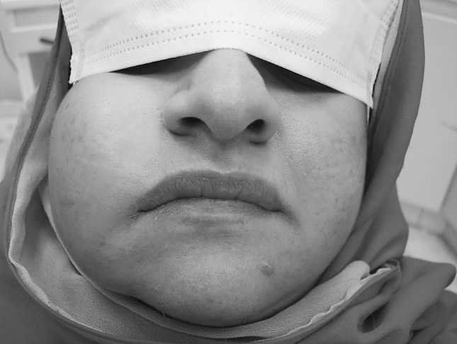
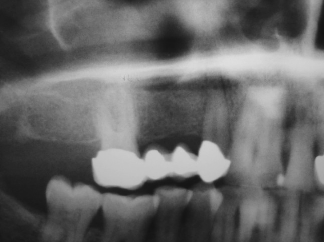
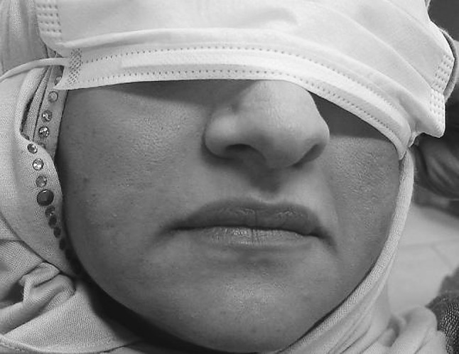
A 48 year old female patient reported to our center (Bneid-Al-Gar Dental Specialty Center, Kuwait) complaining of a swelling in her right cheek of 2 days duration. The appearance of this swelling was sudden in onset and increased progressively in size over this period (Fig. 1). She was in excellent health and taking no medications. On examination she had a diffuse swelling in her right cheek, fluctuant and tender. Oral examination did not reveal any obvious dental or oral cause for this swelling. On further questioning the patient it was determined that 2 days before the development of this swelling she had a fixed partial denture in the upper right quadrant recemented and she also mentioned that the upper right canine was slightly tender on percussion. Her past history revealed that she had received filler injection in her cheeks bilaterally about 6 years before. She was unaware of the nature of filler materials used. Radiographic examination showed widening of the periodontal ligament space in relation to the upper right canine (Fig. 2). Drainage was established through an incision intraorally through the buccal mucosa. A yellowish white discharge was obtained which was sent for microbiological examination. Culture and sensitivity test reported as staphylococcus aureus and sensitive to cloxacillin. The patient was admitted and started on IV antibiotics. The response to the antibiotics was slow and pus continued to drain though in decreasing quantity. At the time of discharge the swelling had decreased considerably but had not resolved completely. She was discharged after a week and was given oral antibiotics for a further period till the swelling subsided completely (Fig. 3).
Fig. 1.

Photograph showing diffuse swelling of the right cheek
Fig. 2.

Radiograph showing widening of periodontal ligament space in relation to the right canine
Fig. 3.

10th day post-operative picture
Discussion
This case has been reported here to highlight the potential risk of dental procedures or infections to dermal fillers. Injectable filler materials for facial rejuvenation has reduced the need for complicated facial surgery but these materials themselves have presented complications like formation of nodules, granulomas, abscess and recurrent infection at the injection site. The causes of these complications are multifactorial and may include foreign body reaction or the emergence of a biofilm infection [2]. Biofilm may be a potential source for many of the late complications like granulomas or abscesses. A biofilm is a quiescent infection by bacteria introduced probably at the time of injection, resulting in the formation of a structured community of micro-organisms adherent to an inert surface and encapsulated by a protective self developed polymeric matrix [3]. It is characterized as a living colony adherent to the foreign implant with surface attachment, structural heterogeneity, genetic diversity, complex community interactions and a protective extracellular matrix of polymeric substance such as polysaccharide [4]. They are able to maintain their integrity against the host immune system by reduced metabolism and growth rate. They become antibiotic resistant by lowering its metabolism and are protected from phagocytosis by the extrapolymeric system membrane. The antibiotic resistance is profound because of the changed biofilm environment and quorum sensing (cell to cell signaling throughout the colony) [5]. It is reported that biofilm allow up to 1,000 times better resistance to antibiotics [6]. This may be the reason the response of our patient to antibiotic therapy was very slow.
Bacteria are the prime source of biofilm [7]. The free floating bacteria in tissues become adherent to the foreign body material and thus develop biofilm. Bacteria from dental work, soft tissue surgery or trauma can activate an infective response of this biofilm. Our patient had tenderness of her upper right canine and dental procedure like recementation of her fixed partial denture was done a few days prior to the development of the swelling. Tenderness of her canine disappeared once the FPD was recemented. No other cause for the buccal infection could be determined. It is possible that this dental procedure could have activated an infective response in the dermal filler. Once the biofilm is activated it becomes an acute infection or may take a sub-acute course and result in a granulomatous response. The active infection can be controlled with antibiotics but may recur due to the underlying biofilm [8]. Chronicity and recurrence is the hallmark of biofilm reactivation. Removal of implant and biofilm is the only solution for recurrent infections. A positive history of use of dermal fillers should caution one when carrying out dental treatment in the vicinity of these filler materials. Prophylactic use of antibiotics is debatable which might need further research.
Conclusion
With the increased use of cosmetic dermal fillers, dental professionals are likely to come across facial infection or inflammation which may mimic a dental infection and the severity of which may be bigger than the dental problem may warrant. Attention to the possibility of a pathological relationship between dental procedures and dermal fillers may help one to recognize and initiate appropriate treatment.
References
- 1.Lowe NJ, Maxmell CA, Patnail R. Adverse reaction to dermal fillers. Review. Dermatol Surg. 2005;31(2):1616–1625. [PubMed] [Google Scholar]
- 2.Monheit GD, Rohrich RJ. The nature of long term fillers and the risk of complications. Dermatol Surg. 2009;35:1598–1604. doi: 10.1111/j.1524-4725.2009.01336.x. [DOI] [PubMed] [Google Scholar]
- 3.Mertz PM. Cutaneous biofilm: friend or foe? Wounds. 2003;15:129–132. [Google Scholar]
- 4.Prigent-combaret C, Vidal O, Dorel C, Lejeune P. Abiotic surface sensing and biofilm-dependent regulation of gene expression in Escherichia coli. J. Bact. 1999;181:5993–6002. doi: 10.1128/jb.181.19.5993-6002.1999. [DOI] [PMC free article] [PubMed] [Google Scholar]
- 5.Jayaram A, Wood TK. Bacterial quorum sensing signals circuits and implications for biofilm and disease. Annu Biomed Eng. 2008;10:145–167. doi: 10.1146/annurev.bioeng.10.061807.160536. [DOI] [PubMed] [Google Scholar]
- 6.Ceri H, Olson ME, Stremick C, et al. The calgary biofilm device: new technologies for rapid determination of antibiotic susceptibilities of bacterial biofilm. J Clin Microbiol. 1999;37:1771–1776. doi: 10.1128/jcm.37.6.1771-1776.1999. [DOI] [PMC free article] [PubMed] [Google Scholar]
- 7.Costerton JW, Steward PS, Greenberg EP. Bacterial biofilm: a common cause of persistent infections. Science. 1999;284:1318–1322. doi: 10.1126/science.284.5418.1318. [DOI] [PubMed] [Google Scholar]
- 8.Dijkema SJ, Van der Lie B, Kibbelaar RE. New-fill injections may induce late-onset foreign body granulomatous reaction. Plast Reconstr Surg. 2005;115:76e–78e. doi: 10.1097/01.PRS.0000157022.61766.5D. [DOI] [PubMed] [Google Scholar]


