Abstract
Neonatal septicemia caused by resistant bacteria in intensive care units can be not only life threatening, but may also cause long lasting sequelae. A case of a 2 month old male child with osteomyelitis with pathological fracture of the mandible following methicillin resistant Staphylococcus aureus septicemia is presented. Surgical management, successful outcome and its long term effect on the growth of mandible and dentition is illustrated.
Introduction
Acute osteomyelitis [1, 8] is rarely encountered these days with the advent of modern antibiotics and better health care protocols. This acute infection of bone and bone marrow is nowadays seen when it is caused by drug resistant strains of pyogenic organisms [2].
Acute osteomyelitis in general occurs due to hematogenous spread, or due to direct inoculation of microorganism. Osteomyelitis in neonates [1, 4] is often seen as a consequence of septicemia due to hematogenous spread. Hematogenous osteomyelitis [6] occurs mostly in children due to bacterial seeding within the bone from a remote source, through the circulating blood. Direct inoculation osteomyelitis in the mandible occurs due to direct introduction of bacteria into bone, such as following trauma or uncontrolled odontogenic infection.
Case Report
A 2 month old male infant was referred by the pediatrician due to a painful swelling on the left side of the jaw since the past 3 days. Pre-natal history was normal, and both parents were healthy. The baby was born in another hospital by normal vaginal delivery, and was admitted in the Neonatal Intensive Care Unit (NICU) for a few days to manage hyperbilirubinemia. The child developed a skin infection (Nappy rash) when he was in the NICU, but later he was discharged uneventfully.
He had to be readmitted on the seventh day of discharge due to fever. Blood culture revealed Staphylococcus aureus and appropriate antibiotics were started. Subsequently he developed a Gluteal abscess which was surgically drained. Culture of the pus showed the same organism.
Three weeks later, while still on antibiotics, he developed fever and a painful swelling in the left angle of the mandible. He was having difficulty in suckling. Examination showed a tender swelling in that region, with a pointing abscess (Fig. 1) A working diagnosis of Submasseteric abscess was made which was drained by submandibular incision under general anesthesia. Significant amount of pus was drained and coagulase positive MRSA was cultured, which was resistant to all antibiotics except Gatifloxacin & Meropenem.
Fig. 1.
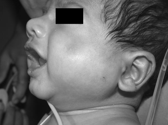
Child with submassteric abscess
Consequently Meropenem was started, and the baby improved significantly enough to be discharged within 1 week. The blood count and C-reactive protein (CRP) titers normalized and he was put on oral Gatifloxacin. The baby was brought back within a week from the date of discharge with fever and swelling in the same region. Computed axial tomography (CT scan) revealed extensive destruction of the left mandibular ramus, with a pathologic fracture at the sub condylar region (Fig. 2).
Fig. 2.
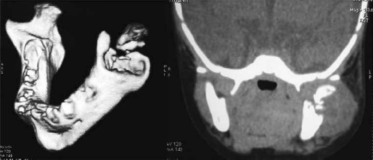
CT showing pathological fracture in subcondylar area & extensive destruction of ramus
A provisional diagnosis of osteomyelitis was made, and pus was drained intra-orally, through a buccal sulcal incision. Thorough curettage was done and the specimen was sent for histopathologic examination and microbiological studies. The bone destruction was so extensive that, only the myo-periosteal scaffold remained in the region.
A continuous closed suction-irrigation system (CSIS) [7] (Fig 3) was placed in infected space, and a water tight closure of the intra oral incision was achieved. There were no readymade devices for suction-irrigation available. So one was custom designed for our situation with materials available to us.
Fig. 3.
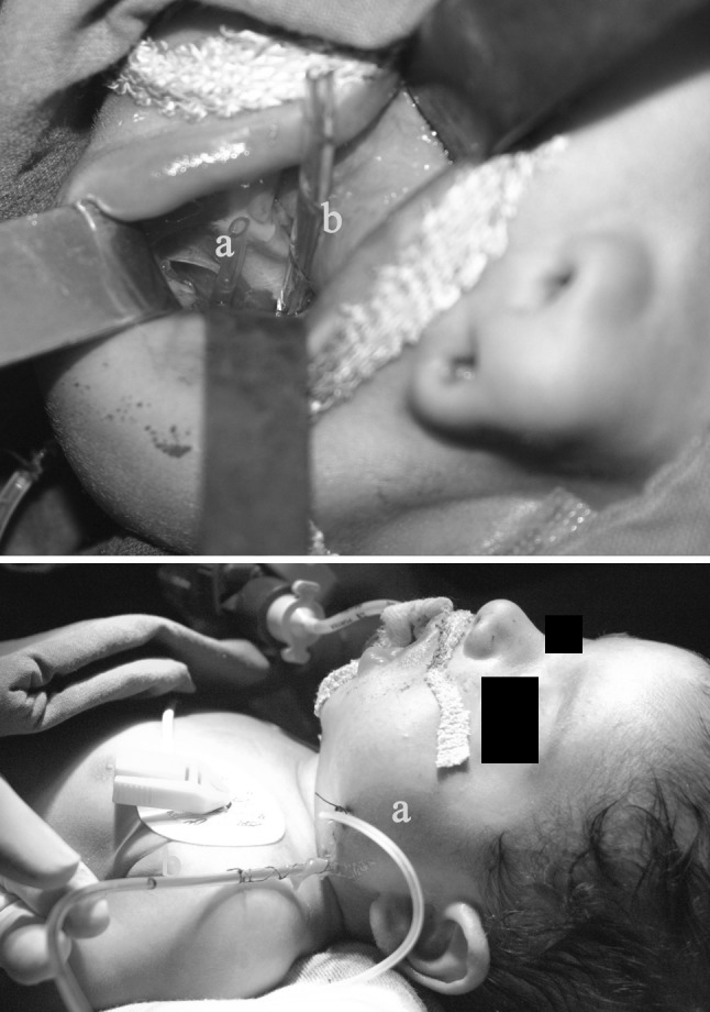
Placement and fixation of closed suction irrigation system (CSIS)
As the name suggests, closed suction-irrigation system (CSIS) has two components—one part for irrigation (Tube ‘a’ in Fig. 3), which was designed using a butterfly Cannula with the needle removed. This is then inserted to the cavity by a percutaneous stab incision. The second component for suction (Tube ‘b’ in Fig. 3) comprised of a small vacuum drain (Removac). Both the tubes were fixed firmly on the skin with silk sutures, and a watertight closure of the intra oral wound was secured so that saliva would not be sucked into the cavity. Alternate Irrigation with saline and antibiotic was done at regular intervals using a 10 cc syringe. The device was retained for 3 days.
Intravenous (I V) Linezolid was started following discussions with the microbiologist. A regular saline with antibiotic irrigation was done through the suction-irrigation system for 3 days. Infection was controlled satisfactorily. I V Linezolid was continued for 10 days and then continued orally for another week. Provisional diagnosis was confirmed by the histopathology report. The child was subsequently discharged, with no restriction on normal oral movements. Parents were advised not lay the child on the affected side. Regular follow up was ensured and close monitoring was done to look for early signs of ankylosis the possibility of future facial deformity was discussed with the parents.
Patient was closely followed up for 1 month. There were no signs of recurrence of infection or Ankylosis during this period. The child was followed up to 3 years at 3 month intervals to look for any discrepancy in the growth of the mandible. Temporomandibular (TMJ) joint movements and development of the dentition were also closely monitored. Orthopantomogram (OPG) was also done at regular intervals. There was no significant alteration in growth of the mandible or deviation in TMJ movements.
Except for a mild flattening of the left angle region which is hardly noticeable, child has near normal mouth opening and facial development after three and half years (Fig. 4) OPG revealed accentuated Antigonial notch on the left side (Fig. 5). Even though significant damage to the teeth buds in the area was expected, since they were the suspected foci of infections, and due to the extensive destruction of the mandibular ramus observed in the CT scan, surprisingly there was only enamel hypoplasia of deciduous teeth on the affected side (Fig. 6). Despite the hypoplasia, the affected teeth were totally asymptomatic.
Fig. 4.
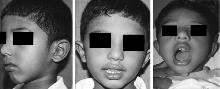
Mouth opening and facial growth after 3 years
Fig. 5.
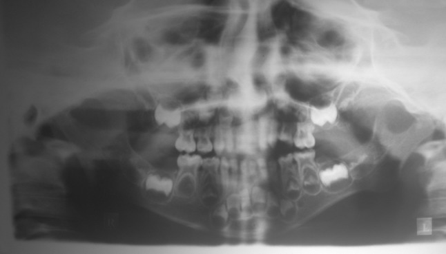
OPG at 3 years
Fig. 6.
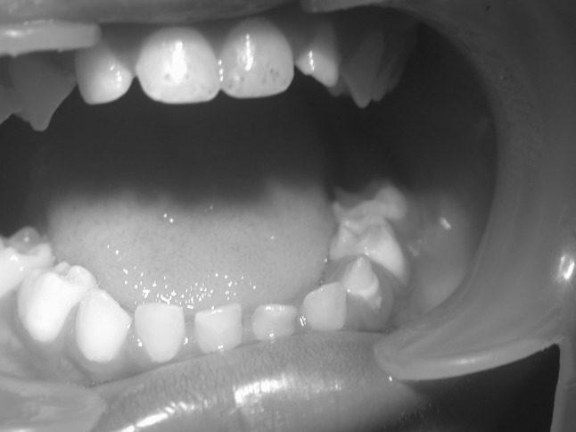
Enamel hypoplasia of primary dentition
Discussion
Nosocomial infections are by and large preventable. Even though MRSA septicemia [1, 3, 4] is common in NICUs, very few cases are reported in the literature, about osteomyelitis of the mandible resulting from MRSA infection in neonates. Since osteomyelitis of the infant mandible has the potential to cause devastating esthetic and functional consequences in the future, its early diagnosis, followed by aggressive surgical & medical management is warranted.
Taking an intraoral periapical X-ray, or an orthopantomogram may not be practical in a two old month infant. The CT scan may be a more practical option for radiographic imaging in such cases [6], even though the amount of radiation may be slightly higher. Appropriate use of modern imaging techniques like three dimensional (3D) CT scans can be helpful to localize and study the extent of the lesion.
Linezolid, a synthetic Antibiotic categorized under the Oxazolidine group [2, 5] is effective against Vancomycin resistant Staphylococcus aureus (VRSA) and MRSA. It can be given for long durations with relatively less adverse effects. It can also be given orally during convalescence.
The Closed suction-irrigation system (CSIS), is common in orthopeadics. It was adopted in this particular case and found effective for continuous irrigation, with the added benefit of local delivery of antibiotic at the infected site and removal of collected fluid. It is especially beneficial in the treatment of osteomyelitis, as local vascularity is compromised.
Healing of the pathologic fracture without stabilization confirms the inherent healing capacity and osteogenic potential of an infants’ mandible. Healing has taken place well without any mode of immobilisation. Moreover, even though there was a large area of bone destruction in the fracture site, the bony segments were not displaced. It was managed as an undisplaced subcondylar fracture, especially because the child did not have any teeth to occlude, and also because the infants diet was entirely milk & liquids!!.
Ankylosis and subsequent facial deformity was a potential late complication that was expected in this case, as the lesion was close to the joint and the pathological fracture at the subcondylar level Parents were advised not to lay the child at the fractured side and to closely observe for any signs of ankylosis. Functional movements of mandible were ascertained during the healing period. Tight bandages and dressings were avoided. Parents were briefed about the about the early signs of Ankylosis so that they could closely observe the child for the signs and inform us accordingly. There was a possiblty of pseudojont formation at the fracture site and deviated jaw movements to the affected side, but the fracture healed perfectly without any deficit.
Conclusion
In this particular case, after 3 years of follow up, it can safely be concluded that the pathologic fracture has healed without any functional or gross esthetic problems. The decision to manage the fracture conservatively without disturbing the condyle, and opting for early mobilization contributed to the successful outcome. The condyle being one of the growth centers, any surgical trauma to the region can lead to significant esthetic & functional problems in the growing mandible.
Here, the dilemma was between aggressive removal of infected tissue and consequent possibility of future deformity, or a conservative approach by maintaining the proximal segment and the risk of incomplete removal of the focus of infection. Three year follow up proves that this balancing act has been successful.
Dealing with recurrent infection at this age, in the same crucial area is very frustrating. We could not find similar situations in the available literature for guidance. The unit that treated the child is a very busy and renouned Neonatal care center in the region. They had encountered & treated many cases of metastatic osteomyelitis in the long bones but never had a case of blood borne osteomyelitis of the jaw bones. This case also highlights the fact that, in fracture healing, we have only less than fifty percent involvement. The rest is done by nature. The surgeons role is only to make the situation condusive for the nature to act. Our hope is that this report will be useful for clinicians who come across similar situations.
Contributor Information
Paul V. Joseph, Phone: +04842424112, Phone: +0483705602, FAX: +04832708353, Email: paulvetticappally@yahoo.com
Ajay Kumar Haridas, Phone: +04942698544, Phone: +0483705602, FAX: +04832708353, Email: ajaykharidas@gmail.com.
References
- 1.Bulbul A, Okan F. Acute osteomyelitis of the iliac bone presenting with gluteal syndrome in a newborn. Eur J Pediatr. 2009;168(12):1529–1532. doi: 10.1007/s00431-009-0948-6. [DOI] [PubMed] [Google Scholar]
- 2.Eckmann C, Dryden M. Treatment of complicated skin and soft-tissue infections caused by resistant bacteria: value of linezolid, tigecycline, daptomycin and vancomycin. Eur J Med Res. 2010;15(12):554–563. doi: 10.1186/2047-783X-15-12-554. [DOI] [PMC free article] [PubMed] [Google Scholar]
- 3.Gelfand MS, Cleveland KO, Heck RK, Goswami R. Pathological fracture in acute osteomyelitis of long bones secondary to community-acquired methicillin-resistant Staphylococcus aureus: two cases and review of the literature. Am J Med Sci. 2006;332(6):357–360. doi: 10.1097/00000441-200612000-00010. [DOI] [PubMed] [Google Scholar]
- 4.Korakaki E, Aligizakis A. Methicillin-resistant Staphylococcus aureus osteomyelitis and septic arthritis in neonates: diagnosis and management. Japn J Infect Dis. 2007;60(2–3):129–131. [PubMed] [Google Scholar]
- 5.Paul WA, Jamshed N, and Horne JP (2002) Latrobe area hospital, Latrobe, Pennsylvania linezolid its role in the treatment of gram-positive, drug-resistant bacterial infections. In: Text Book of Clin Pharmacol, vol 65, pp 633–670 [PubMed]
- 6.Gold R (1991) Professor of pediatrics, diagnosis of osteomyelitis. Pediatr Rev 12(10):292–7 [DOI] [PubMed]
- 7.Rohmiller MT, Akbarnia BA (2010) Closed suction irrigation for the treatment of postoperative wound infections following posterior spinal fusion and instrumentation, vol 35. Lippincott Williams & Wilkins, Spine, pp 642–646 [DOI] [PubMed]
- 8.Sivapathasundaram B. Diseases of the pulp & periapical tissues. In: Rajendran R, Sivapathasundaram B, editors. Shafer’s text book of oral pathology. 6. Amsterdam: Elsevier; 2009. pp. 492–498. [Google Scholar]


