Abstract
Neurogenic neoplasms presenting as parotid gland tumors are extremely uncommon. We present the case of a solitary neurofibroma of the facial nerve within the parotid gland occurring in a 36-year-old male who presented with a painless enlargement in the right-side region of the parotid gland over a period of 1 year with normal facial nerve function. Magnetic resonance imaging revealed a well-demarcated round mass within the right parotid. At surgery, a tumor was found involving the main trunk of the facial nerve, histopathologically representing a neurofibroma.
Keywords: Parotid tumour, Neurofibroma, Facial nerve
Introduction
The intra-parotid tumors of facial nerve origin are rare and their estimated incidence ranges from 0.2 to 1.5 % [1]. More specifically, 79 cases have been reported in the literature involving the intraparotid segment of the facial nerve [2]. The common tumors which arise from nerve tissue are Neurofibroma and schwannoma [3]. Neurofibromas constitute only 0.4 % of all salivary neoplasms [4].
Rarity of this tumor precludes its inclusion in the differential diagnosis of benign parotid tumors at the provisional diagnosis stage. Preoperative diagnosis is extremely difficult due to the variation in clinical presentation and its dependency upon the nerve site involved. Almost half of the intraparotid neurofibromas involve the main trunk of facial nerve and usually present as a painful or painless parotid mass of long standing duration with or without facial paresis, paralysis or spasm [5]. Such a presentation is similar to any other benign parotid tumor making it difficult to diagnose solitary neurofibromas at the clinical stage. Radiological findings of intraparotid neurofibromas are contributory only to the diagnosis of a benign parotid tumor. Sectional imaging helps to determine the extent of the tumor. Recently, a case of solitary neurofibroma involving the whole facial nerve continuously from geniculate ganglion to parotideal segment has been reported [6]. The main basis of diagnosis of neurofibroma is histopathological investigations. These tumors present a challenge because of their risk of malignant transformation and their high probability of operative facial nerve compromise.
We present a case which was diagnosed as a benign parotid tumor preoperatively and following surgical excision and histopathological examination, was diagnosed as a neurofibroma. The diagnostic problems, the biological behavior and therapeutic management of this infrequent tumor entity are discussed.
Case Report
A 36-year-old male patient reported with swelling of the right side of face of 1 year duration. The swelling was spontaneous in onset and had progressed slowly to the present size over the last 1 year. The swelling was not associated with pain or fever. The patient did not report of any loss of function, particularly, facial paresis or paralysis.
General physical examination did not reveal any significant findings. On local examination there was a large (9 × 5 cm), firm, ovoid, nodular mass present in right preauricular region extending to both anterior and posterior triangles of neck. The surface of the swelling was tense and glossy with localized surface erosion present. The edge of this sessile swelling was clearly defined. On palpation, inspectory findings of size, shape, borders and extent were confirmed. The swelling was non-tender, lobulated and of variable consistency. Fluctuation and transillumination were negative. The swelling was neither compressible nor reducible. The swelling was not associated with cervical lymphadenopathy and facial nerve and intra-oral examinations were normal.
Laboratory investigations showed that total leucocyte count was 5,100 mm3, hemoglobin was 10.5 g % and other routine blood and urine investigations were within normal limits. Postero-anterior (PA) view of skull did not show any mandibular changes. Chest radiograph was normal. Fine needle aspiration cytology (FNAC) of the lesion revealed few clumps of spindle shaped cells in background of disintegrated erythrocytes (Fig. 1).
Fig. 1.
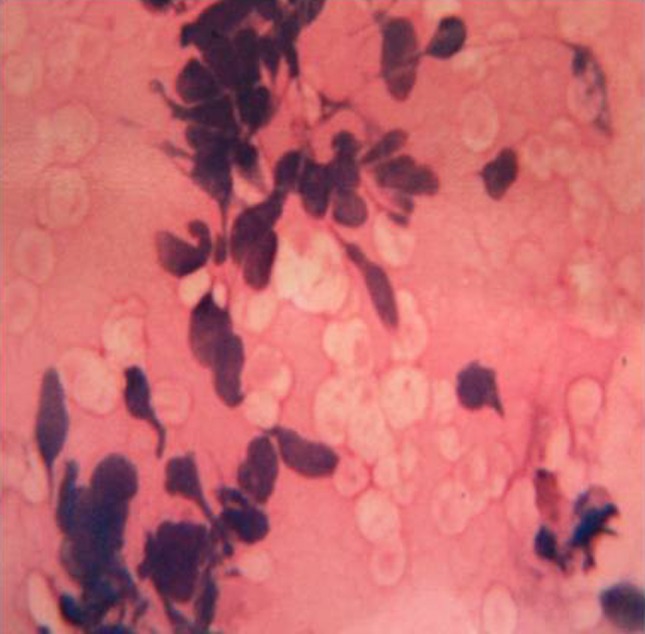
×40 magnification of fine needle aspiration cytology of the lesion revealed few clumps of spindle shaped cells in background of disintegrated erythrocytes
Magnetic resonance imaging (MRI) revealed a 9.3 × 4.9 cm well defined soft tissue lesion involving the right parotid gland extending to anterior and posterior triangle of neck. The lesion was isodense on T1 W and hyperdense on T2 W sequences (with respect to neck muscles) and showed marked enhancement following contrast. Focal areas of necrosis and cystic changes were appreciable (Fig. 2).
Fig. 2.
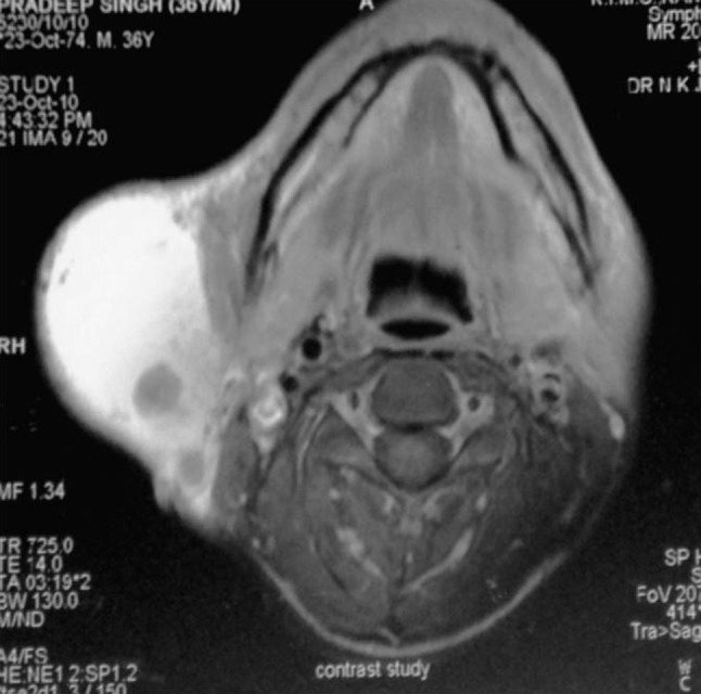
Axial gadolinium-enhanced T1-weighted image (spin echo; TR/TE, 725/14) shows a mass in the right parotid gland with heterogeneous enhancement and areas of focal necrosis
Management
The lesion was surgically removed and sent for histopathologic examination (Fig. 3). An S-shaped cervico-mastoid-facial incision was made and facial nerve identified. The main trunk of the facial nerve was adherent to the dorsal side of the lesion. Nonetheless, although management of neurofibromas tend to involve en bloc resection of the tumor together with the involved nerve due to its incorporation, considering that during the surgery it was possible to resect the tumor from the nerve rather easily, we decided to preserve the nerve (Fig. 4). Superficial parotidectomy was performed. Histopathological examination indicated a benign spindle cell tumor composed of uniformly sized spindle cells with benign slender oval nuclei with pointed ends which was consistent with neurofibroma (Fig. 5). Due to financial constraints by the patient, immunohistochemistry was not possible and the diagnosis was made purely on histological grounds. There was transient facial nerve paresis due to neuropraxia in the post-operative period (Fig. 6). However the patient completely recovered at 3 months follow-up.
Fig. 3.
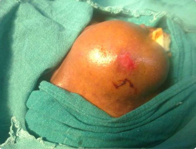
Clinical photograph shows draped pre-operative view of the parotid swelling
Fig. 4.
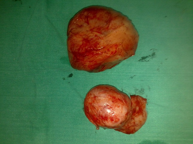
Photograph of the excised surgical specimen
Fig. 5.
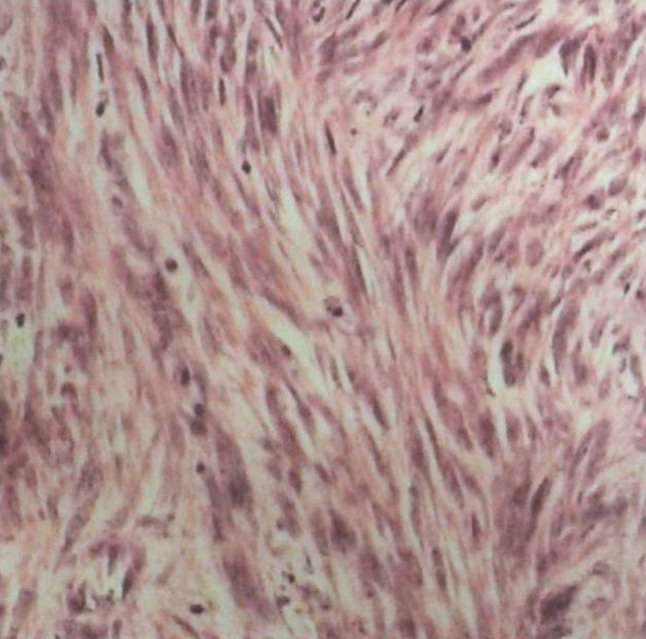
×40 view of histopathological examination showing uniformly sized spindle cells with benign slender oval nuclei with pointed ends
Fig. 6.
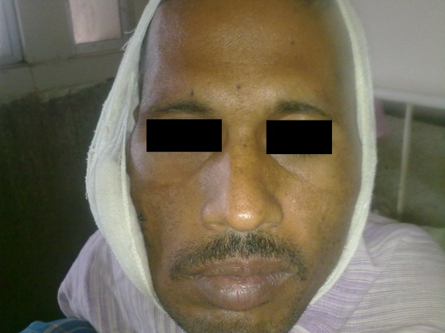
Post-operative clinical photograph showing inability of patient to completely close his right eye
Discussion
Tumors of parotid gland are usually benign and the most common tumor is pleomorphic adenoma of parotid [7]. Tumors of nerve tissue origin presenting as intraparotid mass are usually noted for their rarity. These benign tumors arise mainly from schwann cells and are mostly of two types- schwannoma, which is more common, and neurofibroma, which is rare [8]. Like schwannoma, neurofibroma are typically benign. The schwann cells in these tumors, however, exhibit balletic inactivation of the neurofibromatosis gene. Another difference between schwannomas and neurofibromas is that the former are made of only schwann cells, whereas neurofibromas are formed with many different types of cells. Grossly, schwannomas are solitary, encapsulated tumors usually attached to or surrounded by nerve. It appears to push axons aside and degenerative changes like cystic alterations or hemorrhagic necrosis are usually present. In contrast, the neurofibroma is nonencapsulated and frequently multiple. Axons pass directly through neurofibromas and regressive changes are less common [8]. Neurofibromas may be solitary or multiple, sporadic or may be associated with neurofibromatosis I or II syndromes in 10 % of cases [9].
Intra-parotid neurofibroma usually presents as a slow growing pre auricular mass of long standing duration. The patients usually do not report of associated symptoms of pain, or facial nerve palsy or paresis [5, 10]. Therefore, these tumors are more commonly diagnosed following the histopathological examination of the surgical excision specimen, rather than a pre-operative diagnosis. In few cases, patients have reported with facial paresis and hearing loss as the chief complaint [8]. In the present case, the patient presented with slowly progressive swelling of 1 year duration in the right pre-auricular region.
Complete work-up of the patient includes imaging techniques like conventional radiographs, ultrasonography (USG), computed tomography (CT), MRI. Also, FNAC may be useful, whereas the final diagnosis is established after histopathologic examination. Further, immunohistochemistry may be used to distinguish closely associated other fibrous tumors of parotid.
Conventional radiographs like PA view of the skull or puff-parotid view usually fail to show any pressure changes or other findings. In USG, CT, MRI the tumor usually presents as sharp edged mass. In particular, the MRI reveals slightly heterogeneous lesions that were isointense to brain on T1- and T2-weighted images [11]. In the present case, the PA view did not reveal any findings whereas on MRI a well-demarcated round mass within the right parotid was seen.
It is generally difficult to get a positive reliable diagnosis on FNAC because of extreme difficulty in getting positive cytology owing to the adhesive nature of cells in this tumor and cell-poor collagen fiber-rich nature of neurofibromas [10]. In the present case, few clumps of spindle shaped cells in background of disintegrated erythrocytes indicated towards a fibrous tumor of nerve tissue origin.
Histopathological examination showed proliferation of all elements of nerve which include axons, Schwann cells and fibroblasts [12]. Neurofibromas are characterized by relatively scant, haphazard arrangement of delicate spindle cells among a loosely textured collagenous matrix, and may also incorporate nerve fibers within the tumour matrix. The cellular pattern of neurofibroma is much looser than that of a schwannoma [5]. Close histopathological differential diagnosis of neurofibromas are fibrous tumors of parotid gland which can be differentiated by immunohistochemistry. In contrast to fibrous tumors which are CD 34 positive, neurofibromas are CD 34 negative and also express S-100 protein [13].
Surgical excision is the treatment of choice. Management of neurogenic tumors of the parotid facial nerve is controversial. Schwannomas tend to displace nerves and thus allow for nerve preservation procedures. Neurofibromas, however, incorporate nerves and are generally resected en bloc with the involved nerve. En bloc resection with cable grafting is recommended when nerve fibers are tenuous and interspersed within the tumor capsule [14]. The treatment of benign tumors with intact facial nerve function remains controversial. Some authors state that the results with facial nerve reconstruction are better when there is no preoperative facial weakness rather than in the presence of a long standing palsy [5]. However, others recommend that resection not be performed when all clinical parameters suggest a benign neuroma: intraoperative tumor appearance, inseparability from the facial nerve, and facial movement elicited by electrical stimulation of the tumor [1].
Conflict of interest
None.
References
- 1.Facial nerve tumor, facial nerve centre, massachusetts eye and ear infirmary, www.facialnervecentre.org. Accessed on 10 May 2014
- 2.Chiang CW, Chang YL, Lou PJ. Multicentricity of intraparotid facial nerve schwannomas. Ann Otol Rhinol Laryngol. 2001;110:871–874. doi: 10.1177/000348940111000912. [DOI] [PubMed] [Google Scholar]
- 3.McGuirt WF, Sr, Johnson PE, McGuirt WT. Intraparotid facial nerve neurofibromas. Laryngoscope. 2003;113:82–84. doi: 10.1097/00005537-200301000-00015. [DOI] [PubMed] [Google Scholar]
- 4.Seifert G, Miehlke A, Haubrich J, et al. Pathology, diagnosis, treatment, facial nerve surgery. Stuttgart: Georg Thieme Verlag; 1986. pp. 171–301. [Google Scholar]
- 5.Sullivan MJ, Babyak JW, Kartush JM. Intraparotid facial neurofibroma. Laryngoscope. 1987;97:219–223. doi: 10.1288/00005537-198702000-00016. [DOI] [PubMed] [Google Scholar]
- 6.Yilmaz F, Gurel K, Gurel S, Sessiz N, Boran C (2007) Geniculo-temporo parotideal neurofibroma of the facial nerve. A case report. Neuroradiol J 31 19(6):792–797 [DOI] [PubMed]
- 7.Speight PM, Barrett AW. Salivary gland tumours. Oral Dis. 2002;8:229–240. doi: 10.1034/j.1601-0825.2002.02870.x. [DOI] [PubMed] [Google Scholar]
- 8.Sethi A, Chopra S, Passey JC, Agarwal AK. Intraparotid facial nerve neurofibroma: an uncommon neoplasm. Int J Morphol. 2011;29(3):1054–1057. doi: 10.4067/S0717-95022011000300066. [DOI] [Google Scholar]
- 9.Seifert G, Oehne H. The mesenchymal (non-epithelial) salivary gland tumors. Laryngol Rhinol Otol. 1986;65:485–491. doi: 10.1055/s-2007-1008020. [DOI] [PubMed] [Google Scholar]
- 10.Maheshwari V, Varshney M, Alam K, Khan R, Jain A, Gaur K, Khan AH. Neurofibroma of parotid. BMJ Case Rep. 2011 doi: 10.1136/bcr.05.2011.4172. [DOI] [PMC free article] [PubMed] [Google Scholar]
- 11.Martin N, Sterkers O, Mompoint D, Nahum H. Facial nerve neuromas: MR imaging—report of four cases. Neuroradiology. 1992;34:62–67. doi: 10.1007/BF00588435. [DOI] [PubMed] [Google Scholar]
- 12.Brettau P, Melchiors H, Krogdahl A. Intraparotid neurilemmoma. Acta Otolaryngol. 1983;95(3–4):382–384. doi: 10.3109/00016488309130957. [DOI] [PubMed] [Google Scholar]
- 13.Sato J, Asakura K, Yokoyama Y, Satoh M. Solitary fibrous tumor of the parotid gland extending to the parapharyngeal space. Eur Arch Otorhinolaryngol. 1998;255:18–21. doi: 10.1007/s004050050015. [DOI] [PubMed] [Google Scholar]
- 14.Kavanaugh KT, Panje WR. Neurogenic neoplasms of the seventh clinical nerve presenting as a parotid mass. Am J Otolaryngol. 1982;3(1):53–56. doi: 10.1016/S0196-0709(82)80033-1. [DOI] [PubMed] [Google Scholar]


