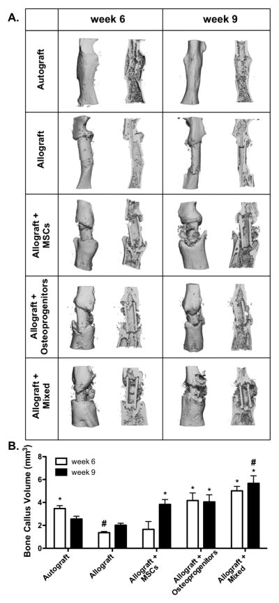
Figure 5.
Micro-computed tomography (μCT) scans taken 6 and 9 weeks post-implantation were reconstructed to assess in vivo bone callus formation (A; intact and sagital cut views). Subsequent quantification revealed that tissue engineered periosteum modified allografts transplanting a mixed cell population significantly increased bone callus volume as compared to transplantation of either MSCs or osteoprogenitors alone, as well as allograft only controls (B) (n=5–6; error bars represent standard error of the mean; p-value of <0.05 indicates significance compared to allograft (*) or autograft (#)).

