Abstract
Glycerol, a product of adipose tissue lipolysis, is an important substrate for hepatic glucose synthesis. However, little is known about the regulation of hepatic glycerol metabolism. Here we show that several genes involved in the hepatic metabolism of glycerol, i.e., cytosolic and mitochondrial glycerol 3-phosphate dehydrogenase (GPDH), glycerol kinase, and glycerol transporters aquaporin 3 and 9, are upregulated by fasting in wild-type mice but not in mice lacking PPARα. Furthermore, expression of these genes was induced by the PPARα agonist Wy14643 in wild-type but not PPARα−null mice. In adipocytes, which express high levels of PPARγ, expression of cytosolic GPDH was enhanced by PPARγ and β/δ agonists, while expression was decreased in PPARγ+/– and PPARβ/δ–/– mice. Transactivation, gel shift, and chromatin immunoprecipitation experiments demonstrated that cytosolic GPDH is a direct PPAR target gene. In line with a stimulating role of PPARα in hepatic glycerol utilization, administration of synthetic PPARα agonists in mice and humans decreased plasma glycerol. Finally, hepatic glucose production was decreased in PPARα-null mice simultaneously fasted and exposed to Wy14643, suggesting that the stimulatory effect of PPARα on gluconeogenic gene expression was translated at the functional level. Overall, these data indicate that PPARα directly governs glycerol metabolism in liver, whereas PPARγ regulates glycerol metabolism in adipose tissue.
Introduction
In most parts of the world, the prevalence of obesity is increasing rapidly. One of the most important secondary ailments of obesity is type 2 diabetes, which affects millions of people worldwide. It is well recognized that elevated plasma free fatty acid levels associated with obesity are a critical intermediate in the pathophysiology of type 2 diabetes (1). Free fatty acids promote diabetes partly by stimulating hepatic gluconeogenesis and glucose output (2–6). However, the mechanism(s) by which free fatty acids achieve this effect remains obscure.
Fatty acids are able to activate the expression of genes via PPARs (7). PPARs are ligand-activated transcription factors that belong to the superfamily of nuclear hormone receptors. Three PPAR isotypes are known: PPARα, PPARβ/δ, and PPARγ. The latter isotype is mainly expressed in adipose tissue and plays an important role in adipocyte differentiation and lipid storage (8). It serves as a target for an important class of antidiabetic drugs, the insulin-sensitizing thiazolidinediones. PPARβ/δ is expressed ubiquitously and thus far has been connected with wound healing, cholesterol metabolism, and fatty acid oxidation (9–11). Finally, PPARα stimulates hepatic fatty acid oxidation and ketogenesis, and regulates production of apolipoproteins. It serves as target for the hypolipidemic fibrate class of drugs, which include fenofibrate and gemfibrozil. Experiments with PPARα-null mice have been invaluable in elucidating the physiologic role of PPARα and have indicated that hepatic PPARα is particularly important during fasting (12–14). Fasted PPARα-null mice suffer from a variety of metabolic defects including hypoketonemia, hypothermia, elevated plasma free fatty acid levels, and hypoglycemia. The mechanism behind the fasting-induced hypoglycemia has so far remained elusive, but it is conceivable that PPARα directly regulates the expression of genes involved in gluconeogenesis. Since fatty acids are ligands for PPARα, the latter mechanism would be able to explain the stimulatory effect of elevated plasma free fatty acids on hepatic gluconeogenesis and glucose output.
In order to ascertain what metabolic steps or pathways are affected by PPARα deletion, we performed microarray analysis with RNA from liver of fasted wild-type and PPARα-null mice. Interestingly, it was found that the expression of several genes involved in gluconeogenesis was decreased in PPARα-null mice compared with wild-type mice. Follow-up analysis indicated that PPARα stimulates the expression of a set of genes involved in the conversion of glycerol to glucose and that at least one of these genes, the cytosolic glycerol 3-phosphate dehydrogenase gene (GPDH) is a direct target of PPARα with a functional PPAR response element in its promoter. Our data demonstrate that PPARα directly regulates glycerol metabolism in liver.
Results
Regulation of gluconeogenic gene expression by PPAR
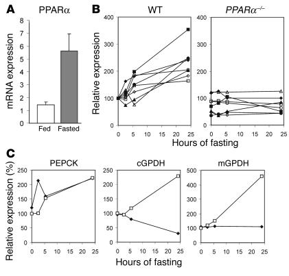
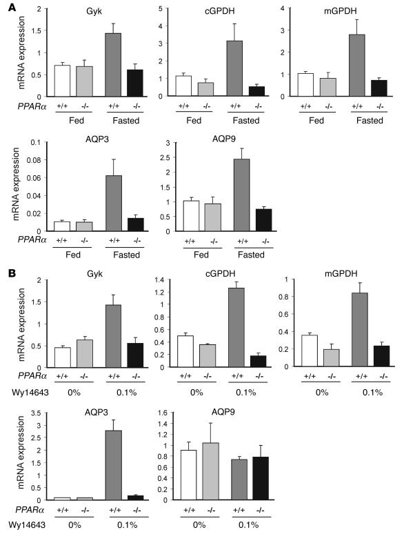
α. In agreement with previous data, hepatic PPARα expression was strongly induced by fasting (Figure 1A). Accordingly, it can be expected that the effects of PPARα on gene expression are especially evident during fasting. To pinpoint novel pathways regulated by PPARα, we compared mRNA of livers of fed and fasted PPARα-null and wild-type mice by oligonucleotide microarray. As expected, the fasting-induced increase in expression of fatty acid oxidative and ketogenic genes was PPARα dependent (Figure 1B). Interestingly, a similar type of regulation was observed for cytosolic GPDH (cGPDH) and mitochondrial GPDH (mGPDH), which are involved in the conversion of glycerol to glucose (Figure 1C). In contrast, phosphoenolpyruvate carboxykinase (PEPCK), which is considered to be the rate-limiting enzyme in gluconeogenesis from lactate/pyruvate, was upregulated during prolonged fasting in a PPARα-independent manner (Figure 1C). Real-time quantitative PCR (Q-PCR) confirmed that cGPDH and mGPDH were upregulated by fasting only in wild-type mice (Figure 2A). Interestingly, a similar type of regulation was observed for glycerol kinase, as well as aquaporin 3 (AQP3) and aquaporin 9 (AQP9). The latter two transporters are involved in the cellular uptake of glycerol (15), which, via the action of glycerol kinase, is phosphorylated to glycerol 3-phosphate, which in turn is converted to the gluconeogenic intermediate dihydroxyacetonphosphate via cGPDH and mGPDH. To establish that the decreased expression in fasted PPARα-null mice is not an indirect consequence of metabolic perturbations in these mice, wild-type and PPARα-null mice were fed for 5 days with the synthetic PPARα ligand Wy14643. Wy14643 consistently upregulated the expression of cGPDH, mGPDH, glycerol kinase, and AQP3 in wild type but not PPARα-null mice (Figure 2B). No induction was observed for AQP9. Taken together, these results indicate that PPARα induces hepatic expression of genes involved in the conversion of glycerol to glucose.
Figure 1.
Oligonucleotide microarray analysis identifies novel putative PPARα target genes. (A) Relative expression of PPARα in liver was determined by Q-PCR in fed and 24-hour-fasted mice (n = 4). The difference was evaluated by Student’s t test (P < 0.01). Error bars represent SEM. (B) Expression of genes involved in fatty acid oxidation and ketogenesis in livers of wild-type and PPARα-null mice, as determined by oligonucleotide microarray (Affymetrix). The average difference (expression) of wild-type at 0 hours was arbitrarily set at 100. Filled diamonds: long-chain fatty acyl-CoA synthetase; open diamonds: carnitine palmitoyltransferase II; filled triangles: long-chain acyl-CoA dehydrogenase; open circles: short-chain acyl-CoA dehydrogenase; open triangles: medium-chain acyl-CoA dehydrogenase; filled circles: dodecenoyl-CoA δ-isomerase; filled squares: HMG-CoA synthase; open squares: HMG-CoA lyase. (C) Hepatic expression of PEPCK (left), cGPDH (middle) and mGPDH (right) after 0, 2.5, 5.5 and 24 hours fasting in wild-type and PPARα-null mice according to oligonucleotide microarray. The average difference (expression) of wild-type at 0 hours was arbitrarily set at 100.
Figure 2.
PPARα upregulates the expression of numerous genes involved in the conversion of glycerol to glucose. (A) Relative expression of glycerol kinase (Gyk), cGPDH, mGPDH, AQP3, and AQP9 were determined by Q-PCR in fed and 24-hour-fasted wild-type and PPARα-null mice. Statistically significant effects were observed by two-way ANOVA for all genes for genotype (P < 0.01), and for the interaction between genotype and feeding status (P < 0.05). (B) Relative expression of Gyk, cGPDH, mGPDH, AQP3, and AQP9 were determined by Q-PCR in wild- type and PPARα-null mice after feeding with Wy14643. Statistically significant effects were observed by two-way ANOVA for all genes for genotype and for Wy14643 treatment, and for the interaction between the two parameters (P < 0.01), except for AQP9. Error bars represent SEM.
PPARγ and PPARβ/δ ligands induce cGPDH expression in adipocytes.
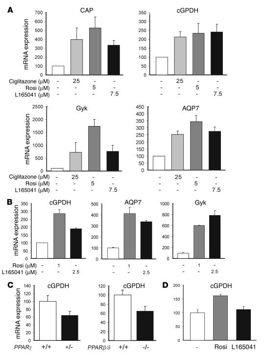
The liver takes up glycerol to convert it into glucose, whereas adipose tissue takes up glucose and converts it into glycerol 3-phosphate, which becomes incorporated into triglycerides. In adipose tissue, the expression of PPARα is low, whereas PPARβ/δ and PPARγ are well expressed. It is well established that the uptake of glucose into adipocytes and its conversion to triglycerides is stimulated by PPARγ (16, 17). To investigate regulation of glycerol metabolism in adipocytes by PPARγ and PPARβ/δ, mature mouse (3T3-L1) and human (SGBS) adipocytes were incubated with PPARγ agonists ciglitazone or rosiglitazone or PPARβ/δ agonist L165041. All ligands, in mouse and human adipocytes, significantly increased expression of cGPDH, glycerol kinase, and AQP7 (Figure 3, A and B). The known PPARγ target c-cbl–associated protein (CAP) was included as a positive control gene. cGPDH is highly expressed in adipocytes, where it functions in the synthesis of glycerol 3-phosphate from glucose (fed state) or gluconeogenic precursors (fasted state) (see Figure 7). It is often used as an adipogenesis marker. Glycerol kinase in adipocytes may catalyze recycling of glycerol, whereas AQP7 encodes a transporter that facilitates export of glycerol from the adipocytes. Supporting a role of PPARγ and PPARβ/δ in regulating cGPDH expression in vivo, cGPDH mRNA was decreased in white adipose tissue (WAT) of PPARγ+/– and PPARβ/δ–/– mice compared with wild-type mice (Figure 3C). Also, rosiglitazone, but not L165041, increased cGPDH mRNA in WAT of wild-type mice (Figure 3D). Taken together, these data suggest that, whereas PPARα induces glycerol utilization in liver, PPARγ and possibly PPARβ/δ seem to be involved in the regulation of intracellular glycerol metabolism in adipose tissue.
Figure 3.
PPARγ and PPARβ/δ agonists induce cGPDH gene expression in adipocytes. (A) 3T3-L1 adipocytes at day 10 of differentiation were treated with the PPARγ agonists ciglitazone (25 μM) or rosiglitazone (Rosi) (5 μM), or the PPARβ agonist L165041 (7.5 μM), and mRNA expression of the indicated genes was determined by Q-PCR. Results are expressed as percentage of control (DMSO). One-way ANOVA indicated that differences in expression were statistically significant for all four genes (P < 0.05). (B) Human SGBS adipocytes at day 13 of differentiation were treated with PPARγ agonist rosiglitazone (1 μM) or PPARβ agonists L165041 (2.5 μM). Expression of the indicated genes was determined by Q-PCR. One-way ANOVA indicated that differences in expression were statistically significant for all three genes (P < 0.05). (C) Expression of cGPDH in WAT of PPARγ+/– and PPARβ/δ–/– mice, as determined by Q-PCR. Differences were statistically significant (Student’s t test, P < 0.05). (D) Expression of cGPDH in WAT of wild-type mice fed 0.01% rosiglitazone or 0.025% L165041, as determined by Q-PCR. The effect of rosiglitazone was statistically significant (Student’s t test, P < 0.01). Error bars represent SEM.
Figure 7.
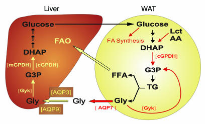
Proposed model integrating the roles of PPARα and PPARγ in glycerol (Gly) metabolism. Adipose tissue releases FFAs and glycerol. FFAs released by adipose tissue ligand-activate PPARα, whose hepatic expression is increased during fasting. Activation of PPARα induces expression of AQP3 and AQP9, which enable glycerol to enter the hepatocytes. Activation of PPARα also induces the expression of glycerol kinase, cGPDH, and mGPDH, which participate in the conversion of glycerol to glucose. In adipose tissue, PPARγ induces the expression of genes promoting the conversion of glucose to FFAs, as well as the conversion of glucose to glycerol 3-phosphate (G3P) from glucose. Glycerol 3-phosphate serves as the direct precursor for triglyceride (TG) synthesis. Moreover, PPARγ stimulates glycerol transport, glyceroneogenesis, and glycerol phosphorylation. Pathways regulated by PPARα are indicated in yellow, whereas those regulated by PPARγ are indicated in red. DHAP, dihydroxyacetonephosphate; Lct, lactate; FAO, fatty acid oxidation. Brackets indicate enzymes.
cGPDH is a direct PPAR target gene.
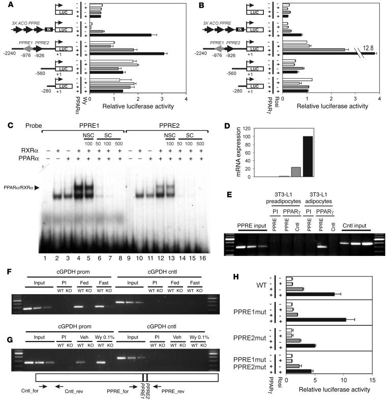
Our data so far suggest that cGPDH is a PPARα target gene in liver and a PPARγ (and possibly PPARβ/δ) target gene in adipose tissue. To determine what genomic region is responsible for PPAR-induced upregulation of cGPDH expression, 2.2 kb of cGPDH promoter sequence immediately upstream of the transcription site was cloned in front of a luciferase reporter, and transactivation studies were carried out in NIH-3T3 cells. It was observed that cotransfections with a PPARα or PPARγ expression vector markedly increased luciferase activity, which was further enhanced by the addition of ligand (Figure 4, A and B). This response to PPARs and ligands was completely abolished in deletion constructs containing 0.5 or 0.25 kb of promoter sequence, suggesting that the PPAR responsive element was located in the region –2.2 to –0.5 kb. Screening of this genomic region yielded two putative PPAR response element (PPREs) about 1 kb upstream of the transcription start site, which differed little from the consensus PPRE (see Supplemental Figure 1; supplemental material available at http://www.jci.org/cgi/content/full/114/1/94/DC1).
Figure 4.
cGPDH is a direct PPARα/γ target gene. Mouse cGPDH reporter constructs containing 2240, 560, or 280 bp of immediate upstream promoter region were transfected into NIH-3T3 cells together with a PPARα (A) or PPARγ (B) expression vector. Normalized activity of the full-length cGPDH reporter in the absence of PPAR and ligands was set at 1. (C) Binding of the PPAR/RXR heterodimer to putative response elements, as determined by electrophoretic mobility shift assay. The double-stranded response elements cGPDH-PPRE1 (lanes 1–8). Fold-excess of specific (SC) or nonspecific (NSC) cold probe is indicated. (D) Expression of cGPDH during 3T3-L1 adipogenesis as determined by Q-PCR. Expression at day 8 was set at 100%. ChIP of PPRE within mouse cGPDH promoter using anti-mPPARγ or anti-mPPARα antibodies. Gene sequences spanning the putative PPREs (+1020 to +782) and a random control sequence (+2519 to +2124) were analyzed by PCR in the immunoprecipitated chromatin of 3T3-L1 preadipocytes and adipocytes (E), fed and fasted wild-type and PPARα-null mice (F), and wild-type and PPARα-null mice treated or not with Wy14643 (G). Preimmune serum was used as a control. (H) Transcriptional activity of site-directed mutants (mut) of the cGPDH promoter. Mouse cGPDH reporter constructs containing double nucleotide changes in PPRE1, PPRE2, or both, were transfected into HepG2 cells together with a PPARγ expression vector. Normalized activity of the reporter in the absence of PPAR and ligand was set at 1. Error bars in A, B, and H represent SEM. Cntl, random control sequence; PI, preimmune serum; prom, promoter; Veh, vehicle; Wy, Wy14643; for, forward primer; rev, reverse primer.
To determine whether these PPREs are able to bind PPAR in vitro, we performed electrophoretic mobility shift assay. In the presence of only PPARα or retinoid X receptor α (RXRα), a single complex was observed, which originated from the reticulocyte lysate (Figure 4C, lanes 2 and 3). An additional, slower moving complex was observed only in the presence of both receptors (Figure 4C, lane 4), indicating that it represented a PPAR/RXR heterodimer. PPRE1 bound the heterodimer PPAR/RXR more efficiently than PPRE2 (Figure 4C, lane 4 vs. lane 12). Specificity of binding was demonstrated by competition with the nonradiolabeled PPRE of the malic enzyme promoter. In contrast, a response element for liver X receptor (LXR) was ineffective in competing for binding with the radiolabeled PPREs. These data demonstrate that the PPREs identified bind the PPAR/RXR heterodimer in vitro, further indicating that cGPDH is a direct PPAR target gene.
To find out whether PPARα and PPARγ are bound to these sequences in vivo, chromatin immunoprecipitation (ChIP) was performed using an anti-mPPARα or anti-mPPARγ antibody. Expression of cGPDH (and PPARγ) is highly upregulated during 3T3-L1 adipogenesis (Figure 4D). Using ChIP, we observed binding of PPARγ to a 238-bp sequence spanning the putative PPREs in differentiated 3T3-L1 adipocytes but not in preadipocytes (Figure 4E). There was no immunoprecipitation of the PPREs with preimmune serum, and no binding of PPARγ to a random control sequence was observed. In the fed and fasted mouse liver, PPARα was specifically bound to the PPRE sequence in wild-type but not PPARα-null mice (Figure 4F). Treatment with Wy14643 enhanced binding of PPARα to the sequence, which was not observed in the PPARα-null mice (Figure 4G). Because of the close proximity between the two PPREs, it was not possible to carry out ChIP for each putative PPRE separately. These data suggest that PPARα binds in vivo to the sequence containing the two PPREs.
Transactivation studies with cGPDH promoter constructs carrying mutations with the PPREs indicated that the most downstream PPRE (PPRE2) was particularly important for PPARγ-mediated promoter activation (Figure 4H). In contrast, mutating PPRE1 did not diminish promoter activation. These data suggest that PPRE2 but not PPRE1 is involved in mediating the effect of PPARγ on cGPDH promoter activity, although it cannot be ruled out that the double nucleotide changes introduced into PPRE1 failed to yield a dysfunctional PPRE. When the region encompassing PPRE1 and/or PPRE2 was placed in front of a heterologous promoter, significant PPARγ-dependent transactivation was observed for PPRE2, and for PPRE1 and PPRE2 together, but not PPRE1 alone (see Supplemental Figure 2). Together, these data suggest that PPRE2 at least partially mediates the PPARγ-dependent increase in cGPDH transcription.
PPARα activation decreases plasma glycerol levels in mice and humans.
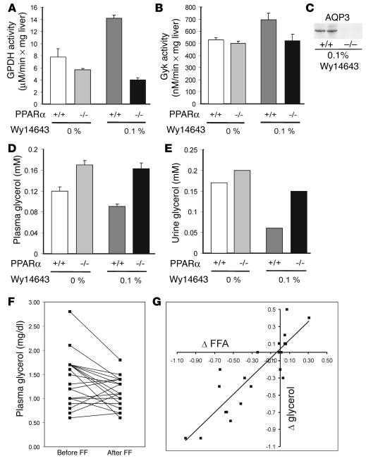
To examine whether induction of genes involved in the conversion of glycerol to glucose by PPARα has any functional consequences, we measured glycerol levels in plasma and urine. Inasmuch as fasting increases glycerol release from adipose tissue and at the same time stimulates hepatic glycerol utilization, it is difficult to interpret the effect of PPARα deletion, which may affect both processes, on glycerol levels in fasted animals. We therefore focused on the effect of PPARα activation by Wy14643. First, it was established that the induction of GPDH, glycerol kinase, and AQP3 gene expression by PPARα was translated at the enzyme activity or protein level (Figure 5, A–C). In line with the mRNA data indicating upregulation of glycerol utilization by PPARα, Wy14643 significantly decreased plasma glycerol concentration in wild-type but not PPARα-null mice (Figure 5D). A similar pattern was observed in urine (Figure 5E). Furthermore, in human atherosclerotic patients, 4-week treatment with fenofibrate caused a mean decrease in plasma glycerol levels of 18% (P < 0.01; Figure 5F). Interestingly, a significant correlation was observed between the fenofibrate-induced decrease in plasma free fatty acids (likely mediated by a PPARα-induced increase in hepatic fatty acid utilization), and the decrease in plasma glycerol, suggesting a common mechanism (Figure 5G). These data provide compelling in vivo evidence that PPARα stimulates hepatic glycerol utilization.
Figure 5.
PPARα activation decreases plasma and urine glycerol levels. Enzyme activity of GPDH (A) or glycerol kinase (B) was determined in liver homogenates of wild-type and PPARα-null mice after feeding with Wy14643 (n = 4 per group). Error bars represent SEM. (C) AQP3 protein was determined by Western blot in the membrane fraction of liver homogenates of wild-type and PPARα-null mice treated with Wy14643. Equal amounts of protein were loaded. Glycerol was determined in plasma (D) (n = 4) and urine (E) (samples in each group were pooled and determined in duplicate) in wild-type and PPARα-null mice after feeding with Wy14643. Significant effects were observed by two-way ANOVA for genotype and for Wy14643 treatment (P < 0.05). (F) Plasma glycerol levels decreased in atherosclerotic patients after 4-week treatment with micronized fenofibrate (FF) (250 mg/day). (P < 0.01, paired Student’s t test) (G) Correlation between changes in plasma free fatty acids (FFA) and glycerol in atherosclerotic patients treated with fenofibrate.
Hepatic glucose production is diminished in PPARα-null mice.
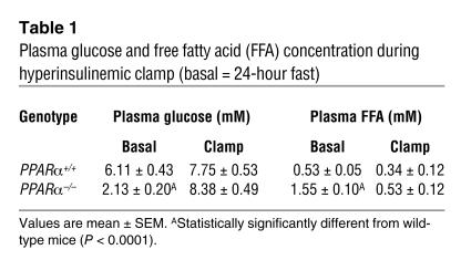
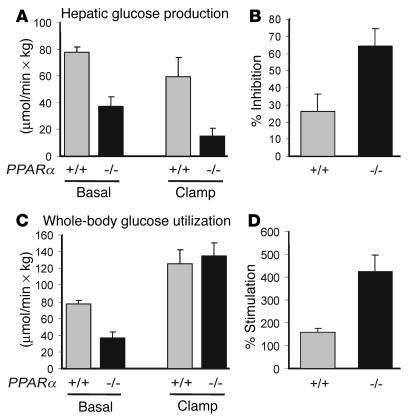
Glycerol is one of the main precursors for hepatic glucose production, particularly during fasting. To find out whether the stimulatory effect of PPARα on hepatic glycerol utilization may translate into decreased hepatic glucose production in PPARα-null mice, hyperinsulinemic clamp experiments were carried out. Both wild-type and PPARα-null mice were fed Wy14643 for 12 days and fasted for 24 hours in order to maximize differences in gluconeogenic gene expression, and thus phenotype, between the two sets of mice. In the basal state (24-hour fast), plasma glucose was almost threefold lower and plasma free fatty acids almost threefold higher in PPARα-null mice (Table 1). Supporting a stimulatory role for PPARα in gluconeogenesis during fasting, hepatic glucose production, which is equal to whole-body glucose utilization in the basal state, was markedly decreased in PPARα-null mice compared with wild-type mice in both the basal and the hyperinsulinemic state (Figure 6A). These data suggest that the fasting-induced hypoglycemia in PPARα-null mice is probably due to impaired gluconeogenesis. Alternatively, the hypoglycemia may be caused by increased whole-body glucose utilization in PPARα-null mice. However, no evidence for this was found as glucose utilization was decreased in PPARα-null mice compared with wild-type mice in the basal state and unchanged in the hyperinsulinemic state (Figure 6C). Overall, PPARα-null mice appeared to be more sensitive to insulin as both the percentage stimulation of whole-body glucose utilization and the percentage inhibition of hepatic glucose output by insulin were augmented compared with wild-type mice (Figure 6, B and D).
Table 1.
Plasma glucose and free fatty acid (FFA) concentration during hyperinsulinemic clamp (basal = 24-hour fast)
Figure 6.
Decreased hepatic glucose production and increased insulin sensitivity in PPARα-null mice. Wild-type and PPARα-null mice administered Wy14643 and fasted were analyzed by hyperinsulinemic clamp technique. (A) Hepatic glucose production under basal and hyperinsulinemic conditions. (B) Percentage of inhibition of hepatic glucose production by insulin. (C) Whole-body glucose utilization under basal and hyperinsulinemic conditions. (D) Percentage of stimulation of whole-body glucose utilization by insulin. Differences between genotypes were statistically significant for all variables except glucose utilization under hyperinsulinemic conditions. P < 0.05, Mann-Whitney U test. Error bars represent SEM.
Discussion
Although PPARα has mostly been connected with fatty acid catabolism, numerous lines of evidence indicate that it influences glucose homeostasis as well. First of all, fasting PPARα-null mice display marked hypoglycemia (12–14). Furthermore, induction of insulin resistance in mice by high-fat feeding is mitigated in the absence of PPARα (18, 19). Paradoxically, in a variety of diabetic animal models, activation of PPARα by synthetic agonists also improves glucose homeostasis (20), possibly by reducing endogenous glucose production (21, 22) and/or increasing glucose disposal (22–24). Recently, it was also observed that induction of the gluconeogenic genes PEPCK and glucose 6-phosphatase by dexamethasone is PPARα dependent (25). However, since PEPCK and glucose 6-phosphatase are not direct target genes of PPARα, the mechanism behind this regulation remains elusive. All together, it can be concluded that, although PPARα has an important influence on glucose metabolism, the mechanisms behind this regulation remain ill defined. Here, it is shown that PPARα decreases plasma glycerol levels in mice and humans by directly upregulating the expression of genes involved in hepatic gluconeogenesis from glycerol, including cGPDH, mGPDH, glycerol kinase, AQP3, and AQP9. The gluconeogenic gene cGPDH is identified as a direct target gene of PPARα with a functional PPAR response element in its promoter. The stimulatory effect of PPARα on gluconeogenic gene expression is associated with elevated hepatic glucose production during fasting. Our data support, extend, and provide a molecular explanation for the largely ignored observation that, in several rodent diabetic models, plasma glycerol levels are decreased by treatment with PPARα agonists (23, 24, 26, 27).
During prolonged fasting, when hepatic glycogen stores are depleted, plasma glucose levels are maintained exclusively by de novo glucose synthesis in liver (gluconeogenesis). The main precursors for hepatic gluconeogenesis are lactate, amino acids, and glycerol, which are converted into glucose via a series of reactions in the cytosol and mitochondria. The contribution of glycerol to hepatic glucose production greatly depends on the nutritional state and may vary from 5% postprandially in humans (28) to being the main gluconeogenic precursor in rodents after prolonged fasting (29). The significance of glycerol as a gluconeogenic precursor is supported by the episodic hypoglycemia observed in patients with isolated glycerol kinase deficiency (30) and by the phenotype of mice lacking both cGPDH and mGPDH (31), which suffer from elevated plasma glycerol concentrations and hypoglycemia before dying within the first week of life. Inasmuch as glycerol is an important gluconeogenic precursor during fasting, and its conversion to glucose in liver is impaired in the absence of PPARα, defective synthesis of glucose from glycerol may explain the fasting-induced hypoglycemia in PPARα-null mice (12–14). Indeed, it was observed that hepatic glucose production was impaired in fasted PPARα-null mice, although the relative importance of defective conversion of glycerol to glucose is hard to estimate.
During feeding, in adipose tissue, PPARγ induces the expression of genes promoting the conversion of glucose to fatty acids, as well as the conversion of glucose to glycerol 3-phosphate (Figure 7). Glycerol 3-phosphate serves as the direct precursor for triglyceride synthesis. Moreover, PPARγ stimulates glycerol transport, glyceroneogenesis, and glycerol phosphorylation (16, 32). During fasting, lipolysis in adipose tissue releases glycerol and fatty acids into the blood, which are carried to the liver for further metabolism. PPARα plays a pivotal role in regulating the metabolism of fatty acids by stimulating hepatic fatty acid oxidation and ketogenesis (7). The present data show that the metabolic fate of glycerol is also under the control of PPARα, which stimulates its conversion to glucose in liver (Figure 7). Together with the previous finding that PPARα suppresses amino acid catabolism and ureagenesis (33), these combined data indicate that PPARα coordinates hepatic nutrient metabolism during fasting. Furthermore, by activating PPARα, fatty acids released from adipose tissue determine not only their own metabolic fate, but also that of other nutrients. Thus, PPARα serves as nutrient sensor that senses changes in feeding status and translates them into metabolic adjustments aimed at maintaining homeostasis. Under conditions of elevated plasma fatty acid concentrations, such as type 2 diabetes and obesity, it can be hypothesized that PPARα becomes permanently activated, resulting in enhanced conversion of glycerol into glucose. Although several aspects of this proposed mechanism remain to be demonstrated in humans, it provides an attractive molecular explanation for the observed link between elevated plasma free fatty acid levels and hepatic glucose production (4).
Our data support previous publications showing that insulin sensitivity is higher in PPARα-null mice (18, 19), at least when the measurement is done under fasting conditions (34). While it is clear from the present study that, after fasting, the expression of gluconeogenic genes is lower in PPARα-null mice, the molecular mechanisms explaining the heightened response to insulin by these mice remain elusive. At the same time, administration of PPARα agonists has also been shown to improve insulin sensitivity in various rodent models of obesity/diabetes (20–24). This situation is comparable to PPARγ, where partial deletion and ligand activation both reduce insulin resistance (35).
It is much harder to reconcile our data with those of Xu et al. (36), which show enhanced hepatic glucose production, as well as enhanced hepatic glucose production from glycerol, in fasted PPARα-null mice compared with fasted wild-type mice. As the fasted PPARα-null mice suffer from severe hypoglycemia, hepatic glucose production can only be enhanced if, at the same time, whole-body glucose utilization is hugely increased. However, the decreased whole-body glucose utilization after a 24-hour fast observed in the present study, combined with a lack of evidence that fatty acid oxidation is impaired in skeletal muscle (37), which would cause higher glucose utilization, indicates that this is unlikely to be the case. In contrast to Xu et al. (36), using a different method, we observed markedly decreased hepatic glucose production in fasted PPARα-null mice. Possible explanations for these seemingly discrepant findings are differences in the background strain of the PPARα-null mice (sv129 vs. C57/B6) and perhaps bias in the method of calculating glucose production by mass isotopomer distribution analysis (MIDA). According to a recent study that employed MIDA, it is possible that decreased hepatic glucose production in PPARα-null mice is also partially due to preferential partitioning of glucose 6-phosphate toward glycogen rather than toward glucose (38).
Previous data have established that AQP7 and probably glycerol kinase are a direct PPARγ target genes in adipocytes (16, 39). Here we confirm upregulation of these genes by PPARγ (and probably PPARβ/δ) and further demonstrate that the cGPDH gene is a direct target of PPARγ in adipocytes. Thus, whereas PPARα controls the hepatic utilization of glycerol, glycerol metabolism in adipocytes is under the control of PPARγ. Remarkably, cGPDH is upregulated by both PPARα and PPARγ, but since the role of cGPDH differs between liver and adipocytes, the effects of this regulation are very different.
PEPCK is often considered to catalyze the rate-limiting step in gluconeogenesis from pyruvate. Numerous transcription factors, including the glucocorticoid receptor, hepatic nuclear factor 3, and the retinoic acid receptor, regulate transcription of the PEPCK gene (40). Recent studies have shown that the PPARγ coactivator 1 (PGC1) stimulates the expression of PEPCK and that this effect is mediated by hepatocyte nuclear factor 4 (41). Since PGC1 is also a coactivator of PPARα, it is no surprise that it can enhance PPARα-mediated transactivation of the cGPDH promoter (unpublished data). In contrast, and in line with previous observations (13), our analysis clearly indicates that the induction of expression of PEPCK during fasting is independent of PPARα.
Since the linkage between PPARα and glycerol metabolism was uncovered by microarray analysis, this study demonstrates the potential of genomics tools to elucidate novel pathways regulated by nuclear hormone receptors. However, to demonstrate a direct involvement of a nuclear hormone receptor in a particular pathway, the analysis should extend beyond merely descriptive data.
In conclusion, although an important role of PPARα in glucose metabolism has been demonstrated by numerous studies, the underlying mechanisms have remained elusive. Based on our study, it can be concluded that PPARα directly stimulates hepatic glycerol metabolism and, via this and other mechanisms, importantly influences hepatic glucose production during fasting. This effect of PPARα may account for the pronounced hypoglycemia in fasted PPARα-null mice.
Methods
Oligonucleotide microarray.
Total RNA was prepared from mouse livers using Trizol reagent (Invitrogen, Breda, The Netherlands). For the oligonucleotide microarray hybridization experiment, 10 μg of total liver RNA pooled from four mice was used for cRNA synthesis. Hybridization, washing and scanning of Affymetrix Genechip Mu6500 probe assays was according to standard Affymetrix protocols (Affymetrix, Santa Clara, California, USA). Fluorimetric data were processed by Affymetrix GeneChip3.1 software, and the gene chips were globally scaled to all the probe sets with an identical target intensity value.
Plasmid and DNA constructs.
Based on sequences available in GenBank, a 2.3 kb fragment of the mouse cGPDH promoter was amplified by PCR from 3T3-L1 genomic DNA. Different-size fragments of the cGPDH promoter were cloned into the KpnI and BglII sites of pGL3 basic vector (Promega Corp., Leiden, The Netherlands). Site-directed mutations were introduced into the PPREs using the QuikChange site-directed mutagenesis kit (Stratagene, Amsterdam, The Netherlands). The sequences of the primers used are provided in Supplemental Table 1. cDNA encoding for mPPARα, mPPARβ, and rPPARγ2 were cloned into pSG5 (Stratagene). Nucleotide fragment surrounding the PPREs within the cGPDH promoter were amplified by PCR and subcloned into the KpnI and BglII sites of pTAL-SEAP (BD Biosciences Clontech, Alphen aan den Rijn, The Netherlands). PPREtkLUC containing three copies of acyl-CoA oxidase PPRE was a generous gift from Ronald Evans (Salk Institute, La Jolla, California, USA).
Animal experiments.
SV129 PPARα-null mice and corresponding wild-type mice were purchased at the Jackson Laboratory (Bar Harbor, Maine, USA). For the fasting experiments, 5-month-old male mice were fasted for 0, 2.5, 5.5 or 24 hours starting at the onset of the light cycle. For the feeding experiments with Wy14643 (Chemsyn, Lenexa, Kansas, USA), 5-month-old female mice were fed 0.1% Wy14643 for 5 days by mixing it in their food. Blood was collected via orbital puncture. Livers were dissected and directly frozen in liquid nitrogen.
For the clamp study, male 3-month-old wild-type (n = 4) and PPARα-null mice (n = 5) were fed 0.1% Wy14643 for 12 days. Mice were fasted for 24 hours prior to the clamp studies. The hyperinsulinemic clamp and assays for blood glucose and plasma free fatty acids were carried out as previously described (42).
The animal experiments were approved by the animal experimentation committee of the Etat de Vaud (Switzerland) or Wageningen University.
Cell culture and transfections.
Mouse fibroblast NIH-3T3 cells or human hepatoma HepG2 cells were grown in DMEM containing 10% FCS. Cells were transfected with PPAR expression and luciferase reporter constructs using PolyFect (QIAGEN Inc., Leusden, The Netherlands) or calcium phosphate precipitation. After transfection, cells were incubated in the presence or absence of PPARs ligands (rosiglitazone 5 μM, Wy14643 10 μM) for 24–48 hours prior to lysis. Promega luciferase assay (Promega Corp.) and standard β-galactosidase assay with 2-nitrophenyl-β-D galactopyranoside were used to measure the relative activity of the promoter. 3T3-L1 fibroblasts were amplified in DMEM plus 10% calf serum and plated for final differentiation in DMEM plus 10% FCS. On day 0, which was two days after reaching confluence, the medium was changed and the following compounds were added: isobutyl methylxanthine (0.5 mM), dexamethasone (1 μM), and insulin (5 μg/ml). On day 3, the medium was changed to DMEM plus 10% FCS and insulin (5 μg/ml). On day 6, the medium was changed to DMEM plus 10% FCS, which was changed every 3 days. SGBS cell culture and induction of adipogenesis were performed exactly as previously published (43). 3T3-L1 adipocytes and SGBS adipocytes were incubated with synthetic PPAR agonists for 36–48 hours prior to RNA extraction.
Isolation of total RNA and Q-PCR.
Total RNA was extracted from cells or tissue with Trizol reagent following the supplier’s protocol. Total RNA 3–5 μg was treated with DNAse I amplification grade and then reverse-transcribed with oligo-dT using Superscript II RT RNase H–. cDNA was PCR amplified with Platinum Taq DNA polymerase. (All these reagents were from Invitrogen.) Primer sequences used in the PCR reactions were chosen based on the sequences available in GenBank. Primers were designed to generate a PCR amplification product of 100–200 bp. Only primer pairs yielding unique amplification products without primer dimer formation were subsequently used for real-time PCR assays. PCR was carried out using Platinum Taq polymerase and SYBR green on an iCycler PCR machine (Bio-Rad Laboratories BV, Veenendaal, The Netherlands). The sequence of primers used is available in Supplemental Table 1. The mRNA expression of all genes reported is normalized to β-actin expression.
cGPDH enzymatic assay.
cGPDH activity was assayed according to the spectrophotometric method of Wise and Green with some modifications (44). Livers were weighted, resuspended, and sheared in 20% homogenization buffer (w/v) (25 mM Tris-HC pH 7.5, 1 mM EDTA and 1 mM 2-mercaptoethanol). After brief sonication, cells were centrifuged at 4°C for 10 minutes at 16,000 g. The supernatant of cell lysate was used for determining the protein concentration by Bio-Rad Protein Assay reagent (Bio-Rad Laboratories BV) and for the enzymatic assay. The same amount of protein was incubated in standard reaction mixture (100 mM triethanolamine, 0.25 mM EDTA, 50 mM 2-mercaptoethanol, and 0.2 mM NADH). The reaction was initiated by the addition of dihydroxyacetone phosphate, and NADH disappearance was followed at 340 nm.
Glycerol kinase assay.
Glycerol kinase activity was assayed according to the spectrophotometric method described by Leclercq et al. with some modifications (45). Whole livers were homogenized in 20% homogenization buffer (w/v) (10 mM Tris pH 7.5, 1 mM EDTA, 0.25 M sucrose plus Complete proteases inhibitor cocktail). Homogenates were then microcentrifuged at 14,000 rpm for 10 minutes at 4°C. Glycerol kinase activity in the supernatant was determined spectrophotometrically at 25°C.
Membrane fractionation and immunoblotting.
Twenty per cent liver homogenates were centrifuged at 4,000 g for 15 min at 4°C, followed by centrifugation of the supernatant at 200,000 g for 1 hour. The pellet was resuspended in homogenization buffer and used for determining the protein concentration by Bio-Rad Protein Assay reagent (Bio-Rad Laboratories BV) or SDS/PAGE. Membrane fractions were resolved by SDS/PAGE on a 12% polyacrylamide gel. Western blotting was carried out as described by Kersten et al. (46). The blot was incubated with rabbit anti-(rat)aquaporin 3 primary antibody (1:400; Chemicon Europe Ltd., Hofheim, Germany) for 16 hours at 4°C.
ChIP.
Pure-bred wild-type or PPARα-null mice on a Sv129 background were used. Mice were fed by gavage with either Wy14643 (50 mg/kg/day) or vehicle (0.5% carboxymethylcellulose) for 5 days. Alternatively, mice were fasted or not fasted for 24 hours. After the indicated treatment, mice were sacrificed by cervical dislocation. The liver was rapidly perfused with pre-warm (37°C) PBS for 5 minutes followed by 0.2% collagenase for 10 minutes. The liver was diced, forced through a stainless steel sieve, and the hepatocytes were collected directly into DMEM containing 1% formaldehyde. After incubation at 37°C for 15 minutes, the hepatocytes were pelleted, and ChIP was carried out using PPARα-specific antibodies as previously described (9).
3T3-L1 cells were differentiated as described above. After cell lysis and sonication, the supernatant was diluted 20-fold in re-ChIP dilution buffer (1 mM EDTA, 20 mM Tris-HCl, pH 8.1, 50 mM NaCl, and 1% Triton-X) prior to incubation with mouse PPARγ antibody. The remainder of the assay was carried out as described previously (9).
Electrophoretic mobility shift assay.
Mouse PPARα and human RXRα proteins were generated from pSG5 expression vectors using a coupled in vitro transcription/translation system (Promega Corp.). The following oligonucleotides were used: GPDHPPRE1for (5′-AGGGAAGGAAGGTCAAAGGCCACTGGTGACAC-3′), GPDHPPRE1rev (5′-GTGTCACCAGTGGCCTTTGACCTTCCTTC-3′), GPDHPPRE2for (5′-GAGATTATCTGAG-GTGAAGGGGCAACCTGTGG-3′) and GPDHPPRE2rev (5′-CCACAGGTTGCCCCTTCACCTCAGATAAT-3′). Oligonucleotides were annealed and labeled by Klenow filling (New England Biolabs (UK) Ltd., Leusden, The Netherlands) using Redivue [α-32P]dCTP (3000 Ci/mmol) (Amersham Biosciences Europe GmBH, Roosendaal, The Netherlands). Binding and electrophoresis was exactly performed as previously described (47), with the exception of unprogrammed lysate, where only 1/6 of the volume was used for binding.
Plasma and urine glycerol.
Levels of glycerol in urine and plasma of mice were determined using the triglyceride assay from Beckman Coulter Nederland B.V. (Mijdrecht, The Netherlands) by omitting the first step (digestion with lipase). Measurements were carried out on a Synchron LX20 analyzer (Beckman Coulter Nederland B.V.).
For measurement of glycerol in human plasma, blood was taken after an overnight fast from 21 male subjects before and after a 4-week treatment with 250 mg of micronized fenofibrate daily. All subjects had significant coronary artery disease as documented by angiography.
Acknowledgments
We would like to thank Marco Alves for the synthesis of L165041 and M. Wabitsch for the gift of the SGBS cell line. This study was supported by the Dutch Diabetes Foundation, with additional support from the Royal Netherlands Academy of Art and Sciences (KNAW), The Netherlands Organization for Scientific Research (NWO), the Wageningen Center for Food Sciences, the Swiss National Science Foundation, and the Human Frontier Science Program.
Footnotes
Nonstandard abbreviations used: aquaporin (AQP); chromatin immuno-precipitation (ChIP); cytosolic GPDH (cGPDH); glycerol 3-phoshate dehydrogenase (GPDH); mass isotopomer distribution analysis (MIDA); mitochondrial GPDH (mGPDH); phosphoenolpyruvate carboxykinase (PEPCK); PPARγ coactivator 1 (PGC1); PPAR response element (PPRE); real-time quantitative PCR (Q-PCR); retinoid X receptor α (RXRα); white adipose tissue (WAT).
Conflict of interest: The authors have declared that no conflict of interest exists.
References
- 1.Bergman RN, Ader M. Free fatty acids and pathogenesis of type 2 diabetes mellitus. Trends Endocrinol. Metab. 2000;11:351–356. doi: 10.1016/s1043-2760(00)00323-4. [DOI] [PubMed] [Google Scholar]
- 2.Chen X, Iqbal N, Boden G. The effects of free fatty acids on gluconeogenesis and glycogenolysis in normal subjects. J. Clin. Invest. 1999;103:365–372. doi: 10.1172/JCI5479. [DOI] [PMC free article] [PubMed] [Google Scholar]
- 3.Gonzalez-Manchon C, Martin-Requero A, Ayuso MS, Parrilla R. Role of endogenous fatty acids in the control of hepatic gluconeogenesis. Arch. Biochem. Biophys. 1992;292:95–101. doi: 10.1016/0003-9861(92)90055-2. [DOI] [PubMed] [Google Scholar]
- 4.Lam TK, et al. Mechanisms of the free fatty acid-induced increase in hepatic glucose production. Am. J. Physiol. Endocrinol. Metab. 2003;284:E863–E873. doi: 10.1152/ajpendo.00033.2003. [DOI] [PubMed] [Google Scholar]
- 5.Rebrin K, Steil GM, Getty L, Bergman RN. Free fatty acid as a link in the regulation of hepatic glucose output by peripheral insulin. Diabetes. 1995;44:1038–1045. doi: 10.2337/diab.44.9.1038. [DOI] [PubMed] [Google Scholar]
- 6.Roden M, et al. Effects of free fatty acid elevation on postabsorptive endogenous glucose production and gluconeogenesis in humans. Diabetes. 2000;49:701–707. doi: 10.2337/diabetes.49.5.701. [DOI] [PubMed] [Google Scholar]
- 7.Kersten S, Desvergne B, Wahli W. Roles of PPARs in health and disease. Nature. 2000;405:421–424. doi: 10.1038/35013000. [DOI] [PubMed] [Google Scholar]
- 8.Rosen ED, Spiegelman BM. Molecular regulation of adipogenesis. Annu. Rev. Cell Dev. Biol. 2000;16:145–171. doi: 10.1146/annurev.cellbio.16.1.145. [DOI] [PubMed] [Google Scholar]
- 9.Di Poi N, Tan NS, Michalik L, Wahli W, Desvergne B. Antiapoptotic role of PPARbeta in keratinocytes via transcriptional control of the Akt1 signaling pathway. Mol. Cell. 2002;10:721–733. doi: 10.1016/s1097-2765(02)00646-9. [DOI] [PubMed] [Google Scholar]
- 10.Leibowitz MD, et al. Activation of PPARdelta alters lipid metabolism in db/db mice. FEBS Lett. 2000;473:333–336. doi: 10.1016/s0014-5793(00)01554-4. [DOI] [PubMed] [Google Scholar]
- 11.Wang YX, et al. Peroxisome-proliferator-activated receptor delta activates fat metabolism to prevent obesity. Cell. 2003;113:159–170. doi: 10.1016/s0092-8674(03)00269-1. [DOI] [PubMed] [Google Scholar]
- 12.Hashimoto T, et al. Defect in peroxisome proliferator-activated receptor alpha-inducible fatty acid oxidation determines the severity of hepatic steatosis in response to fasting. J. Biol. Chem. 2000;275:28918–28928. doi: 10.1074/jbc.M910350199. [DOI] [PubMed] [Google Scholar]
- 13.Kersten S, et al. Peroxisome proliferator-activated receptor alpha mediates the adaptive response to fasting. J. Clin. Invest. 1999;103:1489–1498. doi: 10.1172/JCI6223. [DOI] [PMC free article] [PubMed] [Google Scholar]
- 14.Leone TC, Weinheimer CJ, Kelly DP. A critical role for the peroxisome proliferator-activated receptor alpha (PPARalpha) in the cellular fasting response: the PPARalpha-null mouse as a model of fatty acid oxidation disorders. Proc. Natl. Acad. Sci. U. S. A. 1999;96:7473–7478. doi: 10.1073/pnas.96.13.7473. [DOI] [PMC free article] [PubMed] [Google Scholar]
- 15.Carbrey JM, et al. Aquaglyceroporin AQP9: solute permeation and metabolic control of expression in liver. Proc. Natl. Acad. Sci. U. S. A. 2003;100:2945–2950. doi: 10.1073/pnas.0437994100. [DOI] [PMC free article] [PubMed] [Google Scholar]
- 16.Guan HP, et al. A futile metabolic cycle activated in adipocytes by antidiabetic agents. Nat. Med. 2002;8:1122–1128. doi: 10.1038/nm780. [DOI] [PubMed] [Google Scholar]
- 17.Picard F, Auwerx J. PPAR(gamma) and glucose homeostasis. Annu. Rev. Nutr. 2002;22:167–197. doi: 10.1146/annurev.nutr.22.010402.102808. [DOI] [PubMed] [Google Scholar]
- 18.Guerre-Millo M, et al. PPAR-alpha-null mice are protected from high-fat diet-induced insulin resistance. Diabetes. 2001;50:2809–2814. doi: 10.2337/diabetes.50.12.2809. [DOI] [PubMed] [Google Scholar]
- 19.Tordjman K, et al. PPARalpha deficiency reduces insulin resistance and atherosclerosis in apoE-null mice. J. Clin. Invest. 2001;107:1025–1034. doi: 10.1172/JCI11497. [DOI] [PMC free article] [PubMed] [Google Scholar]
- 20.Lee HJ, et al. Fenofibrate lowers abdominal and skeletal adiposity and improves insulin sensitivity in OLETF rats. Biochem. Biophys. Res. Commun. 2002;296:293–299. doi: 10.1016/s0006-291x(02)00822-7. [DOI] [PubMed] [Google Scholar]
- 21.Chou CJ, et al. WY14,643, a peroxisome proliferator-activated receptor alpha (PPARalpha) agonist, improves hepatic and muscle steatosis and reverses insulin resistance in lipoatrophic A-ZIP/F-1 mice. J. Biol. Chem. 2002;277:24484–24489. doi: 10.1074/jbc.M202449200. [DOI] [PubMed] [Google Scholar]
- 22.Kim H, et al. Peroxisome proliferator-activated receptor-alpha agonist treatment in a transgenic model of type 2 diabetes reverses the lipotoxic state and improves glucose homeostasis. Diabetes. 2003;52:1770–1778. doi: 10.2337/diabetes.52.7.1770. [DOI] [PubMed] [Google Scholar]
- 23.Koh EH, et al. Peroxisome proliferator-activated receptor (PPAR)-alpha activation prevents diabetes in OLETF rats: comparison with PPAR-gamma activation. Diabetes. 2003;52:2331–2337. doi: 10.2337/diabetes.52.9.2331. [DOI] [PubMed] [Google Scholar]
- 24.Ye JM, et al. Peroxisome proliferator-activated receptor (PPAR)-alpha activation lowers muscle lipids and improves insulin sensitivity in high fat-fed rats: comparison with PPAR-gamma activation. Diabetes. 2001;50:411–417. doi: 10.2337/diabetes.50.2.411. [DOI] [PubMed] [Google Scholar]
- 25.Bernal-Mizrachi C, et al. Dexamethasone induction of hypertension and diabetes is PPAR-alpha dependent in LDL receptor-null mice. Nat. Med. 2003;9:1069–1075. doi: 10.1038/nm898. [DOI] [PubMed] [Google Scholar]
- 26.Larsen PJ, et al. Differential influences of peroxisome proliferator-activated receptors gamma and -alpha on food intake and energy homeostasis. Diabetes. 2003;52:2249–2259. doi: 10.2337/diabetes.52.9.2249. [DOI] [PubMed] [Google Scholar]
- 27.Rizvi F, et al. Antidyslipidemic action of fenofibrate in dyslipidemic-diabetic hamster model. Biochem. Biophys. Res. Commun. 2003;305:215–222. doi: 10.1016/s0006-291x(03)00721-6. [DOI] [PubMed] [Google Scholar]
- 28.Baba H, Zhang XJ, Wolfe RR. Glycerol gluconeogenesis in fasting humans. Nutrition. 1995;11:149–153. [PubMed] [Google Scholar]
- 29.Peroni O, Large V, Beylot M. Measuring gluconeogenesis with [2-13C] glycerol and mass isotopomer distribution analysis of glucose. Am. J. Physiol. 1995;269:E516–E523. doi: 10.1152/ajpendo.1995.269.3.E516. [DOI] [PubMed] [Google Scholar]
- 30.Sjarif DR, Ploos van Amstel JK, Duran M, Beemer FA, Poll-The BT. Isolated and contiguous glycerol kinase gene disorders: a review. J. Inherit. Metab. Dis. 2000;23:529–547. doi: 10.1023/a:1005660826652. [DOI] [PubMed] [Google Scholar]
- 31.Brown LJ, Koza RA, Marshall L, Kozak LP, MacDonald MJ. Lethal hypoglycemic ketosis and glyceroluria in mice lacking both the mitochondrial and the cytosolic glycerol phosphate dehydrogenases. J. Biol. Chem. 2002;277:32899–32904. doi: 10.1074/jbc.M202409200. [DOI] [PubMed] [Google Scholar]
- 32.Tordjman J, et al. Regulation of glyceroneogenesis and phosphoenolpyruvate carboxykinase by fatty acids, retinoic acids and thiazolidinediones: potential relevance to type 2 diabetes. Biochimie. 2003;85:1213–1218. doi: 10.1016/j.biochi.2003.11.010. [DOI] [PubMed] [Google Scholar]
- 33.Kersten S, et al. The peroxisome proliferator-activated receptor alpha regulates amino acid metabolism. FASEB J. 2001;15:1971–1978. doi: 10.1096/fj.01-0147com. [DOI] [PubMed] [Google Scholar]
- 34.Haluzik M, Gavrilova O, LeRoith D. Peroxisome proliferator-activated receptor-alpha deficiency does not alter insulin sensitivity in mice maintained on regular or high-fat diet: hyperinsulinemic-euglycemic clamp studies. Endocrinology. 2004;145:1662–1667. doi: 10.1210/en.2003-1015. [DOI] [PubMed] [Google Scholar]
- 35.Yamauchi T, et al. The mechanisms by which both heterozygous peroxisome proliferator-activated receptor gamma (PPARgamma) deficiency and PPARgamma agonist improve insulin resistance. J. Biol. Chem. 2001;276:41245–41254. doi: 10.1074/jbc.M103241200. [DOI] [PubMed] [Google Scholar]
- 36.Xu J, et al. Peroxisome proliferator-activated receptor alpha (PPARalpha) influences substrate utilization for hepatic glucose production. J. Biol. Chem. 2002;277:50237–50244. doi: 10.1074/jbc.M201208200. [DOI] [PubMed] [Google Scholar]
- 37.Muoio DM, et al. Fatty acid homeostasis and induction of lipid regulatory genes in skeletal muscles of peroxisome proliferator-activated receptor (PPAR) alpha knock-out mice. Evidence for compensatory regulation by PPAR delta. J. Biol. Chem. 2002;277:26089–26097. doi: 10.1074/jbc.M203997200. [DOI] [PubMed] [Google Scholar]
- 38.Bandsma RH, et al. Hepatic de novo synthesis of glucose 6-phosphate is not affected in peroxisome proliferator-activated receptor alpha-deficient mice but is preferentially directed toward hepatic glycogen stores after a short term fast. J. Biol. Chem. 2004;279:8930–8937. doi: 10.1074/jbc.M310067200. [DOI] [PubMed] [Google Scholar]
- 39.Kishida K, et al. Enhancement of the aquaporin adipose gene expression by a peroxisome proliferator-activated receptor gamma. J. Biol. Chem. 2001;276:48572–48579. doi: 10.1074/jbc.M108213200. [DOI] [PubMed] [Google Scholar]
- 40.Croniger C, Leahy P, Reshef L, Hanson RW. C/EBP and the control of phosphoenolpyruvate carboxykinase gene transcription in the liver. J. Biol. Chem. 1998;273:31629–31632. doi: 10.1074/jbc.273.48.31629. [DOI] [PubMed] [Google Scholar]
- 41.Rhee J, et al. Regulation of hepatic fasting response by PPARgamma coactivator-1alpha (PGC-1): requirement for hepatocyte nuclear factor 4alpha in gluconeogenesis. Proc. Natl. Acad. Sci. U. S. A. 2003;100:4012–4017. doi: 10.1073/pnas.0730870100. [DOI] [PMC free article] [PubMed] [Google Scholar]
- 42.Voshol PJ, et al. Increased hepatic insulin sensitivity together with decreased hepatic triglyceride stores in hormone-sensitive lipase-deficient mice. Endocrinology. 2003;144:3456–3462. doi: 10.1210/en.2002-0036. [DOI] [PubMed] [Google Scholar]
- 43.Wabitsch M, et al. Characterization of a human preadipocyte cell strain with high capacity for adipose differentiation. Int. J. Obes. Relat. Metab. Disord. 2001;25:8–15. doi: 10.1038/sj.ijo.0801520. [DOI] [PubMed] [Google Scholar]
- 44.Wise LS, Green H. Participation of one isozyme of cytosolic glycerophosphate dehydrogenase in the adipose conversion of 3T3 cells. J. Biol. Chem. 1979;254:273–275. [PubMed] [Google Scholar]
- 45.Leclercq P, et al. Inhibition of glycerol metabolism in hepatocytes isolated from endotoxic rats. Biochem. J. 1997;325:519–525. doi: 10.1042/bj3250519. [DOI] [PMC free article] [PubMed] [Google Scholar]
- 46.Kersten S, et al. Characterization of the fasting-induced adipose factor FIAF, a novel peroxisome proliferator-activated receptor target gene. J. Biol. Chem. 2000;275:28488–28493. doi: 10.1074/jbc.M004029200. [DOI] [PubMed] [Google Scholar]
- 47.IJpenberg A, Jeannin E, Wahli W, Desvergne B. Polarity and specific sequence requirements of peroxisome proliferator-activated receptor (PPAR)/retinoid X receptor heterodimer binding to DNA. A functional analysis of the malic enzyme gene PPAR response element. J. Biol. Chem. 1997;272:20108–20117. doi: 10.1074/jbc.272.32.20108. [DOI] [PubMed] [Google Scholar]


