Abstract
Objective:
To compare stress distribution in a tooth restored with metal and fiber posts of varying diameters (1.2 and 1.4 mm) by means of three-dimensional finite element analysis (3D-FEA).
Materials and Methods:
Four 3D-FEA models were constructed: (1) fiber post (1.2 and 1.4 mm) and (2) metal post (1.2 and 1.4 mm). The material properties were assigned and a force of 100 N was applied at 45° angle to the longitudinal axis of the tooth onto the palatal surface incisal to the cingulum. Analysis was run and stress distribution pattern was studied.
Results:
Maximum stresses in the radicular tooth structure for fiber post were higher than that for metal post. In the former models, stresses in the tooth structure were slightly reduced with increase in fiber post diameter.
Conclusions:
To reduce stress in the remaining radicular tooth structure, it is better to use a fiber post of a large diameter.
Keywords: Fiber post, finite element analysis, metal post, post diameter
INTRODUCTION
Endodontically treated teeth are generally weakened as a result of loss of tooth structure due to decay, previous restorative procedures and endodontic access preparation. Further destruction of these teeth can be prevented by a protective restoration. Post and core system is a widely used method for treatment of structurally weakened teeth. The primary objective of post and core procedure is replacement of the lost tooth structure in order to facilitate crown support and retention.[1]
Certain factors become important in the success of posts, such as remaining coronal tooth structure, choice of material, properties of materials like modulus of elasticity, stress distribution, etc. On stress measurement and analysis, a wide range of methods are available, namely the strain gauge method, the loading test, the photoelastic method and the finite element method.[2] Using these methods, several studies on fracture strength and fracture pattern within the tooth structure have been conducted.[3] These techniques are homologous, two-dimensional, and difficult to reproduce.[4] A valid index of stress distribution at the root structures has been difficult to create based solely on experimental and clinical observation.[5] Only a few studies have focused on the influence of post diameter on the stress distribution in the remaining radicular tooth structure. The purpose of this study was to compare stress distribution in a tooth restored with metal and fiber posts of varying diameters by means of three-dimensional finite element analysis (FEA).
MATERIALS AND METHODS
A freshly extracted, caries-free maxillary central incisor with straight root canal was randomly collected. The tooth was placed in a block of wax. A CT scan of the tooth was taken and axial sections at a distance of 1mm from each other were obtained.
The image obtained from the CT scan was imported into AutoCAD software (Autodesk, Inc., California, USA) and outline of each individual layer was traced. The layers were then stacked one on top of the other to obtain outlines of the surface contours of the tooth and a digital model was obtained using Pro/Engineer software (Parametric Technology Corporation, USA). The periodontal ligament was modeled as a layer 0.3 mm thick around the root surface.[6] Finally bone was modeled around the tooth.
Two different types of posts were compared in this study. They were metal post made of gold and glass fiber post. Post length inside canal and apical gutta percha were assumed to be 9 mm and 5 mm, respectively. Two configurations of post diameter were compared-1.2 mm (gold and fiber post) and 1.4 mm (gold and fiber post). The model was then altered to accommodate 2 gold posts (1.2 mm and 1.4 mm diameters) and 2 fiber posts (1.2 mm and 1.4 mm diameters), core, apical 5 mm of gutta percha and all ceramic crown and 4 models were obtained.
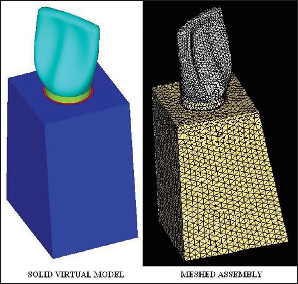
The digital tooth models with post, core and crown were imported to Hypermesh software (Altair Engineering, Michigan, USA). The models of the surface contours of the tooth were converted into solid models [Figure 1]. These models were then meshed [Figure 1] using tetrahedral elements with Hypermesh software as a neutral file using the STL (stereo lithography) format. All materials were assumed to be homogenous, isotropic, and linear elastic.
Figure 1.

Solid virtual model of maxillary central incisor and meshed assembly
The cement layer between the post and the dentin was too thin to adequately model in the finite element simulation, but the cement was treated as part of dentin because the mechanical properties of dentin and cement were similar. No significant error was expected from omission of the cement layer in the model.[7]
Nodes were assigned and elements were designed for stress analysis. Due to the complicated geometry of the tooth, free meshing technique was adopted. The completed models consisted of 62,138 three-dimensional tetrahedral elements and 17,189 nodes for models with 1.2 mm posts and 58,525 three-dimensional tetrahedral elements and 16,451 nodes for models with 1.4 mm posts.
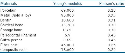
The completed models were then converted into an Ansys input file and imported into Ansys software (Ansys, Inc., Pennsylvania, USA). Material properties were prescribed to each of the components as tabulated in Table 1.
Table 1.
Mechanical properties of materials in finite element analysis model[3]

Boundary conditions and loading
Nodes have a tendency to move in different directions and also can rotate based on the direction of the load applied. On loading, the shape gets changed which is called as deformation and there will be no stress accumulation. Hence, to get the stresses model need to be fixed. The bottom portion of the cortical bone was fixed and a load of 100 N was applied on the ceramic crown at 45° angle to the long axis of the tooth onto the palatal surface incisal to the cingulum.
The maximum stresses generated in the tooth and post were calculated as von Mises stress.
RESULTS
Results are shown in terms of colored contour patterns. Red indicates the highest range and dark blue the least.
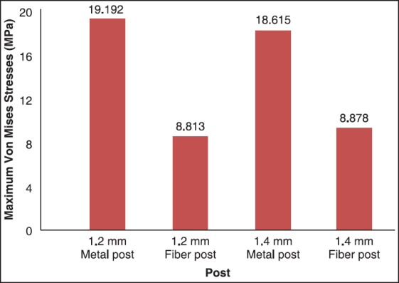
Maximum stress (15.37 MPa for 1.2 mm fiber post and 15.33 MPa for 1.4 mm fiber post) in the remaining radicular tooth structure for fiber post and core models were observed on the inner side of the proximal wall at the level of the cervical region irrespective of post diameter [Figure 2a and b]. As for the metal post and core models, maximum stress (15.018 MPa for 1.2 mm metal post and 14.925 MPa for 1.4 mm metal post) was observed on the inner side around the post apex irrespective of post diameter [Figure 2c and d]. The fiber post and core models showed larger maximum von Mises stress values occurring in the remaining radicular tooth structure than that of the metal post and core, and the maximum stress was slightly reduced with an increase in fiber post diameter [Graph 1].
Figure 2.

Distribution of von Mises stresses in the remaining radicular tooth structure with (a) 1.2 mm fiber post, (b) 1.4 mm fiber post, (c) 1.2 mm metal post, (d) 1.4 mm metal post, Distribution of von Mises stresses in the internal area of posts (e) 1.2 mm fiber post, (f) 1.4 mm fiber post, (g) 1.2 mm metal post, (h) 1.4 mm metal post
Graph 1.

Maximum von Mises stresses occurring in the remaining radicular tooth structure
For the fiber post and core models, stress concentration (8.813MPa for 1.2 mm fiber post and 8.878 MPa for 1.4 mm fiber post) in the post region was observed at the junction between the fiber post and the core [Figure 2e and f]. For the metal post and core, stress concentration was observed at the outer layer of the post [Figure 2g and h]. In the fiber post and core models, the maximum von Mises stress in the fiber post slightly increased with an increase in fiber post diameter [Graph 2].
Graph 2.

Maximum von Mises stresses occurring in the internal area of the post
DISCUSSION
Various methods are available on stress measurement and analysis, namely the strain gauge method, the loading test, the photoelastic method and the finite element method.[2]
The finite element method (FEM) is an innovative theoretic method used for resolving unworkable engineering problems.[4] FEM was first developed in 1943 by A. Hrennikoff and Richard Courant to solve complex elasticity and structural analysis problems in civil and aeronautical engineering. Davy and co-workers applied FEM to the study of post and core restorations.[8] FEA is an accurate numerical method in stress analysis and has been applied to dental biomechanics.[7]
Young's modulus of elasticity and Poisson's ratio of the modeled material are specified for each element. A system of simultaneous equations is generated and solved to yield predictable stress distributions in each element throughout a structure. The variables may be manipulated with computer precision, which eliminates variation resulting from sampling error. FEM analysis repeated any number of times will yield identical results 100% of the time. Thus it is certain that the results are always caused by manipulation of the variables and not by chance. For this reason, conventional inferential statistical analysis is not normally included in FEA.[4]
Stresses are produced as a result of mastication forces imposed on a structure. The distribution or pattern of these stresses is the result of the angle of the load and the geometry of the object. In addition, notches or imperfections present within the material may cause localized increase in the magnitude of the stresses, known as stress concentrations. These stress concentrations can contribute to the failure of the material through crack formation and an increased likelihood of fatigue failure.[9]
Using FEA, the stress generated can be analyzed accurately by assessing the stress concentration areas. In addition, FEA allows the investigation of a single variable in a complex structure. This means that FEA is less time-consuming as no time is wasted on standardization issues or on preparing multiple specimens as in mechanical tests. Because of these advantages, this method has been used to investigate the mechanical behavior of endodontically treated teeth subjected to different techniques and restorative materials.[10,11,12] At this point, a few limitations need to be addressed regarding the present study. The structures and materials used in this study were considered to be linear elastic, homogeneous, and isotropic. This meant that the computational simulation was not absolutely same as that of natural tooth structure and supporting tissues. The elastic modulus and Poisson's ratio values applied for the structures and materials in this study were used as thus determined from study published earlier.[3] Static load was applied in this study. Root fractures generally occur due to dynamic loading, which may then result in a fatigue process as in clinical conditions which are different from static loading.[2]
Very few studies have investigated on the effect of post diameter on stress distribution in a tooth.[3] Therefore, this in-vitro study was undertaken to compare stress distribution in a tooth restored with metal and fiber posts of varying diameters (1.2 and 1.4 mm) by means of three-dimensional FEA (3D-FEA).
FEA shows that maximum stresses in the tooth structure for fiber post and core are higher than that for gold-cast post and core. Stresses in the tooth structure are slightly reduced with increase in fiber post diameter.[8,13] So to reduce stress in the remaining radicular tooth with a large coronal defect, it is recommended to accompany a composite resin core with a fiber post of a large diameter within clinical limits.[3,14] Increase in fiber post diameter leads to an increase in surface area of the post, which in turn might aid in reducing stress development in the remaining radicular tooth structure by stress dispersion.
Glass fiber post shows the lowest peak stresses inside the root. Except for the force concentration at the cervical margin, the glass fiber post induces a stress field quite similar to that of the natural tooth.[15] With fiber post and core, the elastic modulus is similar to that of root dentin. Hence, their flexural property leads to stress concentration on the inner side of the proximal wall at the level of the cervix. This suggests that the elastic modulus influence maximum stress development in the remaining radicular tooth structure.
Cast metal post causes a large stress to concentrate around the base of the post while glass fiber post produces the lowest stress concentration and is hence effective in preventing stress concentration in the restored endodontically treated teeth.[2]
FEA shows that stress on the fiber post is distributed more evenly in the post tip area, whereas for metal post high stress is concentrated around the post tip.[9,16,17] This was because the metal post with a high elastic modulus is more rigid and reduces the strain on dentin, resulting in stress distribution to the apical third of root canal and into the post leading to fracture of root.
Another FEA reports that stress is accumulated within the cast post core system and transmission of stress to the tooth and the supportive structures is low. The fiber post system transfers stress to the tooth and the supportive structures while stress accumulation within post system is low.[18]
Fiber posts show more homogenous stress distribution and a better biomechanical performance than metallic posts since stiffness of fiber post is similar to dentin.[1,5,19,20]
FEA reports that stainless steel posts present the highest level of stress concentration followed by carbon fiber posts.[10,21] Steel posts are most dangerous to the root, potentially leading to its fracture, whereas glass reconstruction system gives most benign stressing condition.[11] Fiber posts failed at a certain compressive force but they were repairable unlike the cast posts where the fracture is irreparable.[22]
Results of this study coincide with that of the above-mentioned studies. However, further studies are required to evaluate the effect of post diameter on stress distribution in a tooth.
CONCLUSION
Within the limitations of this study, it has been concluded that:
The maximum von Mises stress in the remaining radicular tooth structure for the fiber post was higher than that for metal post and core. However, it became slightly reduced with the increase in fiber post diameter. Hence to reduce stress in the remaining radicular tooth structure, it is better to use a fiber post of a large diameter within clinical limits.
The location of maximum stress development in the remaining radicular tooth structure was on the inner side of the proximal wall at the level of cervical region for the fiber post. For the metal post and core, it was on the inner side around the post apex. Hence fracture is induced at the cervical region of tooth restored with fiber post and composite core allowing the repair, whereas vertical root fracture is induced in tooth restored with metal post making the tooth irreparable.
ACKNOWLEDGMENT
The authors like to express sincere thanks to M. S. Ramaiah School of Advanced Studies, Bengaluru, for the help rendered in doing the Three Dimensional Finite Element Analysis.
Footnotes
Source of Support: Nil
Conflict of Interest: None declared.
REFERENCES
- 1.Adanir N, Belli S. Stress analysis of a maxillary central incisor restored with different posts. Eur J Dent. 2007;1:67–71. [PMC free article] [PubMed] [Google Scholar]
- 2.Yamamoto M, Miura H, Okada D, Komada W, Masuoka D. Photoelastic stress analysis of different post and core restoration methods. Dent Mater J. 2009;28:204–11. doi: 10.4012/dmj.28.204. [DOI] [PubMed] [Google Scholar]
- 3.Okamoto K, Ino T, Iwase N, Shimizu E, Suzuki M, Satoh G, et al. Three-dimensional finite element analysis of stress distribution in composite resin cores with fiber posts of varying diameters. Dent Mater J. 2008;27:49–55. doi: 10.4012/dmj.27.49. [DOI] [PubMed] [Google Scholar]
- 4.Holmes DC, Diaz-Amold AM, Leary JM. Influence of post dimension on stress distribution in dentin. J Prosthet Dent. 1996;75:140–7. doi: 10.1016/s0022-3913(96)90090-6. [DOI] [PubMed] [Google Scholar]
- 5.Silva NR, Castro CG, Santos-Filho PC, Silva GR, Campos RE, Soares PV, et al. Influence of different post design and composition on stress distribution in maxillary central incisor: Finite element analysis. Indian J Dent Res. 2009;20:153–8. doi: 10.4103/0970-9290.52888. [DOI] [PubMed] [Google Scholar]
- 6.Jeon PD, Turley PK, Moon HB, Ting K. Analysis of stress in the periodontium of the maxillary first molar with a three-dimensional finite element model. Am J Orthod Dentofacial Orthop. 1999;115:267–74. doi: 10.1016/s0889-5406(99)70328-8. [DOI] [PubMed] [Google Scholar]
- 7.Ho MH, Lee SY, Chen HH, Lee MC. Three-dimensional finite element analysis of the effects of posts on stress distribution in dentin. J Prosthet Dent. 1994;72:367–72. doi: 10.1016/0022-3913(94)90555-x. [DOI] [PubMed] [Google Scholar]
- 8.Davy DT, Dilley GL, Krejci RF. Determination of stress patterns in root-filled teeth incorporating various dowel designs. J Dent Res. 1981;60:1301–10. doi: 10.1177/00220345810600070301. [DOI] [PubMed] [Google Scholar]
- 9.Cailleteau JG, Rieger MR, Akin JE. A comparison of intracanal stresses in a post-restored tooth utilizing the finite element method. J Endod. 1992;18:540–4. doi: 10.1016/S0099-2399(06)81210-0. [DOI] [PubMed] [Google Scholar]
- 10.de Castro Albuquerque R, Polleto LT, Fontana RH, Cimini CA. Stress analysis of an upper central incisor restored with different posts. J Oral Rehabil. 2003;30:936–43. doi: 10.1046/j.1365-2842.2003.01154.x. [DOI] [PubMed] [Google Scholar]
- 11.Lanza A, Aversa R, Rengo S, Apicella D, Apicella A. 3D FEA of cemented steel, glass and carbon posts in a maxillary incisor. Dent Mater. 2005;21:709–15. doi: 10.1016/j.dental.2004.09.010. [DOI] [PubMed] [Google Scholar]
- 12.Genovese K, Lamberti L, Pappalettere C. Finite element analysis of a new customized composite post system for endodontically treated teeth. J Biomech. 2005;38:2375–89. doi: 10.1016/j.jbiomech.2004.10.009. [DOI] [PubMed] [Google Scholar]
- 13.Asmussen E, Peutzfeldt A, Sahafi A. Finite element analysis of stresses in endodontically treated, dowel-restored teeth. J Prosthet Dent. 2005;94:321–9. doi: 10.1016/j.prosdent.2005.07.003. [DOI] [PubMed] [Google Scholar]
- 14.Peters MC, Poort HW, Farah JW, Craig RG. Stress analysis of a tooth restored with a post and core. J Dent Res. 1983;62:760–3. doi: 10.1177/00220345830620061501. [DOI] [PubMed] [Google Scholar]
- 15.Pegoretti A, Fambri L, Zappini G, Bianchetti M. Finite element analysis of a glass fibre reinforced composite endodontic post. Biomaterials. 2002;23:2667–82. doi: 10.1016/s0142-9612(01)00407-0. [DOI] [PubMed] [Google Scholar]
- 16.Reinhardt RA, Krejci RF, Pao YC, Stannard JG. Dentin stresses in post reconstructed teeth with diminishing bone support. J Dent Res. 1983;62:1002–8. doi: 10.1177/00220345830620090101. [DOI] [PubMed] [Google Scholar]
- 17.Hsu ML, Chen CS, Chen BJ, Huang HH, Chang CL. Effects of post materials and length on the stress distribution of endodontically treated maxillary central incisors: A 3D finite element analysis. J Oral Rehabil. 2009;36:821–30. doi: 10.1111/j.1365-2842.2009.02000.x. [DOI] [PubMed] [Google Scholar]
- 18.Eskitascioglu G, Belli S, Kalkan M. Evaluation of two post core systems using two different methods (fracture strength test and a finite elemental stress analysis) J Endod. 2002;28:629–33. doi: 10.1097/00004770-200209000-00001. [DOI] [PubMed] [Google Scholar]
- 19.Barjau-Escribano A, Sancho-Bru JL, Forner-Navarro L, Rodriguez-Cervantes PJ, Perez-Gonzalez A, Sanchez-Marin FT. Influence of prefabricated post material on restored teeth: Fracture strength and stress distribution. Oper Dent. 2006;31:47–54. doi: 10.2341/04-169. [DOI] [PubMed] [Google Scholar]
- 20.Coelho CS, Biffi JC, Silva GR, Abrahao A, Campos RE, Soares CJ. Finite element analysis of weakened roots restored with composite resin and posts. Dent Mater J. 2009;28:671–8. doi: 10.4012/dmj.28.671. [DOI] [PubMed] [Google Scholar]
- 21.Kaur A, Meena N, Shubhashini N, Kumari A, Shetty A. A comparative study of intra canal stress pattern in endodontically treated teeth with average sized canal diameter and reinforced wide canals with three different post systems using finite element analysis. J Conserv Dent. 2010;13:28–33. doi: 10.4103/0972-0707.62639. [DOI] [PMC free article] [PubMed] [Google Scholar]
- 22.Hegde J, Ramakrishna, Bashetty K, Srirekha, Lekha, Champa An in vitro evaluation of fracture strength of endodontically treated teeth with simulated flared root canals restored with different post and core systems. J Conserv Dent. 2012;15:223–7. doi: 10.4103/0972-0707.97942. [DOI] [PMC free article] [PubMed] [Google Scholar]


