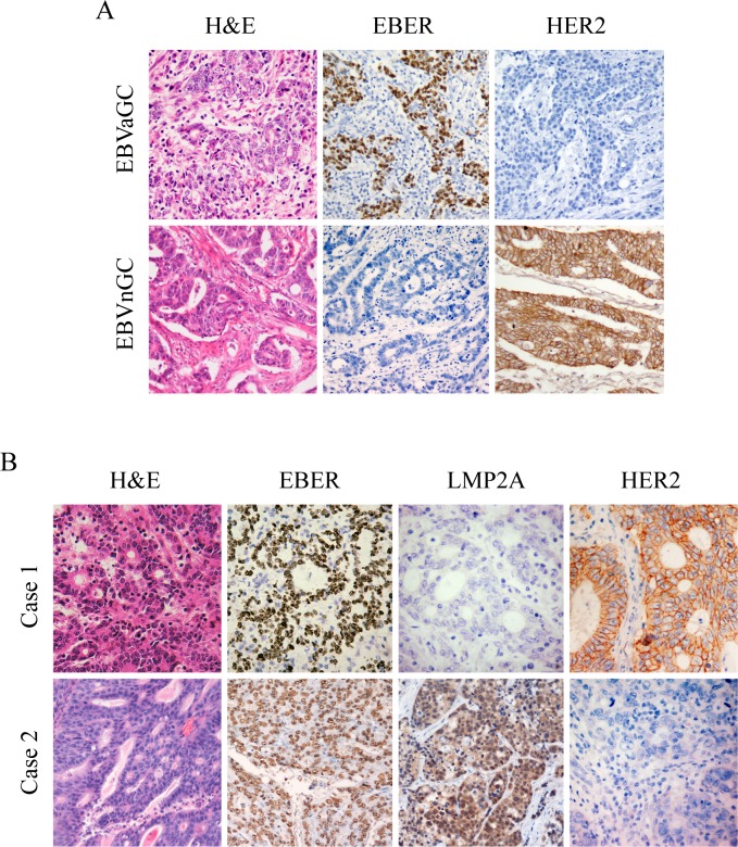
Figure 1. IHC images for expression of HER2, EBER and LMP2A in gastric tumor specimens.
Each of the gastric carcinoma cases was stained by H&E, IHC and EBER-1 in situ hybridization, respectively. For EBER-1 in situ hybridization, the positive signals were restricted only to the tumor nuclei but not in surrounding non-tumor cells. For HER2 IHC staining, positive staining can be seen in the membrane of the tumor cells. (A) Representative images for expression of HER2 in EBVaGC and EBVnGC samples. (B) Representative images of LMP2A and HER2 expression in EBVaGC samples. Case 1 showed negative expression of LMP2A with overexpression of HER2 (3+), and case 2 exhibited positive expression of LMP2A with no apparent expression of HER2 (0). (Original magnification ×200).

