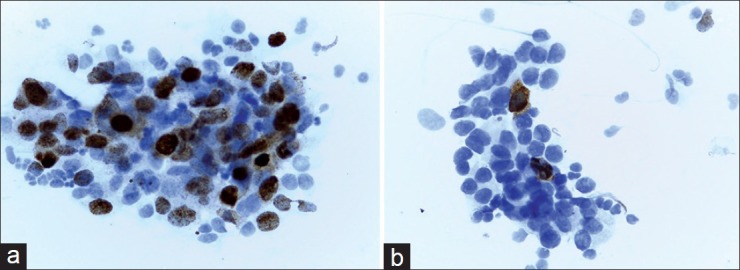
Figure 1.

(a) Positive staining of pAkt in a high-grade breast tumor. About 40% of tumor cells demonstrate an obvious positive nuclear staining. In this case, cytoplasmic staining was noted as negative (×400). (b) Negative expression of pAkt in a breast tumor. Positive staining is limited to sporadic cells. Note coexistence of nuclear and cytoplasmic expression (×400)
