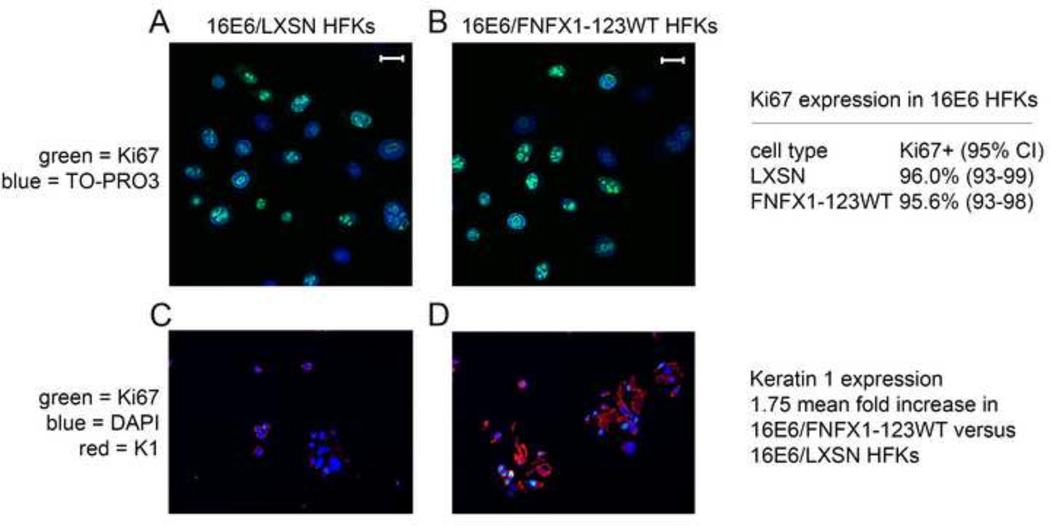
Figure 7. Ki67 expression unchanged with overexpression of FNFX1-123WT and increased Keratin 1.
Immunofluorescent staining was done in 16E6 HFKs transduced with overexpressed FLAG-tagged NFX1-123 wild type (FNFX1-123WT) or LXSN vector control. (A) Ki67 staining of 16E6/LXSN HFKs and (B) 16E6/FNFX1-123WT HFKs. Bar = 10 micron; Green = Ki67; Blue = TO-PRO3, nuclear stain. Quantification of percent positive Ki67 expression by immunofluorescent staining was done in three independent 16E6 HFK cell lines with increased NFX1-123 (FNFX1-123WT) versus three vector control (LXSN) HFKs. Expression pattern of the indicated protein was assessed by dual immunohistochemical (IHC) analysis of pelleted, fixed, paraffin embedded cells. (C) 16E6/LXSN HFKs and (D) 16E6/FNFX1-123WT HFKs stained for Ki67 and Keratin1 (K1). Green = Ki67; Blue = DAPI; Red = Keratin 1. Similar results were observed in three independent experiments using three distinct 16E6 HFK cell lines.

