Abstract
Purpose
The aim of this study is to evaluate the incidence of de novo malignancy after liver transplantation (LT) and compare with those among the general Chinese population.
Methods
A total of 466 patients who had a minimum follow-up time of 6 months were enrolled in the study. All data of medical records and follow up were retrospectively reviewed.
Results
The incidence rate of de novo malignancy was 3.0% (14 in 466 patients). The median elapsed time from transplant to the diagnosis of de novo malignancy was 42 months (range, 6 to 106 months). The cumulative risk for development of de novo malignancy was 1.6%, 2.7%, and 8.2% at 3, 5 and 10 years after LT, respectively. The patients were all male. The types of de novo tumors included digestive system tumor (8 in 14), lung cancer (2 in 14), urologic neoplasm (2 in 14), and hematologic malignant tumor (2 in 14). Over a mean follow-up of 24 months after diagnosis of de novo malignancy, 7 patients (50.0%) died; the overall 5-year patient survival rate was 54.5%. The relative risk of malignancy following LT was 9.5 folds higher than the general Chinese population.
Conclusion
The relative risk of malignancy following LT was much higher than the general Chinese population. Digestive system tumor is the most common type of de novo malignancy after LT in China.
Keywords: Liver transplantation, Transplant recipients, Neoplasms, Incidence
INTRODUCTION
Since the first liver transplantation (LT) was performed in 1963, there has been much progress in immunosuppressive therapy, surgical technique and perioperative treatment [1,2]. With the incidence of infection, bleeding, rejection and other early complications decreasing steadily, more and more liver transplant patients can achieve long-term survival. Beyond allograft-related complications, hepatocellular carcinoma (HCC) recurrence, metabolic syndrome, cardiovascular disease, and renal dysfunction, de novo neoplasms has been one of the leading causes of morbidity and mortality in this recipient population [3,4,5,6]. In the United States and European countries, many authors summarized the clinical data of de novo malignancy recipients. Immunosuppressive drugs are considered the most important cause [7,8]. Posttransplant lymphoproliferative disorders (PTLD) and skin cancer were the top two types of de novo malignancies [9,10,11].
In China, great advances have been made in the past decade in clinical LT. Up to now, more than 20 thousand LTs have been done all over the country. The recipients' survival rates were 76.46%, 63.76%, and 59.25% at 1, 3, and 5 years after LT, respectively. However, few doctors reported their experiences in treating de novo malignancy and most did so in the form of case reports [12,13,14,15]. In our center, the number of de novo malignancies was also relatively less than the literature. So we retrospectively analyzed the patients' data and compared the incidence of de novo malignancy with those among the general Chinese population.
METHODS
Patients
From May 2000 to December 2012, a total of 547 cases of LT were performed in Peking University People's Hospital. All data were collected from the China Liver Transplant Registry. Excluding cases of early death and loss to follow up, a total of 466 patients were included in this study. Three hundreds and eighty-eight patients were male and 78 patients were female. The youngest patient was 15 months old and the oldest was 72 years old. Indications for transplantation were 371 patients with posthepatitis B cirrhosis, 29 with acute liver failure, 15 with alcoholic cirrhosis, 13 with posthepatitis C cirrhosis, 14 with primary biliary cirrhosis, 9 with Wilson disease, 3 with congenital biliary atresia and 12 others. There were 230 patients combined with HCC. All patients' preoperative examination excluded malignant tumors outside of the liver. The recipients had an average follow-up time of 48.0±30.6 months (the minimum follow-up time was 6 months; the longest follow-up time was 144 months). The general characteristics of the 466 patients were listed in Table 1.
Table 1. Demographic and clinicopathologic features of patients (n = 466).
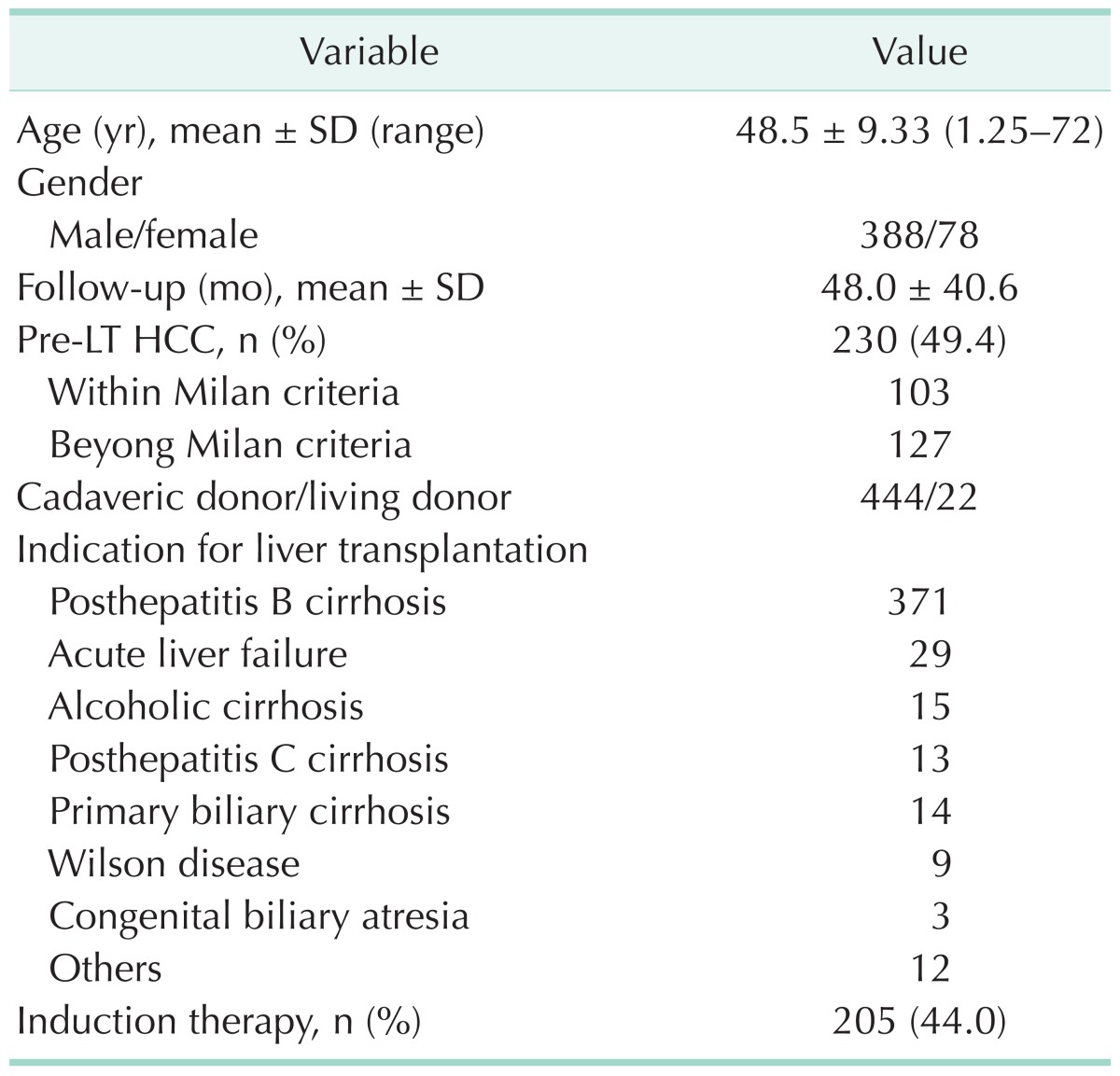
SD, standard deviation; LT, liver transplantation; HCC, hepatocellular carcinoma.
The grafts included 444 cases of cadaveric donor (95.3%) and 22 cases of living donor (4.7%). All operations were orthotopic LT, including classic LT in 193 cases, piggyback LT in 271 cases and combined liver-kidney transplantation in 2 cases.
Ethics statement
Informed written consent was obtained from patients in accordance with the Declaration of Helsinki. The deceased donor livers were obtained through both social and legal donation. All data were analyzed anonymously.
Immunosuppressive therapy
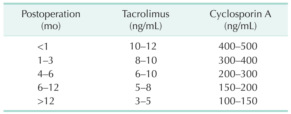
Before the graft reperfusion during the surgery, all patients routinely received methylprednisolone 500 mg. The patients combined with renal dysfunction were administered interleukin-2 receptor antagonists (Simulect or Zenapax) as induction therapy. Calmodulin inhibitor-based triple immunosuppressive therapy was administered to all recipients. Calmodulin inhibitor was tapered to a small dose maintenance therapy and the target concentration of calmodulin inhibitors for different periods was shown in Table 2. Liver function and plasma concentrations of calmodulin inhibitor were tested periodically.
Table 2. The target concentration of calmodulin inhibitors in different periods after liver transplantation.
Recipients who suffered from infection and those with liver cancer exceeding the Milan criteria were administered glucocorticoid for not more than one week. The other patients' glucocorticoid dosages were gradually reduced until withdrawal in three months after the operation. The specific usage was as follows: during the first seven days, intravenous methylprednisolone was administered, the dose was 240 mg, 160 mg, 120 mg, 80 mg, 40 mg, 20 mg, respectively; 8 to 30 days of oral prednisone 15 mg/day; 31 to 60 days of oral prednisone 10 mg/day; 61 to 90 days of oral prednisone 5 mg/day. Mycophenolate mofetil (MMF) was also withdrawn 3 months after operation. The specific usage was as follows: the first month 0.75 g every 12 hours, the second month 0.5 g every 12 hours, the third month 0.25 g every 12 hours. For patients with bone marrow suppression or diarrhea, the dosage of MMF was properly adjusted.
Follow-up assessment
The follow-up interval for the LT recipients was 3 months. The focus of check-ups was the monitoring of liver and kidney function and the plasma concentrations of the calmodulin inhibitor. Carcinoembryonic antigen, alpha-fetoprotein and other tumor markers should be checked each year and chest x-rays, liver ultrasounds and abdominal CTs should also be performed yearly. Gastroscopies and colonoscopies were not routinely recommended if the patient did not display clinical symptoms.
In LT recipients, the diagnostic criteria of de novo malignant tumors included two items. First, the malignant tumor must have emerged after the LT operation. Second, reoccurrence and metastasis of the HCC should be ruled out.
Therapeutic schedule
Treatment of de novo malignancy was based on the guidelines for tumors in general patients. Surgical treatment was offered to all patients who had resectable tumors with no disease spread at the time of diagnosis. Adjuvant treatments were based on tumor guidelines. Palliative treatment was offered when patients were diagnosed at advanced stages.
Statistical analysis
Statistical analyses were performed by SPSS ver. 17.0 (SPSS Inc., Chicago, IL, USA). The categorical variables were compared by Fisher exact test. The Kaplan-Meier method was used to estimate the cumulative probability of de novo malignancies after LT and patient survival rates after the diagnosis of de novo malignancy. P < 0.05 was considered to be statistically significant.
The incidence rates of malignancy in LT recipients versus the general Chinese population were summarized. Information on the incidence rates of major cancers in the general Chinese population was obtained from the National Office for Cancer Prevention and Control [16].
RESULTS
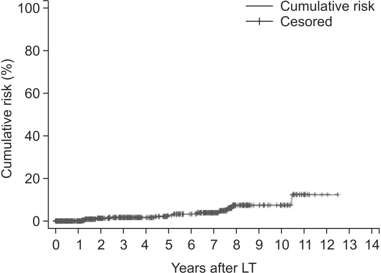
There were 14 patients diagnosed with de novo malignancy after LT and the incidence rate was 3.0%. All the patients who developed de novo malignancy were male. The youngest was 12 years old and the oldest was 70 years old. The median time between liver transplant operation and diagnosis of a de novo malignant tumor was 42 months. The minimum interval was 6 months and the maximum interval was 106 months. The cumulative risk for development of de novo malignancy was 1.6%, 2.7%, and 8.2% at 3, 5, and 10 years after LT, respectively (Fig. 1).
Fig. 1. Cumulative risk of de novo malignancies after liver transplantation (LT).
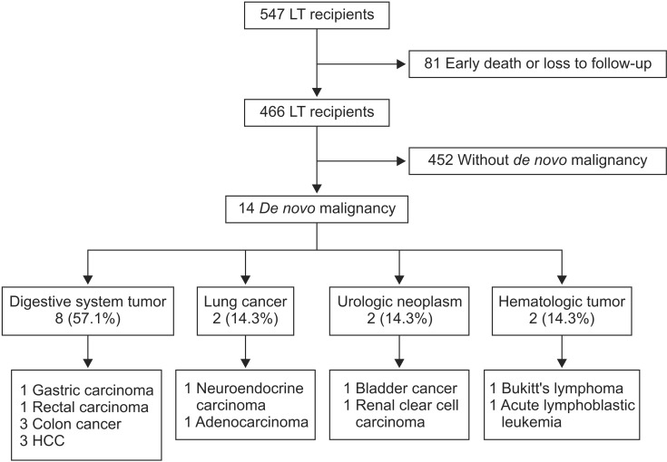
There were 8 digestive system tumors, 2 lung cancers, 2 urologic neoplasms, and 2 hematologic malignant tumors (Fig. 2). Nine patients came to see the doctor for clinical symptoms. Five patients were diagnosed during periodic check-ups. These patients underwent aggressive treatment, including surgery, chemotherapy, and TACE (transhepatic arterial chemotherapy and embolization), except for one patient with an aggressive primary liver cancer. Each patient's details can be visualized in Table 3.
Fig. 2. Clinical characteristics of the study population. LT, liver transplantation; HCC, hepatocellular carcinoma.
Table 3. Demographic and clinicopathologic features of the 14 patients with de novo malignancy.
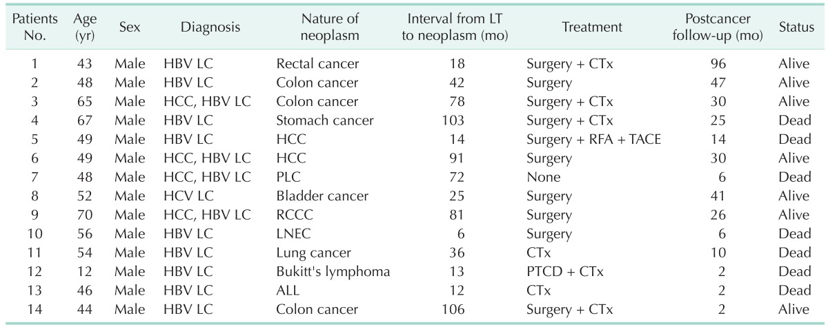
LT, liver transplantation; LC, liver cirrhosis; CTx, chemotherapy; HCC, hepatocellular carcinoma; RFA, radiofrequency ablation; TACE, transhepatic arterial chemotherapy and embolization; PLC, primary liver carcinoma; RCCC, renal clear cell carcinoma; LNEC, lung neuroendocrine carcinoma; PTCD, percutaneous transhepatic cholangial drainage; ALL, acute lymphoblastic leukemia.
During a mean follow-up period of 24±25 months (range, 2 to 96 months) after the diagnosis of de novo malignancy, 7 patients (50.0%) died. Survival analysis showed 1-, 3-, and 5-year survival rates of 62.3%, 54.5%, and 54.5%, respectively.
The development of de novo malignancy has no statistically significant association with recipient age, gender, type of blood, pre-LT HCC, type of graft, induction therapy, type of calmodulin inhibitors (Table 4).
Table 4. Analysis of possible risk factors associated with the development of a de novo malignancy after LT.
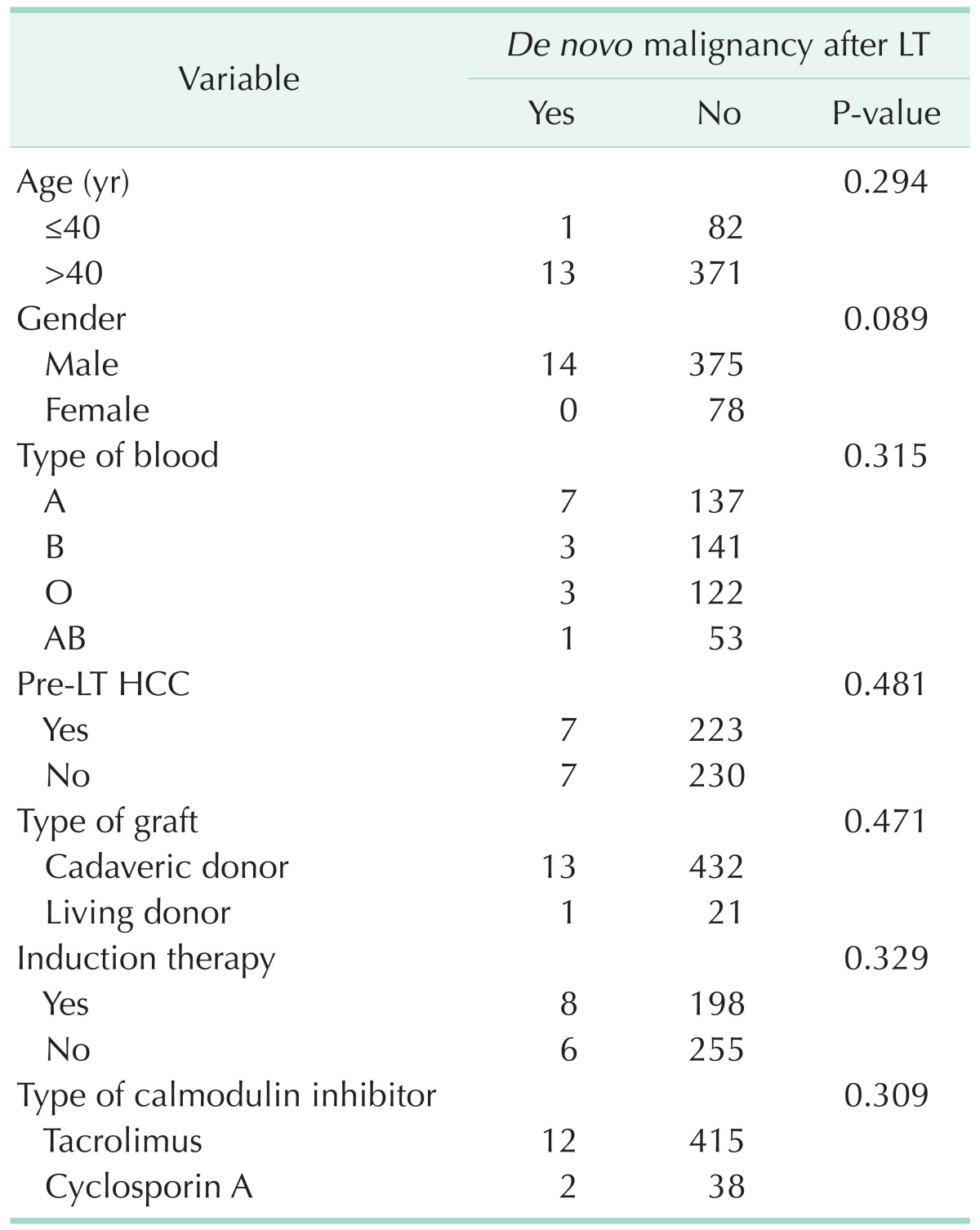
LT, liver transplantation; HCC, hepatocellular carcinoma
The incidence rates of malignancy in LT recipients versus the general Chinese population were summarized in Table 5. The relative risk of malignancy following LT was 9.5 folds higher than the general Chinese population (Table 5).
Table 5. Incidence rates of common malignancies in adult liver transplant patients and the Chinese general population (per 100,000 persons).
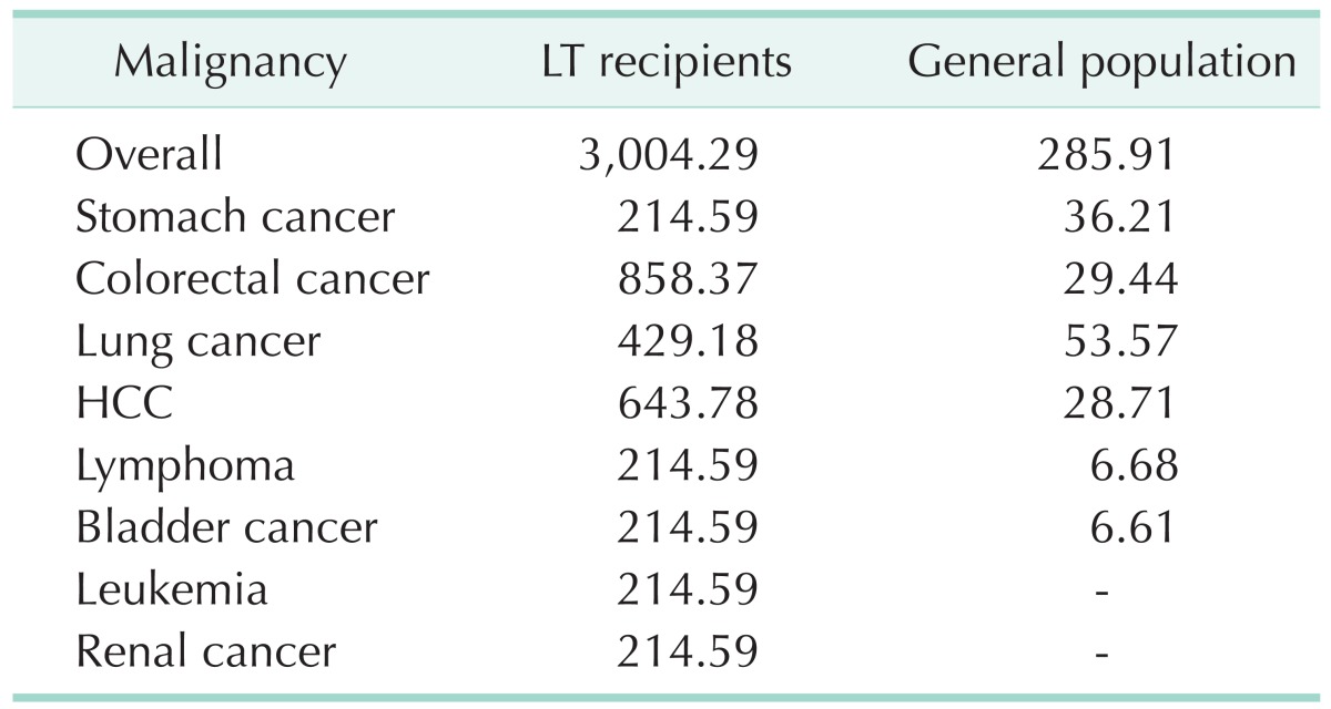
LT, liver transplantation; HCC, hepatocellular carcinoma.
DISCUSSION
As reported, de novo malignancy has been a major cause of death in organ transplantation recipients [17]. The morbidity of de novo malignancy is 1.5% to 15% as reported [18,19]. In China, Zhu et al. [12] reported an incidence rate of 0.9% and Zhang et al. [13] reported an occurrence rate of 0.6%. In our sample, the prevalence rate of de novo malignancy after LT was 3.0% at a mean follow-up of 24 months. Based on the results of this single-center study, the relative risk of overall malignancy following LT was 9.5 folds higher than the general Chinese population.
The cumulative risk for development of de novo malignancy was 1.6%, 2.7%, and 8.2% at 3, 5 and 10 years after LT, respectively. The lower morbidity rate and cumulative rate in our center compared to global levels may be due to the relative lower maintaining concentration of calcineurin inhibitor. In our center, 3 ng/mL is the recommended maintaining concentration of tarcrolimus in long-term survival recipients, which is much lower than the recommended concentration of western countries. No acute rejection was found in long-term survival recipients.
De novo malignancy development after organ transplantation can be influenced by many factors, such as environment, genetics and tumor-associated viral infections. In LT recipients, the immunosuppressant may be the most important risk factor [7,8,19,20]. The application of immunosuppressants successfully prevents rejection and improves survival rates, but in the longrun it places the body in an immunocompromised state (particularly regarding cellular immunity). Cellular mutations are more likely to evade the immune system's surveillance. In order to prevent organ rejection in LT recipients, we chose to minimize the amount of calmodulin inhibitor and withdraw glucocorticoid as soon as possible to reduce the risk of de novo malignancy.
Viral diseases after LT can also induce cancer. As reported in the literature, PTLD were related to Epstein-Barr viral infections, and skin cancers have been related to herpesvirus 8 infections [21,22,23]. In our group, we did not find skin cancer and the patient suffering from Bukitt's lymphoma had no history of Epstein-Barr viral infection.
Peyregne et al. [24] reported there were gender differences for the incidence of de novo malignancy post LT and the incidence in males was significantly higher than that in females. This group of patients had similar results. All the patients diagnosed with de novo malignancy were male, suggesting that gender might indeed be correlated with the occurrence of de novo malignancy. In view of the significantly higher number of male recipients in our sample and the statistic analysis result, the relation between gender and de novo malignancy still requires further analysis.
In western countries, the most common type of de novo malignancy after LTs are skin cancer and PTLD [3,7,8,9,10,11], while solid organ tumors are relatively rare. The occurrence rate of skin cancer is reported as 0.5% to 8.7% [3,11]. In China, most de novo malignancies after kidney transplantation are urologic tumors, and skin cancer and PTLD are rare [25,26]. Our sample mainly included digestive system neoplasms and there was no skin cancer. In view of the incidence of skin cancer in Western countries being significantly higher than that in China and the central importance of recreational sun exposure to the development of skin cancer [27], we thought that genetic differences as well as differences in lifestyle might be the prime causes of the difference in skin cancer occurrence between China and Western countries.
To make a diagnosis of de novo malignancy after LT, we should confirm the diagnosis of malignant tumors and exclude pretransplant lesions and the recurrence of liver cancer. The imaging and pathology results were very useful. In our sample, all the patients were diagnosed by clinical examination and 13 of them received their pathological diagnosis through surgical resection or biopsy.
For the patients with liver cancer before LT, when the liver lesion was found during a follow-up, we tried to identify whether the lesion was a tumor recurrence or a de novo tumor. As reported in the literature, most HCC recurrence occurred in the first 2 years and de novo malignancy was more common in more than 5 years after LT [4]. We thought that the interval between LT and the lesion's diagnosis, the AFP levels before and after LT and the pathological examinations, were useful tools to differentiate the tumors' origin. There was one patient with a high AFP level who received an LT because of HCC. During the first 5 years, there were no signs of recurrence. But in the sixth year, a CT scan revealed a massive HCC, while the serum AFP level remained normal. Because the lesion was not suitable for resection, we did not get a pathological confirmation. Taking the onset time of the tumor and the AFP level into account, we made the diagnosis of de novo liver cancer. The other patient with pretransplant HCC revealed a lesion in the graft after 91 months. Hepatectomy has been done and the pathological examination confirmed the diagnosis of de novo HCC.
As with ordinary tumors, the de novo solid tumor after LT should be removed by operation if there is an opportunity. Reducing the dosage of immunosuppressive agents is an important remedy, which may be useful to improve the antineoplastic immune effect. As reported in the literature, rapamycin has antitumor effects [28]. When the diagnosis of de novo malignancy was confirmed, we could administer rapamycin to the patients to replace tacrolimus or cyclosporine. In our study, there were 10 patients who accepted to undergo surgical resection. Seven patients did not show any sign of recurrence up to now and another 3 patients died of tumor recurrence.
In conclusion, although the cumulative risk of de novo malignancy is lower in our center than that of western countries, the LT recipients had a significantly higher risk of malignancy than the general Chinese population. Digestive system tumor is the most common type of de novo malignancy after LT in China. The onset risk increased with longer survival. Therefore, regular serological and radiographic screening for early diagnosis should be recommended for long-term survival patients. Early treatment might be the only way to improve the prognosis.
ACKNOWLEDGEMENTS
This study was supported by Beijing Medicine Research and Development Fund, No. 20092029; and the Health Industry Scientific Research Fund of China (No. 201002015).
Footnotes
CONFLICTS OF INTEREST: No potential conflict of interest relevant to this article was reported.
References
- 1.Kim JM, Kwon CH, Yun IJ, Lee KW, Yu HC, Suh KS, et al. A multicenter experience with generic mycophenolate mofetil conversion in stable liver transplant recipients. Ann Surg Treat Res. 2014;86:192–198. doi: 10.4174/astr.2014.86.4.192. [DOI] [PMC free article] [PubMed] [Google Scholar]
- 2.Kim JD, Choi DL, Han YS. The paracholedochal vein: a feasible option as portal inflow in living donor liver transplantation. Ann Surg Treat Res. 2014;87:47–50. doi: 10.4174/astr.2014.87.1.47. [DOI] [PMC free article] [PubMed] [Google Scholar]
- 3.Sapisochin G, Bilbao I, Dopazo C, Castells L, Lazaro JL, Rodriguez R, et al. Evolution and management of de novo neoplasm post-liver transplantation: a 20-year experience from a single European centre. Hepatol Int. 2011;5:707–715. doi: 10.1007/s12072-010-9231-1. [DOI] [PMC free article] [PubMed] [Google Scholar]
- 4.Sampaio MS, Cho YW, Qazi Y, Bunnapradist S, Hutchinson IV, Shah T. Posttransplant malignancies in solid organ adult recipients: an analysis of the US National Transplant Database. Transplantation. 2012;94:990–998. doi: 10.1097/TP.0b013e318270bc7b. [DOI] [PubMed] [Google Scholar]
- 5.Hegab B, Khalaf H, Allam N, Azzam A, Al Khail FA, Al-hamoudi W, et al. De novo malignancies after liver transplantation: a single-center experience. Ann Saudi Med. 2012;32:355–358. doi: 10.5144/0256-4947.2012.355. [DOI] [PMC free article] [PubMed] [Google Scholar]
- 6.Park HW, Hwang S, Ahn CS, Kim KH, Moon DB, Ha TY, et al. De novo malignancies after liver transplantation: incidence comparison with the Korean cancer registry. Transplant Proc. 2012;44:802–805. doi: 10.1016/j.transproceed.2012.01.027. [DOI] [PubMed] [Google Scholar]
- 7.Penn I. Occurrence of cancers in immunosuppressed organ transplant recipients. Clin Transpl. 1998:147–158. [PubMed] [Google Scholar]
- 8.Penn I. Post-transplant malignancy: the role of immunosuppression. Drug Saf. 2000;23:101–113. doi: 10.2165/00002018-200023020-00002. [DOI] [PubMed] [Google Scholar]
- 9.Soltys KA, Mazariegos GV, Squires RH, Sindhi RK, Anand R SPLIT Research Group. Late graft loss or death in pediatric liver transplantation: an analysis of the SPLIT database. Am J Transplant. 2007;7:2165–2171. doi: 10.1111/j.1600-6143.2007.01893.x. [DOI] [PubMed] [Google Scholar]
- 10.Tiao GM, Bobey N, Allen S, Nieves N, Alonso M, Bucuvalas J, et al. The current management of hepatoblastoma: a combination of chemotherapy, conventional resection, and liver transplantation. J Pediatr. 2005;146:204–211. doi: 10.1016/j.jpeds.2004.09.011. [DOI] [PubMed] [Google Scholar]
- 11.Belloni-Fortina A, Piaserico S, Bordignon M, Gambato M, Senzolo M, Russo FP, et al. Skin cancer and other cutaneous disorders in liver transplant recipients. Acta Derm Venereol. 2012;92:411–415. doi: 10.2340/00015555-1316. [DOI] [PubMed] [Google Scholar]
- 12.Zhu ZJ, Li L, Zhang YM, Zheng H, Jiang WT, Zhang JJ, et al. The diagnosis and treatment of de novo malignancy after liver transplantion. Chin J Oncol. 2007;29:237–238. [PubMed] [Google Scholar]
- 13.Zhang T, Fu BS, Yi HM, Yi SH, Xu C, Wang GS, et al. The clinical analysis of de novo malignant tumors after liver transplantation: report of four cases. Chin J Organ Transpl. 2010;31:356–359. [Google Scholar]
- 14.Zhang T, Zhang YQ, Fu BS, Yang Y, Cai CJ, Lu MQ, et al. De novo lung cancer after liver transplantation: report of one case and literature review. Organ Transpl. 2010;1:281–286. [Google Scholar]
- 15.Lv Y, Hu LS, Liu C, Yu L, Liu XM, Wang B, et al. Suffering from secondary laryngeal neuroendocrine carcinoma after liver retransplantation, one case report. J Mod Oncol. 2008;16:1210–1211. [Google Scholar]
- 16.Chen W, Zheng R, Zhang S, Zhao P, Li G, Wu L, et al. Report of incidence and mortality in China cancer registries, 2009. Chin J Cancer Res. 2013;25:10–21. doi: 10.3978/j.issn.1000-9604.2012.12.04. [DOI] [PMC free article] [PubMed] [Google Scholar]
- 17.Sheiner PA, Magliocca JF, Bodian CA, Kim-Schluger L, Altaca G, Guarrera JV, et al. Long-term medical complications in patients surviving > or = 5 years after liver transplant. Transplantation. 2000;69:781–789. doi: 10.1097/00007890-200003150-00018. [DOI] [PubMed] [Google Scholar]
- 18.Boin I, Leonardi MI, Stucchi RB, Ataide EC, Almeida JR, Barros RH, et al. De novo posttransplantation nonlymphoproliferative malignancies in liver transplant recipients. Transplant Proc. 2007;39:3284–3286. doi: 10.1016/j.transproceed.2007.07.084. [DOI] [PubMed] [Google Scholar]
- 19.Haagsma EB, Hagens VE, Schaapveld M, van den Berg AP, de Vries EG, Klompmaker IJ, et al. Increased cancer risk after liver transplantation: a populationbased study. J Hepatol. 2001;34:84–91. doi: 10.1016/s0168-8278(00)00077-5. [DOI] [PubMed] [Google Scholar]
- 20.Romero-Vargas ME, Flores-Cortes M, Valera Z, Gomez-Bravo MA, Barrera-Pulido L, Pareja-Ciuro F, et al. Cancers of new appearance in liver transplant recipients: incidence and evolution. Transplant Proc. 2006;38:2508–2510. doi: 10.1016/j.transproceed.2006.08.028. [DOI] [PubMed] [Google Scholar]
- 21.Jain AB, Yee LD, Nalesnik MA, Youk A, Marsh G, Reyes J, et al. Comparative incidence of de novo nonlymphoid malignancies after liver transplantation under tacrolimus using surveillance epidemiologic end result data. Transplantation. 1998;66:1193–1200. doi: 10.1097/00007890-199811150-00014. [DOI] [PubMed] [Google Scholar]
- 22.Guthery SL, Heubi JE, Bucuvalas JC, Gross TG, Ryckman FC, Alonso MH, et al. Determination of risk factors for Epstein-Barr virus-associated posttransplant lymphoproliferative disorder in pediatric liver transplant recipients using objective case ascertainment. Transplantation. 2003;75:987–993. doi: 10.1097/01.TP.0000057244.03192.BD. [DOI] [PubMed] [Google Scholar]
- 23.Euvrard S, Kanitakis J. Skin cancers after liver transplantation: what to do. J Hepatol. 2006;44:27–32. doi: 10.1016/j.jhep.2005.10.010. [DOI] [PubMed] [Google Scholar]
- 24.Peyregne V, Ducerf C, Adham M, de la Roche E, Berthoux N, Bancel B, et al. De novo cancer after orthotopic liver transplantation. Transplant Proc. 1998;30:1484–1485. doi: 10.1016/s0041-1345(98)00326-1. [DOI] [PubMed] [Google Scholar]
- 25.Fei JG, Chen LZ, Zhao JQ, Wang CX, Qiu J, Deng SX, et al. Characteristics and risk factors of malignant tumor in kidney transplantation recipients. China Oncol. 2008;18:223–226. [Google Scholar]
- 26.Fan Y, Shi BY, Chang JY, Bo HW, Wang Z, Ou YJ, et al. Analysis on the occurrence of malignant tumors after kidney transplantation. Med J Chin People Lib Army. 2007;32:529–530. [Google Scholar]
- 27.Terhorst D, Drecoll U, Stockfleth E, Ulrich C. Organ transplant recipients and skin cancer: assessment of risk factors with focus on sun exposure. Br J Dermatol. 2009;161(Suppl 3):85–89. doi: 10.1111/j.1365-2133.2009.09454.x. [DOI] [PubMed] [Google Scholar]
- 28.Campistol JM. Minimizing the risk of posttransplant malignancy. Transplant Proc. 2008;40(10 Suppl):S40–S43. doi: 10.1016/j.transproceed.2008.10.015. [DOI] [PubMed] [Google Scholar]


