

Abstract
In the title complex, [Hg(NCS)2(CH4N2S)2], the HgII atom is four-coordinated having an irregular four-coordinate geometry composed of four thione S atoms of two thiocyanate groups and two thiourea groups. The S—Hg—S angles are 172.02 (9)° for the trans-thiocyanate S atoms and 90.14 (5)° for the cis-thiourea S atoms. The molecular structure is stabilized by an intramolecular N—H⋯S hydrogen bond, which forms an S(6) ring motif. In the crystal, molecules are linked by a number of N—H⋯N and N—H⋯S hydrogen bonds, forming a three-dimensional framework. The first report of the crystal structure of this compound appeared in 1966 [Korczynski (1966 ▸). Rocz. Chem. 40, 547–569] with an extremely high R factor of 17.2%, and no mention of how the data were collected.
Keywords: crystal structure, thiourea, thiocyanate, mercury(II), molecular complex, hydrogen bonding
Related literature
For literature on thiourea- and thiocyanate-based metal–organic crystalline materials and their derivatives, see: Ramesh et al. (2012 ▸); Shihabuddeen Syed et al. (2013 ▸). For the concept of hard and soft acids and bases, see: Ozutsumi et al. (1989 ▸); Bell et al. (2001 ▸). For the crystal structures of similar compounds, see: Nawaz et al. (2010 ▸); Safari et al. (2009 ▸); Shihabuddeen Syed et al. (2013 ▸). For the first report of the crystal structure of the title compound, see: Korczynski (1966 ▸).
Experimental
Crystal data
[Hg(NCS)2(CH4N2S)2]
M r = 468.99
Orthorhombic,

a = 8.5359 (5) Å
b = 9.0337 (5) Å
c = 15.7575 (10) Å
V = 1215.07 (12) Å3
Z = 4
Mo Kα radiation
μ = 13.33 mm−1
T = 293 K
0.20 × 0.20 × 0.15 mm
Data collection
Bruker Kappa APEXII CCD diffractometer
Absorption correction: multi-scan (SADABS; Sheldrick, 2004 ▸) T min = 0.176, T max = 0.240
19798 measured reflections
2397 independent reflections
2158 reflections with I > 2σ(I)
R int = 0.058
Refinement
R[F 2 > 2σ(F 2)] = 0.029
wR(F 2) = 0.075
S = 1.15
2397 reflections
137 parameters
1 restraint
H-atom parameters constrained
Δρmax = 1.44 e Å−3
Δρmin = −1.03 e Å−3
Absolute structure: Flack (1983 ▸), 1149 Freidel pairs.
Absolute structure parameter: 0.034 (12)
Data collection: APEX2 (Bruker, 2004 ▸); cell refinement: APEX2 and SAINT (Bruker, 2004 ▸); data reduction: SAINT and XPREP (Bruker, 2004 ▸); program(s) used to solve structure: SHELXS97 (Sheldrick, 2008 ▸); program(s) used to refine structure: SHELXL97 (Sheldrick, 2015 ▸); molecular graphics: ORTEP-3 for Windows (Farrugia, 2012 ▸) and PLATON (Spek, 2009 ▸); software used to prepare material for publication: WinGX (Farrugia, 2012 ▸) and PLATON.
Supplementary Material
Crystal structure: contains datablock(s) global, I. DOI: 10.1107/S2056989015000584/su5058sup1.cif
Structure factors: contains datablock(s) I. DOI: 10.1107/S2056989015000584/su5058Isup2.hkl
. DOI: 10.1107/S2056989015000584/su5058fig1.tif
A view of the molecular structure of the title complex, with atom labelling. Displacement ellipsoids are drawn at the 50% probability level. The intramolecular N—H⋯S hydrogen bond is shown as a double dashed line (see Table 1 for details).
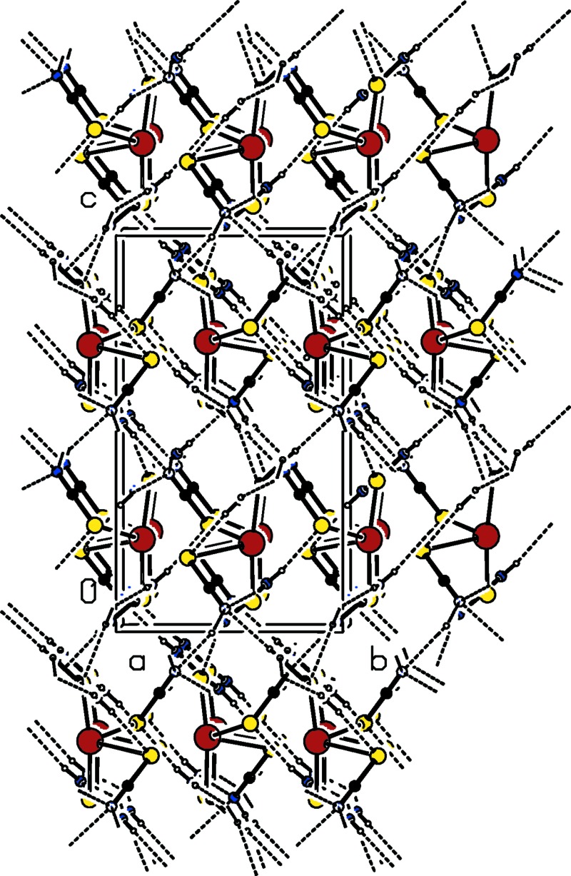
a . DOI: 10.1107/S2056989015000584/su5058fig2.tif
The crystal packing of the title complex, viewed along the a axis. Hydrogen bonds are shown as dashed lines (see Table 1 for details).
CCDC reference: 1043131
Additional supporting information: crystallographic information; 3D view; checkCIF report
Table 1. Hydrogen-bond geometry (, ).
| DHA | DH | HA | D A | DHA |
|---|---|---|---|---|
| N3H3BS1 | 0.86 | 2.55 | 3.404(7) | 174 |
| N3H3AN2i | 0.86 | 2.37 | 3.103(10) | 143 |
| N4H4AN2i | 0.86 | 2.17 | 2.952(10) | 151 |
| N4H4BN1ii | 0.86 | 2.56 | 3.085(11) | 121 |
| N5H5AN1iii | 0.86 | 2.26 | 3.025(10) | 149 |
| N5H5AS4iv | 0.86 | 2.80 | 3.384(7) | 126 |
| N5H5BN2v | 0.86 | 2.21 | 3.025(10) | 158 |
| N6H6AN1iii | 0.86 | 2.25 | 3.019(10) | 149 |
| N6H6BS2vi | 0.86 | 2.56 | 3.419(8) | 172 |
Symmetry codes: (i)  ; (ii)
  ; (iii)
  ; (iv)
  ; (v)
; (v)  ; (vi)
  .
.
Acknowledgments
The authors are grateful to the SAIF, IIT, Madras, India, for the data collection. KR thanks the University Grants Commission, Government of India, for financial support granted under a Major Research Project [F. No. 41–1008/2012 (SR)].
supplementary crystallographic information
S1. Synthesis and crystallization
A mixture of thiourea, ammonium thiocyanate and mercury (II) chloride were dissolved in aqueous solution in the molar ratio 2:2:1 and thoroughly mixed for 1 h to obtain a homogeneous mixture. The solution was allowed to evaporate slowly at ambient temperature. Colourless block-like crystals were obtained in a week.
S2. Refinement
All the H atoms were positioned geometrically with N—H = 0.86 Å and constrained to ride on their parent atoms with Uiso(H) = 1.2Ueq(N).
S3. Comment
This work is part of a research project concerning the investigation of thiourea (N2H4CS) and thiocyanate (SCN) based metal organic crystalline materials and their derivatives (Ramesh et al., 2012; Shihabuddeen Syed et al., 2013). Transition metal thiourea and thiocyanate coordination complexes are candidate materials for device applications including their nonlinear optical properties. As ligands, both thiourea and thiocyanate are interesting due to their potential formation of metal coordination complexes as they exhibit multifunctional coordination modes due to the presence of 'S' and 'N' donor atoms. With reference to the hard and soft acids and bases) concept (Ozutsumi et al., 1989; Bell et al., 2001), the soft cations show a pronounced affinity for coordination with the softer ligands, while hard cations prefer coordination with harder ligands. Several crystallographic reports about mercury(II) complexes usually consist of discrete monomeric molecules with tetrahedral (somewhat distorted) coordination environments around mercury(II) (Nawaz et al., 2010). Herein, we report on the synthesis and crystal structure of the title complex.
The title monomeric complex is composed of two thiocyanate and two thiourea ligands coordinated to the Hg atom via the softer thione S atom (Fig. 1). The four-coordinate mercury atom adopts a severely distorted tetrahedral geometry. The S—Hg—S angles are S3-Hg1-S4 = 172.02 (9) ° for the trans thiocyanate S atoms and S1-Hg1-S2 = 90.14 (5) ° for the trans thiourea S atoms. The bond distances Hg1-S3 and Hg1-S4 are 2.390 (3) and 2.381 (3) Å, respectively, while bond distances Hg1—S1 and Hg1—S2 are 3.064818) and 3.0836 (18) Å, respectively. Bond distances and angles are in agreement with those reported for related compounds (Shihabuddeen Syed et al., 2013; Safari et al., 2009; Nawaz et al., 2010). The SCN moiety is planar [to within 0.007 (1) Å with the C-N and C-S bond lengths corresponding to the values intermediate between single and double bonds. The S2-C2-N2 and S1-C1-N1 units are nearly linear with bond angles of 178.5 (7) and 179.4 (8)°, respectively. The compound is closely related with (thiocyanato-kS)tris(thiourea-kS)mercury(II) chloride (Shihabuddeen Syed et al., 2013). The molecular structure is stabilized by intramolecular an N-H···S hydrogen bond, which forms an S(6) ring motif (Fig. 1 and Table 1).
In the crystal, molecules are connected via N-H···N hydrogen bonds, involving the thiourea NH2 H atoms and the thiocyanate N atom (Fig. 2 and Table 1). This gives rise to the formation of a three-dimensional framework which is reinforced by N-H···S hydrogen bonds (Fig. 2 and Table 1).
The first report of the crystal structure of the title compound appeared in 1966 (Korczynski, 1966) with an extremely high R factor of 17.2 %, and no mention of how the data were collected.
Figures
Fig. 1.

A view of the molecular structure of the title complex, with atom labelling. Displacement ellipsoids are drawn at the 50% probability level. The intramolecular N—H···S hydrogen bond is shown as a double dashed line (see Table 1 for details).
Fig. 2.
The crystal packing of the title complex, viewed along the a axis. Hydrogen bonds are shown as dashed lines (see Table 1 for details).
Crystal data
| [Hg(NCS)2(CH4N2S)2] | F(000) = 872 |
| Mr = 468.99 | Dx = 2.564 Mg m−3 |
| Orthorhombic, Pbc21 | Mo Kα radiation, λ = 0.71073 Å |
| Hall symbol: P 2c -2b | Cell parameters from 2397 reflections |
| a = 8.5359 (5) Å | θ = 2.4–31.2° |
| b = 9.0337 (5) Å | µ = 13.33 mm−1 |
| c = 15.7575 (10) Å | T = 293 K |
| V = 1215.07 (12) Å3 | Block, colourless |
| Z = 4 | 0.20 × 0.20 × 0.15 mm |
Data collection
| Bruker Kappa APEXII CCD diffractometer | 2397 independent reflections |
| Radiation source: fine-focus sealed tube | 2158 reflections with I > 2σ(I) |
| Graphite monochromator | Rint = 0.058 |
| ω and φ scans | θmax = 26.0°, θmin = 2.4° |
| Absorption correction: multi-scan (SADABS; Sheldrick, 2004) | h = −10→10 |
| Tmin = 0.176, Tmax = 0.240 | k = −11→11 |
| 19798 measured reflections | l = −19→19 |
Refinement
| Refinement on F2 | Hydrogen site location: inferred from neighbouring sites |
| Least-squares matrix: full | H-atom parameters constrained |
| R[F2 > 2σ(F2)] = 0.029 | w = 1/[σ2(Fo2) + (0.0237P)2 + 4.6956P] where P = (Fo2 + 2Fc2)/3 |
| wR(F2) = 0.075 | (Δ/σ)max = 0.001 |
| S = 1.15 | Δρmax = 1.44 e Å−3 |
| 2397 reflections | Δρmin = −1.02 e Å−3 |
| 137 parameters | Extinction correction: SHELXL97 (Sheldrick, 2008), Fc*=kFc[1+0.001xFc2λ3/sin(2θ)]-1/4 |
| 1 restraint | Extinction coefficient: 0.0082 (3) |
| Primary atom site location: structure-invariant direct methods | Absolute structure: Flack (1983), 1149 Freidel pairs. |
| Secondary atom site location: difference Fourier map | Absolute structure parameter: 0.034 (12) |
Special details
| Geometry. Bond distances, angles etc. have been calculated using the rounded fractional coordinates. All su's are estimated from the variances of the (full) variance-covariance matrix. The cell esds are taken into account in the estimation of distances, angles and torsion angles |
| Refinement. Refinement of F2 against ALL reflections. The weighted R-factor wR and goodness of fit S are based on F2, conventional R-factors R are based on F, with F set to zero for negative F2. The threshold expression of F2 > σ(F2) is used only for calculating R-factors(gt) etc. and is not relevant to the choice of reflections for refinement. R-factors based on F2 are statistically about twice as large as those based on F, and R- factors based on ALL data will be even larger. |
Fractional atomic coordinates and isotropic or equivalent isotropic displacement parameters (Å2)
| x | y | z | Uiso*/Ueq | ||
| Hg1 | 0.25569 (4) | 0.89378 (3) | 0.72851 (8) | 0.0374 (1) | |
| S1 | 0.4564 (2) | 1.1691 (2) | 0.69569 (13) | 0.0336 (6) | |
| S2 | −0.0283 (2) | 1.0924 (2) | 0.76929 (13) | 0.0334 (6) | |
| S3 | 0.2017 (3) | 0.8900 (3) | 0.57967 (14) | 0.0329 (7) | |
| S4 | 0.3068 (3) | 0.8610 (3) | 0.87589 (15) | 0.0353 (7) | |
| N1 | 0.6053 (9) | 1.0000 (9) | 0.5700 (5) | 0.041 (3) | |
| N2 | 0.0984 (9) | 1.2628 (8) | 0.9017 (5) | 0.037 (3) | |
| N3 | 0.6051 (8) | 0.9346 (7) | 0.8444 (5) | 0.034 (2) | |
| N4 | 0.5630 (8) | 0.7508 (9) | 0.9391 (4) | 0.039 (2) | |
| N5 | −0.0507 (8) | 1.0248 (7) | 0.5262 (4) | 0.031 (2) | |
| N6 | −0.0982 (7) | 0.8404 (8) | 0.6204 (5) | 0.032 (2) | |
| C1 | 0.5444 (9) | 1.0700 (9) | 0.6213 (5) | 0.026 (2) | |
| C2 | 0.0478 (9) | 1.1927 (9) | 0.8463 (5) | 0.025 (2) | |
| C3 | 0.5073 (8) | 0.8485 (9) | 0.8853 (5) | 0.026 (2) | |
| C4 | 0.0009 (9) | 0.9200 (8) | 0.5772 (4) | 0.022 (2) | |
| H3A | 0.70430 | 0.92670 | 0.85300 | 0.0410* | |
| H3B | 0.57020 | 0.99910 | 0.80900 | 0.0410* | |
| H4A | 0.66240 | 0.74400 | 0.94710 | 0.0460* | |
| H4B | 0.50010 | 0.69370 | 0.96640 | 0.0460* | |
| H5A | −0.14960 | 1.04050 | 0.52150 | 0.0370* | |
| H5B | 0.01460 | 1.07740 | 0.49760 | 0.0370* | |
| H6A | −0.19710 | 0.85630 | 0.61570 | 0.0380* | |
| H6B | −0.06470 | 0.77190 | 0.65370 | 0.0380* |
Atomic displacement parameters (Å2)
| U11 | U22 | U33 | U12 | U13 | U23 | |
| Hg1 | 0.0319 (2) | 0.0600 (2) | 0.0205 (2) | −0.0029 (1) | −0.0073 (2) | 0.0032 (3) |
| S1 | 0.0359 (11) | 0.0344 (10) | 0.0304 (10) | −0.0005 (9) | −0.0003 (8) | −0.0009 (9) |
| S2 | 0.0319 (11) | 0.0370 (10) | 0.0314 (11) | 0.0017 (9) | 0.0016 (9) | −0.0026 (9) |
| S3 | 0.0231 (10) | 0.0548 (14) | 0.0209 (11) | 0.0038 (9) | −0.0006 (9) | −0.0002 (9) |
| S4 | 0.0243 (11) | 0.0616 (14) | 0.0200 (11) | 0.0005 (10) | 0.0013 (9) | −0.0005 (10) |
| N1 | 0.035 (4) | 0.051 (5) | 0.038 (4) | 0.000 (4) | 0.003 (4) | −0.005 (4) |
| N2 | 0.039 (4) | 0.035 (4) | 0.037 (5) | 0.002 (3) | −0.002 (3) | 0.002 (3) |
| N3 | 0.026 (4) | 0.033 (4) | 0.044 (4) | −0.001 (3) | −0.005 (3) | 0.011 (3) |
| N4 | 0.041 (4) | 0.044 (4) | 0.031 (4) | 0.010 (3) | −0.005 (3) | 0.009 (3) |
| N5 | 0.031 (4) | 0.031 (4) | 0.030 (4) | 0.000 (3) | −0.003 (3) | 0.006 (3) |
| N6 | 0.026 (3) | 0.036 (4) | 0.033 (4) | −0.001 (3) | −0.007 (3) | 0.007 (3) |
| C1 | 0.024 (4) | 0.032 (4) | 0.022 (4) | −0.004 (3) | −0.004 (3) | 0.008 (3) |
| C2 | 0.025 (4) | 0.036 (4) | 0.015 (4) | 0.003 (3) | 0.005 (3) | 0.011 (3) |
| C3 | 0.028 (4) | 0.031 (4) | 0.019 (4) | 0.008 (3) | −0.002 (3) | −0.010 (3) |
| C4 | 0.028 (4) | 0.026 (4) | 0.012 (4) | 0.001 (3) | −0.005 (3) | −0.001 (3) |
Geometric parameters (Å, º)
| Hg1—S1 | 3.0641 (18) | N4—C3 | 1.313 (11) |
| Hg1—S2 | 3.0836 (18) | N5—C4 | 1.318 (9) |
| Hg1—S3 | 2.390 (3) | N6—C4 | 1.302 (10) |
| Hg1—S4 | 2.381 (3) | N3—H3A | 0.8600 |
| S1—C1 | 1.655 (8) | N3—H3B | 0.8600 |
| S2—C2 | 1.648 (8) | N4—H4A | 0.8600 |
| S3—C4 | 1.736 (8) | N4—H4B | 0.8600 |
| S4—C3 | 1.722 (7) | N5—H5A | 0.8600 |
| N1—C1 | 1.151 (11) | N5—H5B | 0.8600 |
| N2—C2 | 1.162 (11) | N6—H6A | 0.8600 |
| N3—C3 | 1.310 (10) | N6—H6B | 0.8600 |
| S1—Hg1—S2 | 90.14 (5) | C3—N3—H3A | 120.00 |
| S1—Hg1—S3 | 87.35 (8) | C3—N3—H3B | 120.00 |
| S1—Hg1—S4 | 99.39 (8) | H3A—N3—H3B | 120.00 |
| S2—Hg1—S3 | 93.51 (8) | S3—C4—N5 | 117.1 (6) |
| S2—Hg1—S4 | 90.75 (8) | S3—C4—N6 | 122.9 (6) |
| S3—Hg1—S4 | 172.02 (9) | N5—C4—N6 | 119.9 (7) |
| Hg1—S1—C1 | 86.2 (3) | C3—N4—H4A | 120.00 |
| Hg1—S2—C2 | 99.4 (3) | C3—N4—H4B | 120.00 |
| Hg1—S3—C4 | 102.1 (2) | H4A—N4—H4B | 120.00 |
| Hg1—S4—C3 | 105.9 (3) | C4—N5—H5A | 120.00 |
| S1—C1—N1 | 179.4 (8) | C4—N5—H5B | 120.00 |
| S2—C2—N2 | 178.4 (8) | H5A—N5—H5B | 120.00 |
| S4—C3—N3 | 123.5 (6) | C4—N6—H6A | 120.00 |
| S4—C3—N4 | 117.4 (6) | C4—N6—H6B | 120.00 |
| N3—C3—N4 | 119.1 (7) | H6A—N6—H6B | 120.00 |
| S2—Hg1—S1—C1 | −145.2 (3) | S2—Hg1—S3—C4 | −26.2 (3) |
| S3—Hg1—S1—C1 | −51.7 (3) | S1—Hg1—S4—C3 | −55.8 (3) |
| S4—Hg1—S1—C1 | 124.0 (3) | S2—Hg1—S4—C3 | −146.1 (3) |
| S1—Hg1—S2—C2 | −56.6 (3) | Hg1—S3—C4—N6 | −53.0 (7) |
| S3—Hg1—S2—C2 | −144.0 (3) | Hg1—S3—C4—N5 | 129.8 (5) |
| S4—Hg1—S2—C2 | 42.8 (3) | Hg1—S4—C3—N4 | −139.9 (6) |
| S1—Hg1—S3—C4 | −116.2 (3) | Hg1—S4—C3—N3 | 42.8 (8) |
Hydrogen-bond geometry (Å, º)
| D—H···A | D—H | H···A | D···A | D—H···A |
| N3—H3B···S1 | 0.86 | 2.55 | 3.404 (7) | 174 |
| N3—H3A···N2i | 0.86 | 2.37 | 3.103 (10) | 143 |
| N4—H4A···N2i | 0.86 | 2.17 | 2.952 (10) | 151 |
| N4—H4B···N1ii | 0.86 | 2.56 | 3.085 (11) | 121 |
| N5—H5A···N1iii | 0.86 | 2.26 | 3.025 (10) | 149 |
| N5—H5A···S4iv | 0.86 | 2.80 | 3.384 (7) | 126 |
| N5—H5B···N2v | 0.86 | 2.21 | 3.025 (10) | 158 |
| N6—H6A···N1iii | 0.86 | 2.25 | 3.019 (10) | 149 |
| N6—H6B···S2vi | 0.86 | 2.56 | 3.419 (8) | 172 |
Symmetry codes: (i) −x+1, y−1/2, z; (ii) x, −y+3/2, z+1/2; (iii) x−1, y, z; (iv) −x, −y+2, z−1/2; (v) x, −y+5/2, z−1/2; (vi) −x, y−1/2, z.
Footnotes
Supporting information for this paper is available from the IUCr electronic archives (Reference: SU5058).
References
- Bell, N. A., Branston, T. N., Clegg, W., Parker, L., Raper, E. S., Sammon, C. & Constable, C. P. (2001). Inorg. Chim. Acta, 319, 130–136.
- Bruker (2004). APEX2, SAINT and XPREP. Bruker AXS Inc., Madison, Wisconsin, USA.
- Farrugia, L. J. (2012). J. Appl. Cryst. 45, 849–854.
- Flack, H. D. (1983). Acta Cryst. A39, 876–881.
- Korczynski, A. (1966). Rocz. Chem. 40, 547–569.
- Nawaz, S., Sadaf, H., Fettouhi, M., Fazal, A. & Ahmad, S. (2010). Acta Cryst. E66, m952. [DOI] [PMC free article] [PubMed]
- Ozutsumi, K., Takamuku, T., Ishiguro, S. & Ohtaki, H. (1989). Bull. Chem. Soc. Jpn, 62, 1875–1879.
- Ramesh, V., Rajarajan, K., Kumar, K. S., Subashini, A. & NizamMohideen, M. (2012). Acta Cryst. E68, m335–m336. [DOI] [PMC free article] [PubMed]
- Safari, N., Amani, V., Abedi, A., Notash, B. & Ng, S. W. (2009). Acta Cryst. E65, m372. [DOI] [PMC free article] [PubMed]
- Sheldrick, G. M. (2004). SADABS. University of Göttingen, Germany.
- Sheldrick, G. M. (2008). Acta Cryst. A64, 112–122. [DOI] [PubMed]
- Sheldrick, G. M. (2015). Acta Cryst. C71, 3–8.
- Shihabuddeen Syed, A., Rajarajan, K. & NizamMohideen, M. (2013). Acta Cryst. E69, i33. [DOI] [PMC free article] [PubMed]
- Spek, A. L. (2009). Acta Cryst. D65, 148–155. [DOI] [PMC free article] [PubMed]
Associated Data
This section collects any data citations, data availability statements, or supplementary materials included in this article.
Supplementary Materials
Crystal structure: contains datablock(s) global, I. DOI: 10.1107/S2056989015000584/su5058sup1.cif
Structure factors: contains datablock(s) I. DOI: 10.1107/S2056989015000584/su5058Isup2.hkl
. DOI: 10.1107/S2056989015000584/su5058fig1.tif
A view of the molecular structure of the title complex, with atom labelling. Displacement ellipsoids are drawn at the 50% probability level. The intramolecular N—H⋯S hydrogen bond is shown as a double dashed line (see Table 1 for details).
a . DOI: 10.1107/S2056989015000584/su5058fig2.tif
The crystal packing of the title complex, viewed along the a axis. Hydrogen bonds are shown as dashed lines (see Table 1 for details).
CCDC reference: 1043131
Additional supporting information: crystallographic information; 3D view; checkCIF report