. 2015 Feb 20;4:e06074. doi: 10.7554/eLife.06074

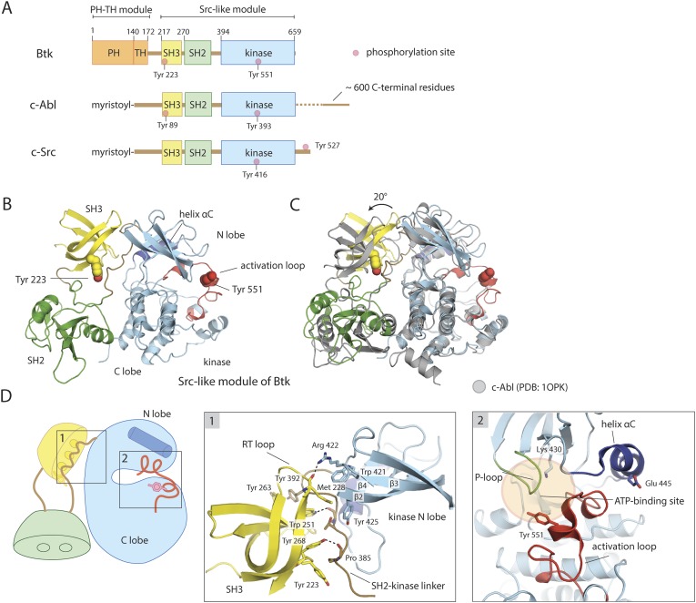
Figure 1. Crystal structure of the Src-like module of Btk.

(A) Domain architectures of Btk, c-Abl, and c-Src. (B) Model for the Src-like module of Btk, based on the crystal structure of the domain-swapped dimer. (C) Comparison of the Src-like modules of Btk and c-Abl (PDB: 1OPK) (Nagar et al., 2003), superimposed on the C lobes of the kinase domains. There is a relative rotation of about 15° for the SH2 domains and 20° for the SH3 domains for the two structures. (D) Details of the SH3/SH2-kinase linker interface and the kinase catalytic cleft in Btk. Left panel: a cluster of hydrophobic residues on the SH3 domain packs against the polyproline type-II helix formed by the SH2-kinase linker. This type of interaction is seen in most SH3-peptide ligand complexes. Right panel: the activation loop of Btk folds into the mouth of the catalytic cleft, blocking part of the ATP-binding cleft. Glu 445, in helix αC, forms an ion pair with Lys 430 in the active conformation but is prevented from doing so in this inactive conformation by the activation loop and helix αC.

DOI: http://dx.doi.org/10.7554/eLife.06074.003

Figure 1—source data 1. Structures of fragments of Tec family kinases in the Protein Data Bank.
elife06074s001.xlsx (45KB, xlsx)
DOI: 10.7554/eLife.06074.017

Figure 1.

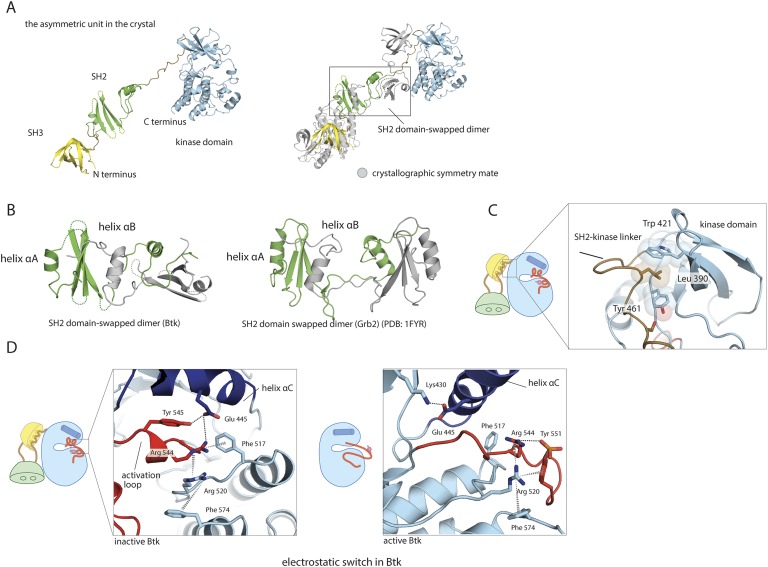
Figure 1—figure supplement 1. Structural details of the Src-like module of Btk.

Figure 1—figure supplement 1.

(A) The Src-like module of Btk forms a domain-swapped dimer in the crystal lattice, with one molecule per asymmetric unit. The SH3 domain and the kinase domain are intact, but the SH2 domain forms a domain-swapped dimer with another molecule. (B) Molecular details of the domain-swapped SH2 dimer in Btk and Grb2 (Schiering et al., 2000). The SH2 domain opens up at a position within the β-sheet, so that helix αB of one polypeptide chain packs against the β-sheet of the other. (C) Interactions between the SH2-kinase linker and the kinase domain. Leu 390 of the linker stabilizes the inactive conformation of the kinase domain by being sandwiched between residues Trp 421 and Tyr 461 in the kinase domain. (D) The ‘electrostatic switch’ in the Btk kinase domain. The active conformation of the Btk kinase domain is modeled from the crystal structure of Lck (PDB: 3LCK) (Yamaguchi and Hendrickson, 1996) using Modeller (Eswar et al., 2006). Activation entails a shuffling of salt-bridge and polar hydrogen-bond partners.