Abstract
During mating, yeast cells must perforate their rigid cell walls at the right place to allow cell–cell fusion. In this issue, Dudin et al. (2015; J. Cell Biol. http://dx.doi.org/jcb.201411124) image mating fission yeast cells with unprecedented spatiotemporal resolution. The authors find that when mating cells come into contact, they form aster-like actin structures that direct cell wall remodeling precisely to the point of contact.
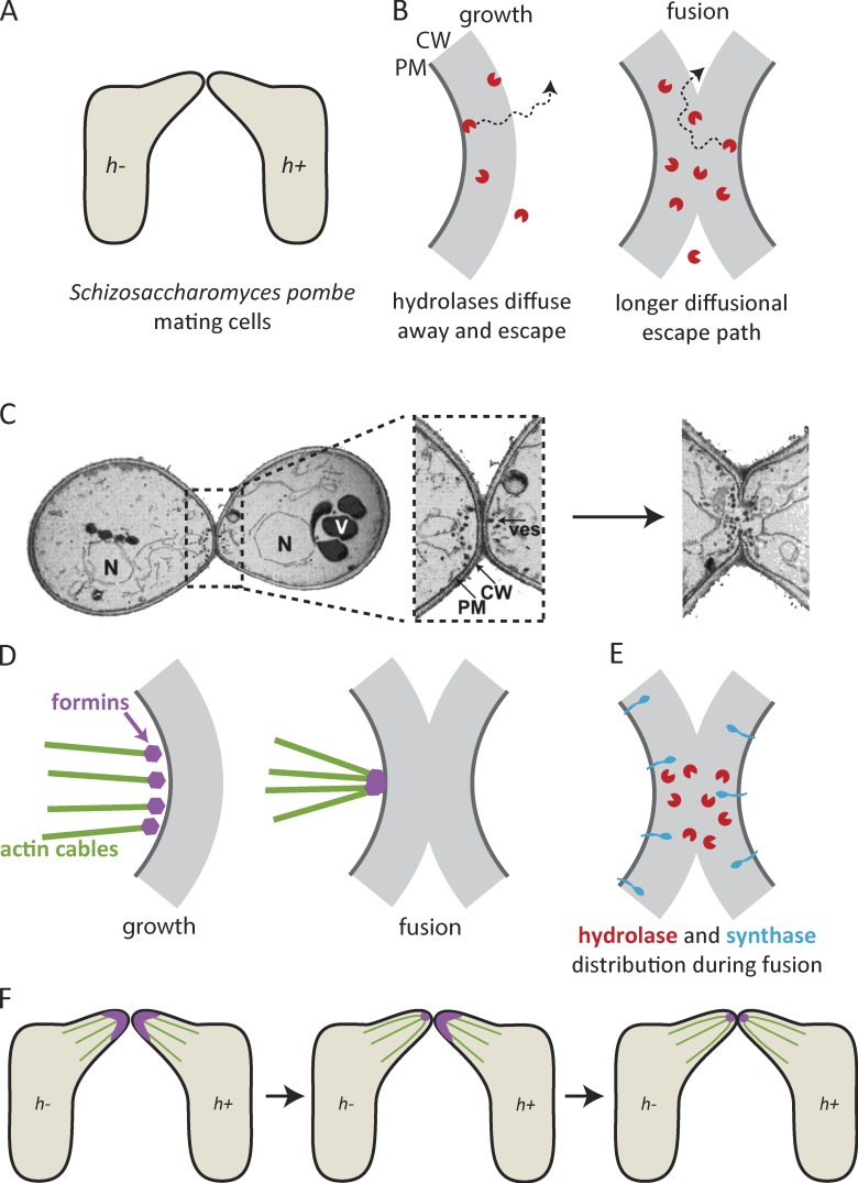
At its core, sex is about the fusion of two haploid cells to form a diploid. For nonmotile cells like yeasts, that requires growth of mating projections to bridge the distance between the mating partners (Fig. 1 A). Yeast cells are protected from osmotic lysis by rigid cell walls, and growth of the mating projection involves local secretion of hydrolases that make the cell wall more elastic at the growing tip (Klis et al., 2006). As the wall expands, new components are added by synthases to maintain a continuous, unbroken wall. The process is orchestrated by a “cell wall integrity” signaling pathway, which monitors cell wall stress and delicately balances hydrolysis and synthesis to guarantee that no holes develop (Levin, 2011). But when it comes to mating, a hole must be made in both partners’ walls at the point of contact to allow cell–cell fusion. Precise positioning is key, as an off-center hole would lead to lysis. How is such precision achieved?
Figure 1.
Cell fusion during yeast mating: focus and communication. (A) Mating fission yeast cells grow projections toward each other and fuse at the point of contact. (B, left) Secreted hydrolases weaken the rigid cell wall to enable expansion, and rapidly diffuse away. (B, right) At a point of cell–cell contact, diffusional escape paths are longer, so hydrolases build up. (C) Focused delivery of secretory vesicles (ves) in mating budding yeast after contact. The image is adapted from Gammie et al. (1998), © The American Society for Cell Biology. (D) Actin cables during growth of the projection (left) and in the fusion focus (right). (E) Distribution of hydrolases and synthases in fusing cells. (F) The fusion focus forms first in the h− mating partner and then in the h+ mating partner. CW, cell wall; PM, plasma membrane; N, nucleus; V, vacuole.
An appealingly simple hypothesis—based on the observation that many hydrolases are secreted enzymes that can only transiently degrade the wall before diffusing away (Fig. 1 B)—is that when the mating projections come into contact, hydrolases from one partner would diffuse into the local wall of the other. Because diffusional escape paths are longer when cells are juxtaposed, hydrolases would be concentrated and make a hole only at the point of contact (Huberman and Murray, 2014). However, this purely geometrical effect cannot be the whole story, as classic genetic studies identified mutants of Saccharomyces cerevisiae that grew mating projections and achieved cell wall contact but failed to degrade the cell wall between mating partners (Kurihara et al., 1994). One set of mutants revealed that fusion requires especially high levels of pheromone secretion, which suggests that mating partners signal to each other to coordinate local wall remodeling (Brizzio et al., 1996). Elegant cytological analyses of another set of mutants have also suggested that vesicles delivering hydrolases are targeted precisely to the site of cell–cell contact (Fig. 1 C; Gammie et al., 1998). These inferences are strongly supported and expanded by a study in this issue (Dudin et al.), which provides a beautifully detailed characterization of mating in the distantly related fission yeast Schizosaccharomyces pombe.
Using time-lapse microscopy and super-resolution imaging to monitor components of the actin cytoskeleton, Dudin et al. (2015) found that actin cables directed myosin V traffic to a broad zone at the tip of the growing mating projection. However, after cell–cell contact, actin cables were tightly focused toward a central “fusion focus” (Fig. 1 D). After focus formation, hydrolases were concentrated in a narrow region, whereas synthases were still distributed broadly (Fig. 1 E). The authors suggest that tightly focused myosin V–mediated delivery of hydrolases overwhelms the local synthases to make a hole in the central cell wall. In the surrounding wall, synthases counteract hydrolases to maintain cell wall integrity.
How does the fusion focus form? A mating-specific formin, Fus1, became tightly localized to a small spot, where it presumably promoted focused actin polymerization and barbed-end anchoring (Dudin et al., 2015). Focus formation could arise from highly focused upstream signaling by formin regulators like Cdc42. Another possibility is suggested by the observation that, as also seen in budding yeast (Sheltzer and Rose, 2009), myosin V was required for focus formation. Thus, one could envision a positive feedback focusing mechanism in which formin-nucleated actin cables enable myosin V–mediated delivery of formins or their activators. Cells in which fusion focus formation was blocked by mutation of Fus1 or myosin V were unable to degrade juxtaposed cell walls and kept growing longer projections, attesting to the importance of the focus in enabling cell wall degradation.
Why does the fusion focus only form upon cell–cell contact? The walls of the mating projections display mating type–specific agglutinins, which help mating partners stick to each other and might conceivably signal that contact has been established. Alternatively, focus formation might be triggered upon perception of a high-threshold pheromone concentration (Brizzio et al., 1996): pheromone levels would rise as the projections approach each other, and might be further increased after contact due to the same geometrical considerations discussed earlier for hydrolases.
Intriguingly, Dudin et al. (2015) found that one of the mating partners, the h− cell, always developed an actin fusion focus before the other, the h+ cell (Fig. 1 F). The basis for this asynchrony is unknown, but if the focus is indeed triggered by a threshold pheromone level, it could be that one pheromone crosses the threshold before the other. The h− cells produce M-factor, whereas h+ cells produce P-factor. If P-factor were to accumulate more rapidly at the contact site, it might reach critical levels and trigger h− cells to make their focus first. The ensuing more focused secretion of M-factor by the h− cell might then trigger and correctly position focus formation by the h+ cell. Whatever the mechanism, the finding that one partner always focuses first makes it attractive to speculate that this asynchrony enables communication between mating partners that allows them to coordinate focus formation directly across from each other.
Acknowledgments
The authors were supported by National Institutes of Health/National Institute of General Medical Sciences grant GM103870.
The authors declare no competing financial interests.
References
- Brizzio V., Gammie A.E., Nijbroek G., Michaelis S., and Rose M.D.. 1996. Cell fusion during yeast mating requires high levels of a-factor mating pheromone. J. Cell Biol. 135:1727–1739 10.1083/jcb.135.6.1727 [DOI] [PMC free article] [PubMed] [Google Scholar]
- Dudin O., Bendezú F.O., Groux R., Laroche T., Seitz A., and Martin S.G.. 2015. A formin-nucleated actin aster concentrates cell wall hydrolases for cell fusion in fission yeast. J. Cell Biol. 10.1083/jcb.201411124 [DOI] [PMC free article] [PubMed] [Google Scholar]
- Gammie A.E., Brizzio V., and Rose M.D.. 1998. Distinct morphological phenotypes of cell fusion mutants. Mol. Biol. Cell. 9:1395–1410 10.1091/mbc.9.6.1395 [DOI] [PMC free article] [PubMed] [Google Scholar]
- Huberman L.B., and Murray A.W.. 2014. A model for cell wall dissolution in mating yeast cells: polarized secretion and restricted diffusion of cell wall remodeling enzymes induces local dissolution. PLoS ONE. 9:e109780 10.1371/journal.pone.0109780 [DOI] [PMC free article] [PubMed] [Google Scholar]
- Klis F.M., Boorsma A., and De Groot P.W.. 2006. Cell wall construction in Saccharomyces cerevisiae. Yeast. 23:185–202 10.1002/yea.1349 [DOI] [PubMed] [Google Scholar]
- Kurihara L.J., Beh C.T., Latterich M., Schekman R., and Rose M.D.. 1994. Nuclear congression and membrane fusion: two distinct events in the yeast karyogamy pathway. J. Cell Biol. 126:911–923 10.1083/jcb.126.4.911 [DOI] [PMC free article] [PubMed] [Google Scholar]
- Levin D.E.2011. Regulation of cell wall biogenesis in Saccharomyces cerevisiae: the cell wall integrity signaling pathway. Genetics. 189:1145–1175 10.1534/genetics.111.128264 [DOI] [PMC free article] [PubMed] [Google Scholar]
- Sheltzer J.M., and Rose M.D.. 2009. The class V myosin Myo2p is required for Fus2p transport and actin polarization during the yeast mating response. Mol. Biol. Cell. 20:2909–2919 10.1091/mbc.E08-09-0923 [DOI] [PMC free article] [PubMed] [Google Scholar]


