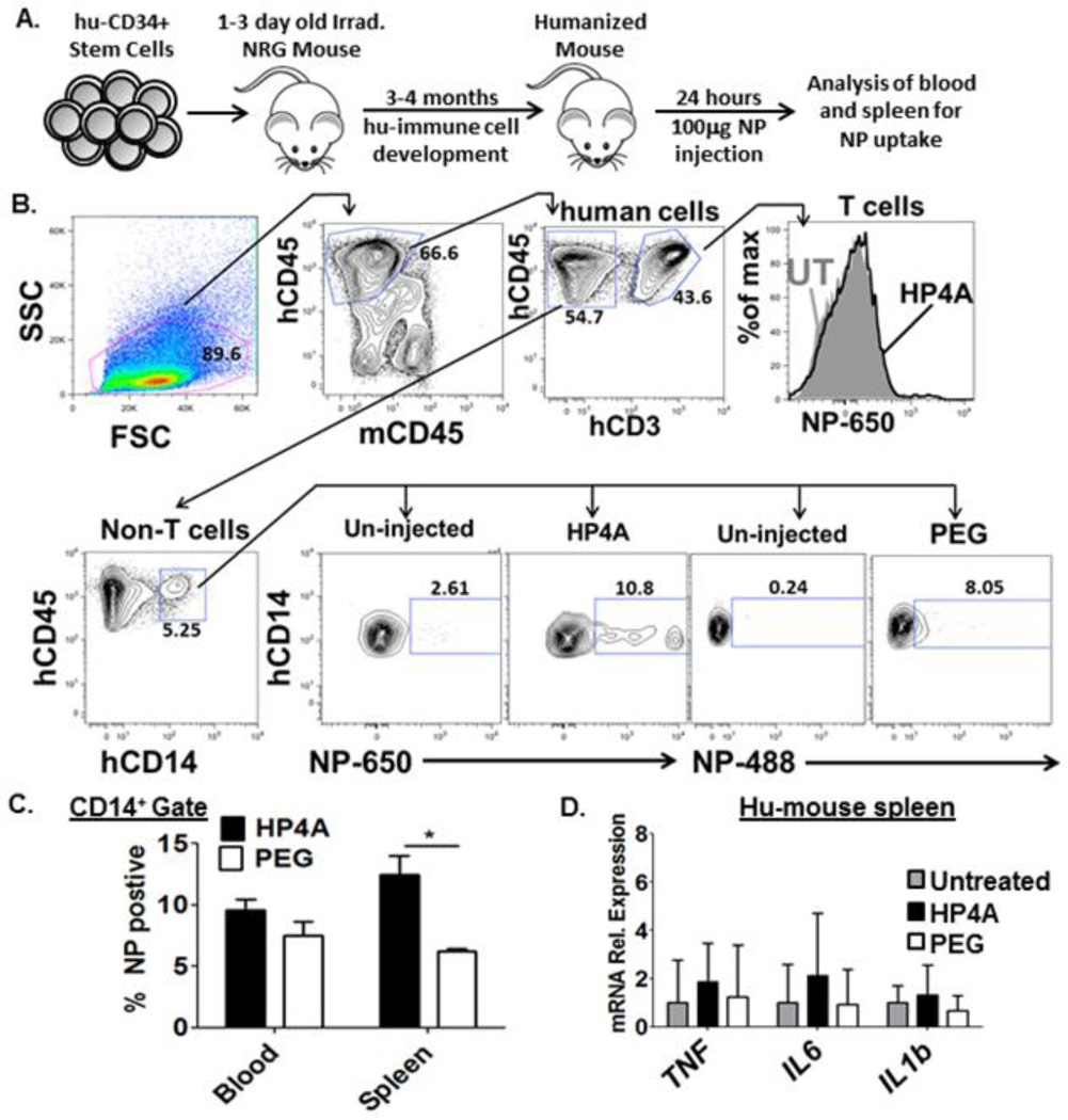
Figure 6. HP4A-based 80×320 nm rod uptake and inflammatory cytokine responses using a humanized mouse model.
(A) Schematic of humanized mouse model. CD34+ cells from hu fetal liver are injected into 1–3 day old irradiated immunodeficient NRG (NOD.Rag1−/−Il2rg−/−) mice. 3–4 months post transplant the mice are injected with NP and human immune cells are analyzed for NP uptake. (B) Gating scheme used to analyze HP4A and HP4A-PEG (PEG) NP uptake by human T cells (anti-human CD45+ CD3+) or monocytes (anti-human CD45+CD14+). Gates for NP+ were set based on un-injected controls. (C) Frequency of NP+ cells in human CD14+ gate from blood and spleen of humanized mice 24 hours after 100 µg NP injection. (D) mRNA expression level of human pro-inflammatory cytokines in spleen of untreated and NP treated humanized mice. n = 4 animals per treatment group. Statistical Analysis by 2-way ANOVA. *P<0.05. Data are representative of two independent experiments.

